Nanomedicine in Ovarian Cancer: Advances in Imaging, Targeted Delivery, and Theranostic Therapeutic Platforms
Dorota Bartusik-Aebisher, Izabella Wilk, David Aebisher

TL;DR
This review explores how nanomedicine is improving ovarian cancer diagnosis and treatment through advanced imaging, targeted drug delivery, and theranostic platforms.
Contribution
The paper provides a comprehensive overview of recent nanomedicine platforms and strategies for ovarian cancer, emphasizing their potential for precision and clinical translation.
Findings
Nanomedicine platforms enhance imaging accuracy and drug delivery in ovarian cancer.
Theranostic systems enable real-time visualization and treatment response monitoring.
Immune-nanomedicine and nucleic acid therapies offer new strategies for overcoming resistance.
Abstract
The major advances in nanomedicine for ovarian cancer are outlined in this review, underlining how engineered lipid, polymeric, inorganic, hybrid and biomimetic platforms improve imaging accuracy, intraperitoneal delivery, and therapeutic precision. Targeted ligands, stimuli-responsive release, and stealth coatings are key design principles which are demonstrated alongside applications in magnetic resonance imaging (MRI), positron emission tomography (PET)/single-photon emission computed tomography (SPECT), near-infrared imaging (NIR), ultrasound, and multimodal imaging. Nucleic acid nanotherapies, photothermal and enzyme-responsive systems, immune-nanomedicine and intraperitoneal depot strategies are also discussed in this article. The importance of standardisation and personalised nanotherapeutic approaches in ovarian cancer is highlighted by the evaluation of biodistribution, safety,…
Genes, proteins, chemicals, diseases, species, mutations and cell lines named across the full text — each resolved to its canonical identifier and authoritative record.
Click any figure to enlarge with its caption.
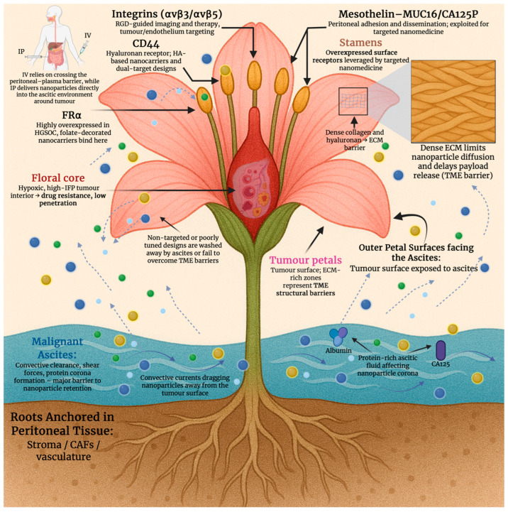 Figure 1
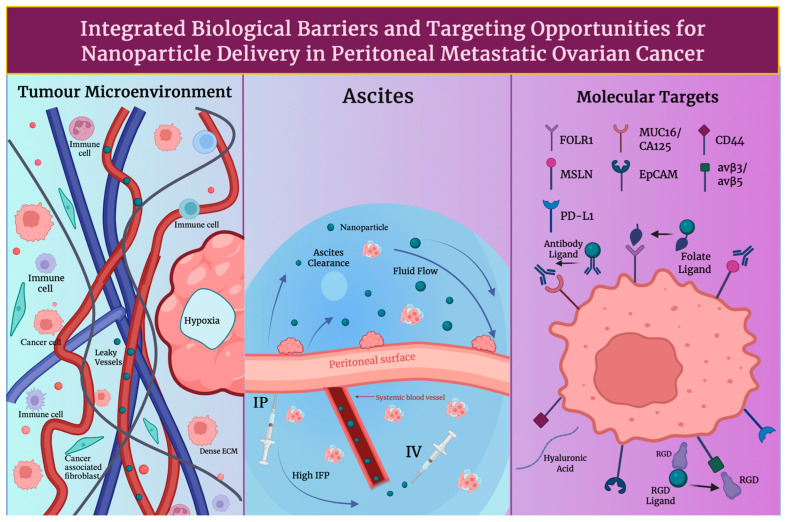 Figure 2
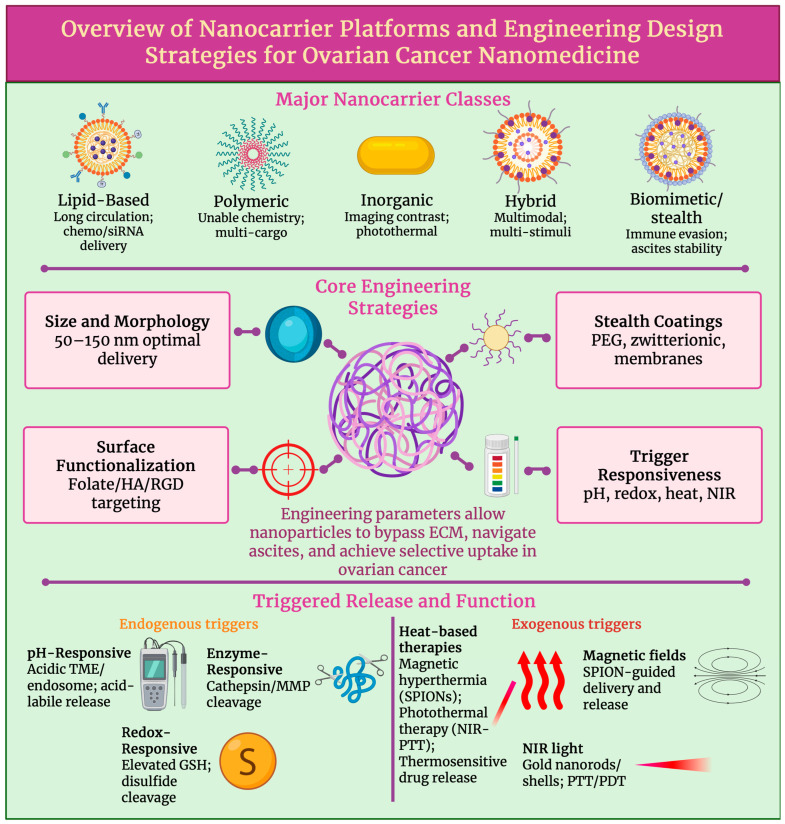 Figure 3
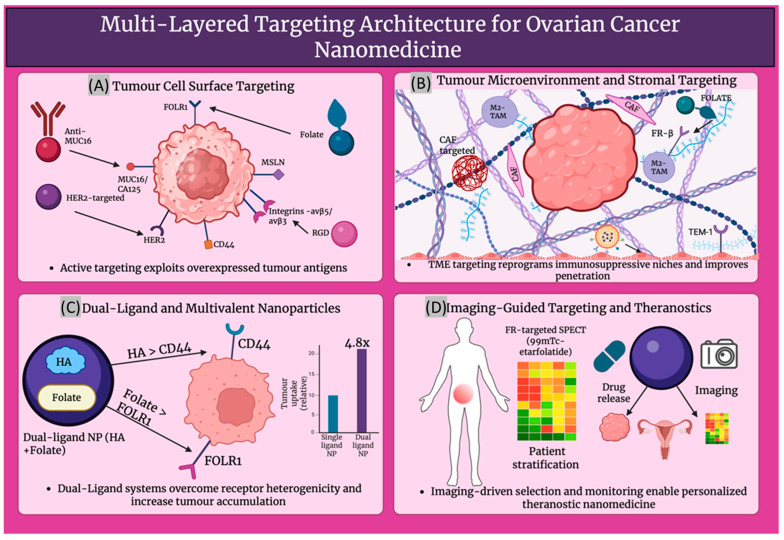 Figure 4
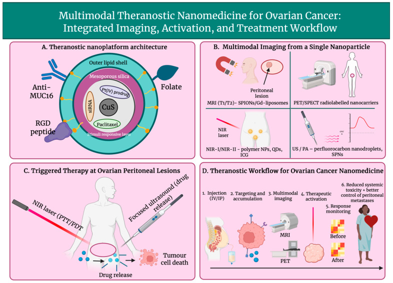 Figure 5
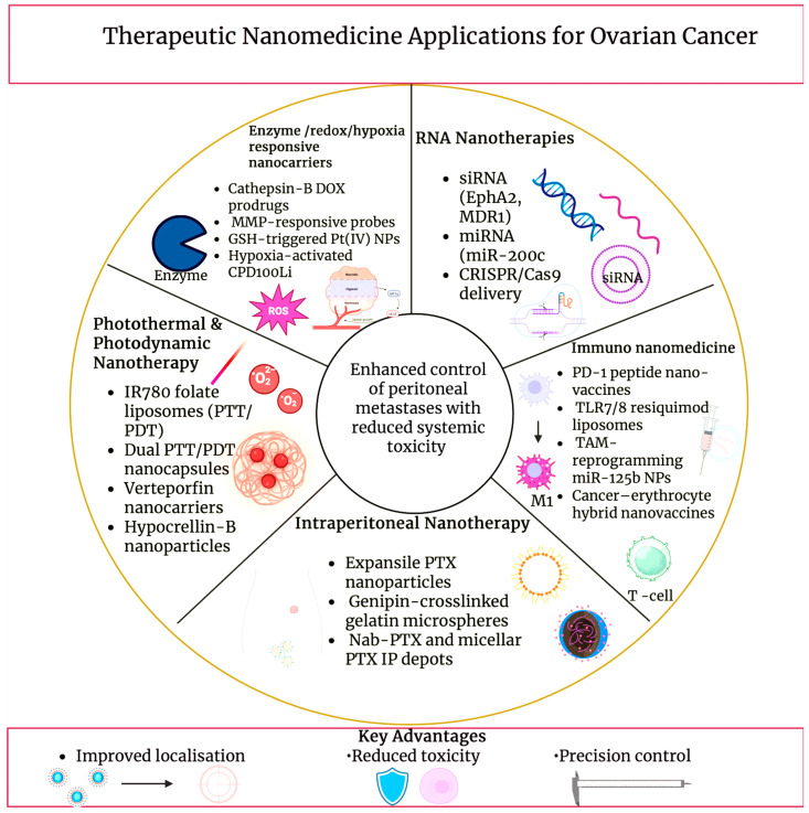 Figure 6
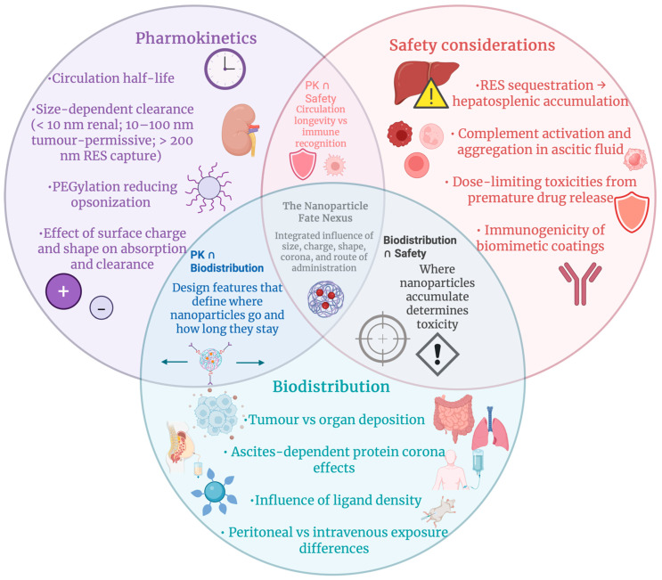 Figure 7
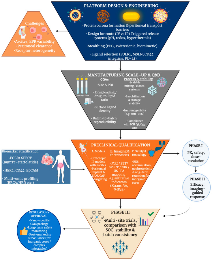 Figure 8
 Figure 9
Peer Reviews
No public reviews on file for this paper yet. If you reviewed it on a platform where reviews are public (OpenReview, ICLR, NeurIPS, ICML), you can paste yours below so the community can read it here.
Videos
No videos yet. Explain this paper in a talk, walkthrough, or lecture? Add one.
Taxonomy
TopicsNanoparticle-Based Drug Delivery · Nanoplatforms for cancer theranostics · Ovarian cancer diagnosis and treatment
1. Introduction
In 2020 ovarian cancer remained a high-mortality disease in Europe, with 39,000 new cases and 27,000 deaths reported, corresponding to an age-standardised incidence rate (ASR) of 15.5/100,000 and an ASR mortality of 10.3/100,000 [1]. Declining mortality trends with a predicted ASR of 4.32 per 100,000 women in 2022, as estimated by subsequent population-based analysis across Europe [2]. Despite these declines, updated estimates based on Global Cancer Observatory (GLOBOCAN) 2022 data show the ovarian cancer burden in Europe to continue to be substantial, with continually high incidence and mortality rates compared to other regions [3]. Ovarian cancer is a primary target for nanomedicine strategies in imaging, targeting and therapy, as these high mortality rates reflect late-stage symptom presentation, with most cases diagnosed at International Federation of Gynaecology and Obstetrics (FIGO) stage III or IV. Peritoneal metastatic spread, typical of HGSOC, and treatment resistance also contribute to this elevated mortality rate [4].
Biomarkers, such as CA-125 and human epididymis protein (HE4), showing low sensitivity and non-specific symptoms in the early stages of disease, remain the primary limitations of early detection of ovarian cancer, despite advances [5]. The window for effective intervention is reduced, as this delay often leads to diagnosis at advanced stages. Around 70% of patients presenting with epithelial ovarian carcinoma relapse following first-line therapy, presenting recurrence to be exceedingly common [6]. Therapeutic effectiveness is concurrently undermined by drug resistance, mediated by mechanisms such as DNA damage repair changes, altered transmembrane drug transport, epigenetic modifications and signalling pathway dysregulation [7]. These barriers pose direct challenges in the context of nanomedicine, as high-sensitivity targeted imaging and biomarker-loaded nanoprobes are demanded for early detection, while resistance and recurrence require nanocarriers that overcome residual disease niches and heterogeneity and are capable of overcoming efflux pumps. Crucial intervention points for imaging, targeting and therapeutic nanomedicine in ovarian cancer are highlighted by the triad of frequent relapse, chemoresistance and late detection [8].
Cytoreductive surgery and platinum-taxane chemotherapy are relied on as the standard treatments of advanced ovarian cancer; although, microscopic peritoneal implants often fail to be completely removed during surgery, and residual disease remains an utmost source of relapse [9]. The development of drug resistance mechanisms, for example, enhanced DNA repair or efflux pumps, in tumour cells, dose-limiting toxicities and non-specific biodistribution hinder systemic chemotherapy [10]. Nanoparticle-based platforms aim to utilise the therapeutic window for nanomedicine by combating these limitations. These platforms intend to penetrate residual microscopic disease, unreachable by standard chemo or surgery, improve tumour-specific delivery, and decrease off-target toxicity [11]. Challenges such as tumour heterogeneity, abnormal peritoneal fluid dynamics, and integration into current surgical/chemotherapeutic workflows, nonetheless, need to be overcome for effective translation of nanomedicine.
The improvement of pharmacokinetics (PK), enabling targeted delivery, and adding real-time imaging are solutions offered by nanomedicine to overcome ovarian cancer’s therapeutic impediments. Reduced cardiotoxicity and sustained activity in relapsed disease can be achieved by pegylated liposomal encapsulation, exemplifying PK gains. Liposomal encapsulation: shrinks volume of distribution, boosts tumour exposure versus free drug and extends circulation half-life [12]. Although recognised limitations of pegylated liposomes, such as the accelerated blood clearance (ABC) phenomenon in particular, counterbalance these benefits following repeated dosing. Additionally, rapid opsonisation and hepatic uptake, which can lead to reduced circulation time, reduced tumour accumulation, and unpredictable therapeutic performance, are promoted by the generation of anti-PEG IgM antibodies. Non-specific biodistribution and dose-limiting toxicity, inherent to conventional chemotherapy, can aim to be overcome by targeting moieties (e.g., peptides, antibodies, and folate derivatives) attached onto nanocarriers, which will concentrate the active compound at ovarian cancer cells or within their microenvironment [13], while imaging-capable nanoparticles allow earlier detection and surgical guidance, beyond delivery. Further, contrast-enhanced platforms for MRI, PET/SPECT, and optical/near-infrared (NIR) modalities, including intraoperative probes designed for peritoneal micrometastases, are catalogued in ovarian cancer-focused reviews [14]. Intra-abdominal tumour visualisation during cytoreductive surgery can be illustrated with preclinical work with erythrocyte-derived NIR nano-constructs in support of the theragnostic concept “see and treat” [15]. Together these advances address three needs: Drugs are kept in circulation and within tumours longer, optimising PK; intratumoral drug-to-healthy-tissue ratios are increased by molecular targeting; and disease is mapped, resection is guided, and response is monitored by longitudinal or real-time imaging. Although clinical translation is still hindered by heterogeneity and peritoneal transport barriers [16], nanomedicine is positioned as a practical complement and, in certain scenarios, an enhancer to systemic chemotherapy and surgery due to the maturation of ovarian-specific nanoplatforms across delivery and diagnostic fronts.
2. Biological and Pathophysiological Context for Nanomedicine Delivery
2.1. Tumour Microenvironment, Peritoneal Dissemination and Ascites
Formidable physical and biochemical barriers to nanomedicine delivery are imposed by the tumour microenvironment (TME) in ovarian cancer. Aberrant, leaky vasculature and inadequate perfusion cause regions of hypoxia that promote drug resistance and limit oxygen-dependent imaging probes by triggering hypoxia-inducible factor 1, alpha subunit (HIF-1a)-mediated changes [17]. Compressed lymphatics and abnormal vessel outflow elevate interstitial fluid pressure (IFP); the extravasation of nanoparticles into the tumour interstitium is further impeded by this, and rather than retention, convective clearance is favoured [18]. Dense stromal architecture, which restricts nanoparticle diffusion and delays payload release, is a consequence of extensive extracellular matrix (ECM) remodelling, as seen in Figure 1. This includes increased collagen deposition, hyaluronic acid accumulation and activation of cancer-associated fibroblasts [19]. Hypoxia, high IFP, and dense ECM must be addressed by nanomedicine design in ovarian cancer. This can be performed by enhancing tumour penetration, imaging contrast, and therapeutic efficiency through the integration of stimuli-responsive release, optimised carrier architecture, and stromal targeting features.
Effective nanomedicine delivery in advanced ovarian cancer is significantly obstructed by peritoneal dissemination and accumulation of malignant ascites. Nanoparticle residence time is reduced, and diffusion into implants is limited by convective clearance generated by tumour nodules bathed in ascitic fluid and seeded on peritoneal surfaces [20]. Nanoparticle attachment and retention on the peritoneal surface is impeded by the fluid-driven shear and movement of ascites. Furthermore, due to hepatic clearance and limited transport across serosal membranes, systemic intravenous delivery faces poor peritoneal penetration [21]. Thus, local concentration can be enhanced by the emergence of intraperitoneal administration of nanoparticles as a strategic route. Rapid drainage and nanoparticle clearance in ascitic fluid, even in this route, require design features such as prolonged retention, large tumoral surface binding, and size optimisation to improve uptake and therapeutic index in disseminated peritoneal disease [22].
2.2. Molecular Targets
Anchors for targeted nanomedicine in EOC are provided by several cell surface biomarkers that are frequently overexpressed. In high-grade serous disease, FOLR1 (FR-α) is enriched, and folate-directed therapeutics and diagnostics are underpinned by it. Subtype distribution is quantified, and assay standardisation needs are highlighted in recent clinic-pathologic studies [23]. While mesothelial adhesion and peritoneal dissemination are mediated by mesothelin (MSLN) binding MUC16/CA125, this is an axis widely leveraged for nanoparticle and antibody/nanobody targeting [24]. Further, epithelial tumour cells are marked by epithelial cell adhesion molecule (EpCAM), and it has been linked to prognosis and chemoresistance. EpCAM is used for EV-based readouts and surface functionalisation with targeting ligands or aptamers [25]. The hyaluronan receptor, CD44, supports hyaluronic acid (HA)-based carriers or dual-target designs, whilst enriching stem-like subpopulations [26]. Whereas RGD-guided imaging/therapy and intraperitoneal targeting in ovarian models are enabled by integrins (avβ3/avβ5) on tumours and antagonistic endothelium [27], as shown in Figure 1. Although its prognostic value is heterogeneous, it is important to mention PD-L1 as a marker variably expressed in EOC. It plays a significant role in immuno-nanomedicine combinations and immune-responsive theranostics [28]. Ligand selection, patient stratification, and payload placement are informed by these biomarkers for imaging, targeting, and therapeutic nanoplatforms in ovarian cancer. Table 1 summarises the key molecular targets seen in ovarian cancer.
2.3. Routes of Administration
The exposure of peritoneal implants is crucially shaped by route selection. The standard route remains as intravenous (IV) therapy, although drug transfer to surface-embedded nodules is limited by the peritoneal plasma barrier and systemic clearance [37], as seen in Figure 2. After IV administration, typically only 0.1–2% of the injected nanomaterial dose accumulates in solid tumours, with noticeable inter-lesional variability. At the peritoneal surface, higher locoregional concentrations can be achieved by intraperitoneal (IP) delivery as a route of administration. IP delivery has also shown survival advantages in meta-analysis and randomised trials [38]. Patient selection and supportive care are informed by common challenges, such as toxicity and abdominal discomfort, as shown by patient-reported outcomes from GOG-172 [39]. However, hyperthermic intraperitoneal chemotherapy (HIPEC) is preferred in a surgical environment; following interval cytoreduction, HIPEC is shown to increase recurrence-free and overall survival in randomised trials. Data from long-term analysis also reinforces its strengths [40]. In primary and certain recurrent diseases, a 2022 meta-analysis also shows HIPEC to be of benefit to patients, while heterogeneity is mentioned in indications and regimens [41]. Long-circulating, stealth carriers with active targeting to reach peritoneal deposits are favoured for IV administration in nanomedicine. Contrastingly, designs that resist ascitic clearance and have the ability to remain stable in mild hyperthermia (41–43 degrees Celsius) are valued in IP/HIPEC [42]. Thus, peritoneal exposure and therapeutic index in ovarian cancer can be improved by prolonged half-life and tumour homing in IV administrative routes and increasing peritoneal surface adhesion, nanoparticle diameter optimisation and thermal stability in IP/HIPEC routes [43].
2.4. Clinical Relevance and Limitations of the EPR Effect in Humans
The enhanced permeability and retention (EPR) effect in humans is real but heterogenous: therefore, rather than being a reliable default, its effectiveness depends on patient-specific and lesion-specific biology, which includes vascular permeability, perfusion and stromal composition. Tumour uptake of nanomedicines has been confirmed by clinical imaging studies, but significant inter- and intra-individual variability is consistently observed [44]. In patients, a 35-fold difference in liposome deposition across metastatic lesions, together with an association between higher deposition and more favourable outcomes, was quantified by PET/CT with ^64^Cu-MM-302 (HER2-targeted pegylated liposomal doxorubicin), which supported the concept of image-guided patient selection [45]. The extent of this variability is particularly relevant in ovarian cancer because the disease often presents with disseminated peritoneal implants showing heterogenous vascularisation and perfusion. Consequently, in poorly perfused lesions, EPR-only platforms may underperform, motivating the integration of active targeting, tumour microenvironment modulation, and theranostic data to personalise delivery strategies [46]. In this regard, nanomedicine strategies in ovarian cancer increasingly pair quantitative imaging modalities (PET/SPECT/MRI) with therapeutic payloads to map nanoparticle deposition, adjust dosing, and combine passive EPR with molecular ligands, such as folate or CD44, or stimuli-responsive release mechanisms [47], and in doing so, overall theranostic performance is significantly improved.
3. Nanocarrier Platforms and Engineering Design
In the context of ovarian cancer, nanocarrier engineering increasingly follows a translational logic in which design choices are evaluated for imaging compatibility, patient stratification, and clinical feasibility, in addition to delivery performance. Several recurring design principles, including particle size control, surface chemistry and stealthing, stimulus-responsive release, and targeting strategy, govern performance in ovarian cancer. While material composition dictates imaging or therapeutic modality compatibility, how these shared parameters are tuned to the peritoneal tumour microenvironment primarily determines translational success.
3.1. Lipid-Based Systems
Polyethylene glycolated (PEGylated) liposomes increase circulation time, decrease cardiotoxicity, and are demonstrated in recurrent ovarian cancer. Pegylated liposomal doxorubicin (PLD), Doxil and Caelyx, showed improved results in platinum-sensitive disease, whilst exceeding topotecan’s effectiveness; PLD carboplatin is also considered an efficient treatment [48]. Intravascular, hyperthermia-induced rapid release with image-guided dosing is enabled by thermosensitive liposomes; lessons acquired from translational research and empirical design guidelines for thermosensitive liposome development are provided in a well-established body of research [49]. Nanocarriers are often manipulated with surface ligands to recognise and bind to molecular biomarkers; for example, folate ligands/antibodies are used to bind to folate receptor α (FAα), a characteristic ovarian cancer molecular marker. Peptides, such as arginine-glycine-aspartic acid (RGD) or aptamers, can be used to increase absorption and facilitate biomarker-guided patient selection [50]. Furthermore, biodistribution and response can be monitored during imaging by PET or NIR probes being co-loaded onto liposomes or lipid nanoparticles (LNPs) [51]. To intercede endosomal escape, ionisable lipids, helper phospholipids, cholesterol, and PEG-lipids are used in LPNs for siRNA and mRNA. Whereas, in ovarian models, dioleoylphosphatidylcholine (DOPC)-based formulation, known as EPHARNA, successfully silenced the EphA2 gene and achieved antitumour activity; here local drug exposure can be maximised by IP administration [52]. Nanocarrier diameter, PEG surface density, TME activators (pH/redox) and administration route have been shown to marginally progress transport across ascites-rich peritoneal environments and to regulate the very variable EPR effect in ovarian cancer models. Although, it is important to mention that these strategies do not uniformly overcome ascites-associated transport barriers, and rather than decreasing EPR, EPR-mediated accumulation aims to be exploited by contemporary nanocarrier designs, where present, while integrating complementary mechanisms, such as intraperitoneal delivery, active targeting, or stimuli-responsive release, in order to account for its heterogenicity and limited reliability in advanced disease [53].
3.2. Polymeric Nanoparticles
Adjustable size, surface chemistry, and stimuli sensitivity for systemic and intraperitoneal delivery are enabled by polymeric nanocarriers, making them fundamental to ovarian cancer nanomedicine. Polylactic-co-glycollic acid (PLGA) nanoparticles provide biodegradability and substantial drug/siRNA co-loading; surface functionalisation, for example, folate or hyaluronan for CD44; as well as peritoneal tumour retention, endosomal escape, and reversion of chemoresistance, for example, HA-PLGA nanoparticles co-loaded with paclitaxel (PTX) and/or small interfering RNA targeting focal adhesion kinase (FAK siRNA) [54,55]. Hydrophobics, such as PTX, can be solubilised by PEG-poly lactic acid (PLA) copolymer micelles. These micelles can also enable dose increase, and they can be modified for pH-induced PEG detachment and endosomal escape. Due to PEG-PLA micellar formulations being clinically established and manufacturable, their translation to ovarian cancer applications is well supported [56]. Poly-amidoamine (PAMAM) is an example of a dendrimer that can offer multivalent, well-defined structures for siRNA/miRNA delivery against resistance mediators, such as twist-related protein 1 (TWIST) and p70S6K. This can be used in combination with PTX to improve antitumour efficiency by being made redox/reactive oxygen species (ROS) reactive for TME activation [57]. Cathepsin or pH-cleavable linkers are used by polymer drug conjugates (PDCs) and antibody polymer drug conjugates for controlled release and can multiplex drugs, for example, gemcitabine and doxorubicin, or they can be used to add immune targeting, such as programmed death ligand 1 (PD-L1). This can lengthen survival in ovarian models and, when paired with imaging labels, supports theranostics [58]. Ascites and peritoneal nodule penetration are improved across platforms targeting moieties, such as FAα, IP delivery and tumour-penetrating peptides. Whilst platinum and taxane resistance is addressed by co-delivery of chemotherapeutic agents with RNA interference (RNAi) or poly (ADP-ribose) polymerase (PARP)/70-kDa ribosomal protein S6 kinase 1 (P70S6K) inhibitors. Design confluence toward compact, stable, and activatable nanocarriers, illustrated in recent PLGA-PTX hybrids and mixed micelles, shows optimisation for ovarian cancer imaging-drug delivery workflows [59].
pH- and redox-responsive release mechanisms are frequently integrated across lipid and polymeric nanocarriers, enabling spatially controlled payload activation in ovarian cancer models, as discussed in Section 3.5.
3.3. Inorganic and Hybrid Systems
Multimodal imaging, targeting, and therapy are allowed by inorganic and hybrid nanoplatforms in ovarian cancer. Simple surface functionalisation for antibody or aptamer targeting is offered by gold nanoparticles, including spheres, rods and shells. Additionally, these nanoparticles provide powerful plasmonics for photoacoustic (PA) or photothermal therapy and drug conjugation; the latest ovarian-focused reviews emphasise progress towards precision oncology and overcoming clinical obstacles [60]. Mesoporous silica nanoparticles (MSNs) supply high capacity and adjustable pores. ‘Wormhole’ MSNs have been successful in achieving TME-targeted theranostics and optoacoustic discovery of ovarian lesions. Further, radioisotope-loaded holmium-166-loaded mesoporous silica nanoparticles (166Ho-MSNs) can show peritoneal metastasis control with restricted off-target dose [61], whereas MRI contrast and magnetic hyperthermia are enabled by iron oxide nanoparticles (IONPs). Heating efficiency can be improved by engineering multicore IONPs and biocompatible nanoclusters, whilst deep stimulation for apoptosis and vascular disturbance can be permitted by alternating magnetic fields [62]. Quantum dots (QDs) can provide a bright and stable fluorescence, useful for intraoperative guidance and selected drug-aptamer conjugates, such as mucin 1, cell surface associated (MUC1) aptamer-doxorubicin, that pair chemotherapy with imaging in ovarian models [63,64]. Lastly, magnetic MSNs, or plasmonic silica shells, are examples of hybrid core-shell designs that can multiplex functions such as loading cytotoxins or siRNA, adding stimuli-sensitive release, and combining PA/MR/fluorescence signals. This supports image-guided therapy and ascites/peritoneal nodule management [65]. The use of antifouling coatings to navigate peritoneal fluid, radiotheranostic agents for intraperitoneal disease, and clinically orientated manufacturing remain as ongoing priorities [66].
3.4. Surface Functionalisation and Stealthing
Suppression of opsonisation, prolonging of circulation time, and improvement of drug delivery to tumours can be achieved by hydrophilic ‘stealth’ coatings in ovarian cancer nanocarrier design. The reason behind this hydrophilic requirement is because a steric and energetic barrier that reduces nonspecific protein absorption and protein corona formation develops when highly hydrated polymer layers bind water molecules. Hydrophilic coatings decrease rapid hepatic and splenic clearance by limiting opsonin binding and subsequent recognition by the mononuclear phagocyte system, and in doing so prolong circulation time and enhance tumour accumulation. PEGylation is the most reliable tool, demonstrating a reduction in protein adsorption and renal clearance. Although the ‘accelerated blood clearance’ effect, as well as anti-PEG antibodies, encourages other options [67]. Carboxybetaines and sulfobetaines are examples of zwitterionic polymers that show elongated circulation, decreased complement stimulation, and sensitive designs appropriate for drugs and genes. Zwitterionic nanoparticles, such as upper critical solution temperature (UCST) type and hyperthermia-activated systems, are used in ovarian cancer models to improve paclitaxel efficiency and retention [68]. In addition, wrapping nanocarrier cores with membranes from platelets, RBCs, leukocytes or sometimes even ovarian cancer cells is called biomimetic cloaking. This process facilitates immune evasion, homotypic binding, and multivalent targeting, ideal for imaging and therapy, by adding ‘self’ markers, such as cluster of differentiation 47 (CD47) and adhesion ligands [69].
Stealthed nanocarriers in combination with epithelial or stromal targets, such as FRα, tumour endothelial marker 1 (TEM1) and luteinising hormone releasing hormone (LHRH), in addition to layer-by-layer polymers, to achieve ‘target and release’, radiation sensitisation and multi-imaging modalities, such as photoacoustics or CT, while alleviating peritoneal clearance, are being used for ovarian cancer [70]. Ovarian cancer nanomedicine reviews emphasise PEG, zwitterion, and membrane-coated platforms that can co-deliver siRNA or chemo, aid image-guided interventions, and strengthen immunotherapy. Key design parameters involve graft density, membrane isolation purity, valency of ligand, and deterioration to balance stealth with tumour penetration and endosomal escape [71].
3.5. Triggered and Controlled Release Mechanisms
Triggered or controlled release designs use tumour-specific cues to achieve site-specific drug release in ovarian cancer. pH, redox and enzymes are considered endogenous triggers. In pH-triggered release designs, acidic TME or endosomes can dissolve acid-labile linkers, or the swelling of ionisable matrices can accelerate release and endosomal escape. Intratumoral deposition and efficiency can be improved by multiple pH-sensitive polymer/lipid systems [72]. Moreover, redox-controlled release designs rely on increased glutathione (GSH) or reactive oxygen species (ROS) to cleave disulphide or thioketal linkers, allowing for on-the-spot drug or siRNA release and combination therapies [73]. Whereas, in enzyme-triggered release designs, cathepsins or matrix metalloproteinases (MMPs) in peritoneal metastases degrade shells or cut peptide linkers during selective unloading [74]. Protein corona-resistant surfaces, such as zwitterionic or antifouling, preserve targeting and release kinetics by maintaining trigger accessibility in ascitic fluid [75].
Stimuli-responsive control is provided by exogenous triggers. In designs that are heat responsive, heat-based treatment is coupled with controlled drug delivery. Heat-based treatment mechanisms extend beyond conventional hyperthermia and include magnetic hyperthermia using superparamagnetic iron oxide nanoparticles (SPIONs), which are exposed to alternating magnetic fields (AMFs); photothermal therapy via NIR-absorbing nanomaterials; and thermosensitive carriers that release payloads above set transition temperatures. Spatially controlled cytotoxicity, heat-enhanced drug diffusion, and synergistic chemo- or phototherapy, particularly relevant for intraperitoneal disease, are enabled by these heat-based strategies. Magnetic field hyperthermia (MFH)-enabled chemotherapy with MRI monitoring in ovarian cancer models [76] and NIR-activated gold nanostructures, which enable spatially precise photothermal ablation or drug release [77], are included in representative examples. Further, carriers that rely on magnetic fields can concentrate drugs and, when combined with AMF, can combine heat-induced release and targeting [78]. Free paclitaxel in ovarian cancer cells is being outperformed by developing UCST zwitterionic nanogels, which can trigger drug release under mild hyperthermia [79].
We can see that precise and on-demand drug release can be achieved by multi-stimuli strategies: by pH, redox and enzymatic (endogenous) and thermal, photonic and magnetic (exogenous) triggers, as shown in Figure 3. This can advance peritoneal retention, response and tumour penetration, while minimising off-target toxicity, which is essential for image-guided, combination treatment in ovarian cancer [80]. Table 2 summarises the key nanocarrier platforms and engineering designs in ovarian cancer nanomedicine.
3.6. Design Scalability and Clinical Translation
Design scalability and clinical translation of ovarian cancer nanocarriers rely on production-ready formulations, such as PLGA and liposomes with modifiable surfaces, size and drug release, while maintaining batch-to-batch reproducibility and pharmacokinetics. In a clinical example, solid tumours were treated with albumin-bound paclitaxel nanoparticles, known as Nab-paclitaxel; this demonstrated that consistent pharmacokinetics and enhanced safety can be achieved by a scalable production process in which nanoparticle size was controlled and albumin coating and reproducible drug loading were involved, therefore acting as a standard for clinical translatability in nanomedicine [88]. Rigorous analytical profiling, for example of hydrodynamic size, polydispersity index (PDI) and drug-to-lipid ratio, correlated with critical quality attributes under the International Council for Harmonisation (ICH) guidelines, specifically Q8, quality by design (QbD), supports regulatory filings and reduces variability [89]. Additionally, standardisation of analytical methods and aligning with developing nanomedicine standards strengthens reproducibility [90]. Opsonisation and off-target toxicity are decreased by biocompatible engineering, such as PEGylation and biodegradable backbones, like PLGA, while allowing tumour localisation in ovarian models [91]. Translation is frequently limited by storage stability; particle integrity and shelf life are preserved by controlled reconstitution and lyophilisation with optimised cryoprotectants. Whereas frozen or lyophilised formats can preserve encapsulation and activity of lipid nanoparticles [92]. Process intensification, which can be performed by inline mixing and the use of closed systems, as well as by authenticated scale-up models connecting minor-scale clinical process parameters (CPPs) to manufacturing-scale batches for scalability, addresses batch-to-batch variation across sites, commonly highlighted in preclinical-to-clinical evaluations [93]. Lastly, regulatory alignment now benefits from clearer guidance globally that is specific to products which are nano-enabled, and complex injectables are more frequently used by regulatory agencies. These are trends which can be seen in recent surveys and overviews in the industry, but they also demand enhanced stability and immunogenicity [94].
Together, these engineering parameters not only control nanocarrier stability and delivery efficiency but also determine compatibility with imaging data, patient stratification strategies, and subsequent clinical translation.
4. Targeting Strategies in Ovarian Cancer Nanomedicine
4.1. Active and TME Targeting Strategies in Ovarian Cancer Nanomedicine
Active targeting in ovarian cancer nanomedicine often makes use of antigens that are evidently overexpressed on tumourous tissue, in comparison to their limited distribution in healthy tissue. On many high-grade serous tumours, folate receptor-α (FRα) is expressed in abundance. FRα has been used to design theranostic nanoemulsions and micelles that can enable MRI monitoring whilst concentrating platinum drugs in peritoneal metastases [95]. When antibody-loaded paclitaxel nanoparticles and anti-MUC16 nanomicelles have been used to target mesothelin and MUC16 (CA125), ovarian cancer targeting ability and tumour uptake were increased, and systemic toxicity was reduced [96,97]. Additionally, non-invasive MRI and optical imaging, with tumour-specific accumulation in human epidermal growth factor receptor 2 (HER2)-positive xenografts, is facilitated by HER2-targeted iron oxide nanoparticles tagged with cisplatin [98]. This concept is extended to circulating or minimal residual disease by EpCAM-directed magnetic particles, which are described in the molecular imaging and contrast agent database (MICAD), as well as by other related imaging probes [99]. Further, siRNA or photosensitisers are delivered to avß3-positive ovarian tumour and endothelial cells by integrin-targeted RGD-labelled nanoparticles, which enable the concurrent attack of tumour cells and angiogenic vasculature [100]. Hyaluronic acid-decorated carriers frequently exploit CD44 to deliver cytotoxins [101] or to deliver MDR1 siRNA selectively to chemoresistant CD44-overexpressing ovarian cancer cells [102]. In addition, complementary TME targeting focuses on tumour-associated macrophages (TAMs), fibroblasts and vasculature. Following accumulation in ovarian TAMs, mRNA or manganese-loaded nanocarriers and large anionic nanoparticles can reprogramme TAMs towards antitumour phenotypes or increase the effectiveness of chemotherapy [103]. On activated macrophages, folate receptor ß is selectively expressed and has been widely used in folate-targeted macrophage nanotherapies, therefore offering a selective entry point for TAM-directed nanomedicine in ovarian cancer [104]. Co-targeting of tumour cells and fibroblasts, remodelling of extracellular matrix and enhancement of immune infiltration in ID8 ovarian models can be achieved by cancer-associated fibroblast mimetic or cancer-associated fibroblast (CAF) hybrid membrane-coated nanoparticles [105]. A route to inhibit angiogenesis is provided by HA-coated siRNA nanocarriers targeting CD44 on tumour endothelium [106]; this integrates vascular, stromal and immune targeting into multifunctional ovarian cancer nanomedicines.
4.2. Overcoming Receptor Heterogeneity Through Dual-Targeted and Theranostic Nanoparticle Platforms
The marked receptor heterogenicity of ovarian cancer is being addressed by increasingly exploited multivalent and dual ligand nanoparticle systems. A significantly higher uptake in SKOV3 ovarian cancer cells and a 4.82-fold higher tumour accumulation in vivo than single ligand control, as shown in Figure 4, was exhibited by dual CD44 and folate receptor-targeted hyaluronic acid-ceramide folic acid nanoparticles, showing synergistic targeting and improved visualisation of intraperitoneal tumours [107]. More broadly, it has been established by dual-ligand gold nanoparticles that multivalent engagement of two receptors can considerably improve cell recognition and differentiation between high- and low-expressing tumour cells, providing conceptual support for dual-receptor nanomedicine in heterogenous ovarian tumours [108]. Targeting is further integrated with imaging data to support image-guided treatment by theranostic platforms. Dual fluorescence and MRI contrast are provided by HER2-targeted gold nanoshell complexes, while NIR photothermal ablation of HER2-overexpressing, drug-resistant OVCAR3 ovarian cancer cells is enabled in vitro [109]. Biomarker-guided companion diagnostics have shown clinical translatability in folate receptor-positive, platinum-resistant ovarian cancer, where patients who are most likely to benefit from the FR-targeted drug conjugate vintafolide are selected using the FR-targeted SPECT tracer ^99m^Tc-etarfolatide [110], demonstrating imaging-based enrichment of nanotherapeutic trials. From a broader molecular profiling viewpoint, the key determinants for guiding personalised, nanoparticle-enabled therapy in ovarian cancer are highlighted as multi-omic profiling of BRCA/HRD, HER2, FOLR1 and other targets [111]. On this basis, Garbuzenko et al. suggest a bank of pre-synthesised, ligand-decorated nanocarriers whose formulation, either containing drugs, siRNAs or targeted peptides, is chosen ex vivo, in accordance with the patient’s tumour gene-protein signature, offering a practical framework for truly personalised ovarian cancer nanomedicine [112]. Table 3 describes the major receptor-ligand axes exploited in ovarian cancer nanomedicine.
5. Imagining and Theranostic Nanomedicine
Imaging nanomedicine in ovarian cancer follows a common theranostic logic across MRI, PET/SPECT, optical, ultrasound, and photoacoustic modalities: nanoparticles are engineered to accumulate at disseminated peritoneal lesions, biodistribution is reported quantitatively or intraoperatively, and where possible triggered therapy is enabled. Therefore, differences between modalities reflect signal physics instead of fundamentally distinct delivery strategies.
5.1. Magnetic Resonance Imaging (MRI)
High-resolution visualisation of ovarian cancer cells within the peritoneal cavity can be enabled by MRI nanoprobes. Strong T_2_ contrast is provided by superparamagnetic iron-oxide nanoparticles (SPIONs), and they can be used for ovarian tumour uptake after engineering. Folate-decorated Fe_3_O_4_ SPIONs generated marked negative contrast in tumours visualised within orthotopic intraperitoneal models. This supported theranostic use by enabling detection of peritoneal implants in preclinical models [125]. In addition to passive contrast, delivery and response by MRI has been simultaneously monitored by theranostic magnetic iron oxide platforms, enabling the detection of differences in accumulation of nanoparticles across heterogenous ovarian lesions and decreased peritoneal metastases in preclinical models. These platforms have also been used to deliver cisplatin to ovarian tumours [98]. Gadolinium (Gd)-labelled liposomes go beyond intravascular residence and intensify reflexivity for T1-weighted, positive-contrast imaging. Vigorous MR signal enhancement, which improved the visualisation of intraperitoneal tumours and complementary NIR signal, was achieved by dual-Gd liposomes co-loaded with indocyanine green (ICG). This enabled multimodal mapping of ovarian cancer, improving detection, treatment monitoring and surgical guidance [126]. Superior tumour MR contrast and adaptability to antibody or peptide functionalisation for ovarian applications have long been demonstrated by Gd-liposomal formulations, often using polychelating amphiphilic polymers, to raise Gd content [127]. Together, SPIONs and Gd-liposomes form the basis of MRI-based mapping of peritoneal metastases and facilitate theranostic workflows that unite lesion localisation with simultaneous delivery and on-treatment monitoring of ovarian cancer.
5.2. Nuclear Imaging (PET/SPECT)
Quantitative, whole-body tracking of the nanoparticle in ovarian cancer is facilitated by radiolabelled nanocarriers for PET and SPECT; this can provide continuous insight into tumour accumulation and circulation and can help spot off-target distribution. Characterisation of pharmacokinetics and optimisation of delivery strategies in nanomedicine have been progressed using liposomes and polymeric nanoparticles tagged with radionuclides such as ^99m^Tc, ^64^Cu or ^89^Zr [51]. Radiolabelled expansile nanoparticles exhibited high peritoneal tumour uptake in intraperitoneal tumour models, imitating advanced ovarian cancer. These nanoparticles approached 30% of the administered dose, and clear distinctions between intravenous and intraperitoneal delivery were revealed. This study highlighted the importance of nuclear imaging for treatment optimisation and biodistribution mapping [128]. Also emerging as theranostic agents are radiolabelled biologics and nanoparticles. PET-centred imaging of ovarian cancer lesions is permitted by ^99^Zr conjugated to anti-Müllerian-inhibiting substance type II receptor (MISRII) antibodies, while combined radionuclide treatment, which enables patient selection and customised dosimetry, is allowed by therapeutic analogues labelled with ^177^Lu or ^213^Bi [129,130]. Radiolabelled nanocarriers are described to function as non-invasive tracers in recent reviews, which state they can display therapeutic nanoparticle biodistribution and monitor response to treatment over time. Therefore, radiolabelled nanocarriers can play a significant role in precision management of ovarian peritoneal metastases [131], which drive most ovarian cancer-related mortality.
5.3. Optical and Near-Infrared (NIR) Imaging
Intraoperative and metastatic localisation in ovarian cancer is increasingly relying on optical and NIR imaging, using NIR-fluorophores and QD nanocarriers. Improved tissue penetration, lowered autofluorescence, and enhanced tumour-to-background contrast can be provided by fluorophores producing emission in the first (NIR-I, 700–900 nm) or the second window (NIR-II, 1000–1700 nm). These features are crucial in enabling the visualisation of the small peritoneal implants, characteristic of advanced ovarian cancer, as shown in Figure 5. For instance, 36 h after injection, a high signal-to-noise detection (S/N > 5) of orthotopic ovarian tumours, local lymph nodes and dispersed peritoneal metastases was enabled by polymer nanoparticles, which emitted in the NIR-II range at 1060 nm. These findings support complete tumour resection in murine models [132]. Coupling with tumour-targeting peptides for fluorescence-guided surgery (FGS), showing submillimetre resolution, is facilitated by multi-functional QDs; they also offer adjustable emission and high quantum yield (38%) [133]. In relation to ovarian carcinoma, NIR-labelled molecular probes, which target G protein-coupled receptors (GnRHR) or folate receptors, have demonstrated the ability to differentiate between peritoneal lesions and neighbouring bowel or muscle. Tumour-to-intestinal ratios of more than 4 have been attainable with these probes, in addition to continued retention for periods of up to 48 h [134]. FGS are already using targeted NIR-fluorophores to help guide cytoreductive surgery of peritoneal carcinomatosis, significantly increasing lesion detection and aiding in the establishment of surgical margins; therefore, their translation into clinical practice is already ongoing [135]. Pairing NIR-fluorophore nanocarriers with therapeutic agents in nanomedicine applications enables concurrent tumour localisation and delivery, hence reinforcing theranostic function, in which visualisation and treatment of disease in the peritoneal cavity are carried out by a single nano-platform.
5.4. Ultrasound and Photoacoustic Imaging
Real-time and non-ionising visualisation of ovarian cancer is offered by ultrasound (US) and PA imaging, and their detection sensitivity and therapeutic effectiveness are decidedly improved by nanomedicine platforms. Typically, acoustic nanodroplets enclose a perfluorocarbon core that, under focused US, can go through acoustic droplet vaporisation, changing into echogenic microbubbles. This effect can promote local drug release while intensifying contrast. Accumulation of the nanodroplets selective to the ovarian tumour and pH-responsive conversion to microbubbles can be demonstrated by folic acid-targeted ferritin nanocages incorporating perfluoropentane (FA-FRT-PFP). Contrast-improved US imaging, together with substantial cytotoxicity, even after only low-intensity focused ultrasound activation, can be achieved using FA-FRT-PFP [136]. Profound intratumoral penetration, ROS-mediated cell killing and US/PA dual imaging that can monitor treatment response ongoingly can be exhibited by dual US-activatable nanodroplets, intended for folate receptor-positive ovarian cancer [137]. In recent studies, nanoparticles integrated with olaparib have been modified for use as ultrasound contrast agents for BRCA1 and BRCA2 (BRCA) proficient ovarian cancer, enabling integrated diagnosis and therapy by pairing PARP inhibitor delivery with contrast-enhanced ultrasound signals [138]. A complementary, completely organic PA platform, defined by strong NIR absorption, great photostability and readily achievable surface functionalisation for precise molecular targeting, can be provided by semiconducting polymer nanoparticles (SPNs) [139]. Strong contrast PA imaging and photoimmunotherapy in orthotopic and metastatic ovarian cancer models has recently been allowed by quinoidal SPNs with NIR-II absorption. This has led to the yielding of strong tumour suppression and inhibition of metastasis, while preserving a clear PA signal generated from deep pelvic lesions [140]. In tandem, dependable separation of exogenous signal from endogenous chromophores, which improved detectability of lesions and helped to guide following therapy, was enabled by a specific spectral signature PA nano-agent, engineered for ovarian cancer [141]. On the whole, activatable US/PA theranostic systems that can map, treat and vigorously monitor ovarian cancer can be established by acoustic nanodroplets and semiconducting polymer nanoparticles.
5.5. Multimodal Imaging Nanoplatforms
Nanosystems designed to carry out two (dual-mode) or three (tri-mode) imaging capabilities within a single construct are called multimodal imaging nanoplatforms. They were developed with the intention of overcoming limitations of traditional imaging of small peritoneal lesions, such as poor sensitivity. Additionally, they were also engineered to incorporate high-resolution structural imaging outputs, like ultrasound (US), CT and MRI, with molecular or functional information that can be provided by imaging modalities such as PA, PET and fluorescence. In a recent review about ovarian cancer nanoparticle imaging agents, the tailoring of platforms for integration into standard imaging modalities, such as fluorescence, US, PA, PET/CT and MRI, is highlighted [14]. Dual-mode anatomical-molecular imaging can be well exemplified by folate-targeted oxygen/indocyanine green lipid nanoparticles (FA-OINPs). By their implementation, both US and PA signals are enhanced in SKOV3 ovarian cancer xenografts, while under NIR laser and US irradiation, photo-sonodynamic and photothermal therapy are provided [142]. PET/CT for quantitative biodistribution and MR temperature imaging is enabled by copper sulphide nanoparticles tagged with ^64^Cu. This facilitates real-time monitoring of photothermal therapy in HeyA8 and SKOV3-ip1 ovarian cancer models, combining PET-based molecular guidance with MR-based thermometry [143]. T1-weighted MRI, fluorescence (FL), and infrared thermal (IRT) imaging in SKOV3 models are provided by biodegradable hollow mesoporous organosilica nanoplatforms (HMON@CuS/Gd_2_O_3_). This enables FL/MRI/IRT-guided mild photothermal therapy that, without systemic toxicity, can destabilise lysosomes and suppress tumour growth [144]. Phase transitional, folate-targeted perfluoropentane nanodroplets, payloaded with Fe_3_O_4_ and 10-hydroxycamptothecin (FA-HCPT-Fe_3_O_4_-PFP NDs), achieved MRI and PA imaging. After low-intensity focused ultrasound stimulated liquid-to-gas transition, a strong ultrasound contrast was generated while concurrently releasing chemotherapeutic agents in mice bearing SKOV3 xenografts. This demonstrated true MRI/PA/US tri-mode theranostics [145]. Lastly, MicroSPECT/CT-based multimodal imaging of SKOV3 xenografts is demonstrated by telodendrimer paclitaxel nanomicelles labelled with ^125^I; this guides drug delivery by quantifying tumour-selective accumulation and pharmacokinetics [146]. Collectively, these platforms show how integrated anatomical-molecular imaging in ovarian cancer nanomedicine encourages precise lesion detection, image-guided intervention, and real-time monitoring of nanotherapy effectiveness.
5.6. Quantitative Imaging Biomarkers
The unifying translational bridge across imaging modalities, enabling nanoparticle accumulation, retention, and therapeutic response to be compared independently of carrier composition, can be provided by quantitative imaging biomarkers. Quantitative imaging biomarkers can achieve this by delivering measurable parameters to track nanocarrier delivery and to foresee or monitor treatment response in ovarian cancer nanomedicine. Nanoparticle studies report increasing amounts of standardised parameters, such as K^trans^ and V_e_, %ID/g and tumour-to-muscle ratios. A notable example is the quantitative dynamic contrast enhanced (DCE)-MRI approach established by Nomani et al. In this study, a nano-sized MRI contrast agent was used in three ovarian cancer tumour xenograft models. Tumours were stratified for responsiveness to nanotherapeutics, and the enhanced permeability and retention (EPR) effect was estimated non-invasively by derived permeability and retention parameters, such as K^trans^ and V_e_ [147]. In relation to PET-based molecular biomarkers, Yang et al. manufactured a ^68^Ga-DOTA-c(NGR)2 probe, which targets CD13 in ovarian cancer xenografts. Tumour uptake as %ID/g and tumour-to-muscle ratios were quantified on microPET. PET-obtained uptake values could be recognised as biomarkers for targeted nanocarrier localisation and candidate therapy selection, due to these metrics correlating with CD13 expression and blocking experiments [148]. Reviews of nanoparticle imaging in ovarian cancer, with a focus on clinical applications, such as by Di Lorenzo et al., describe how radiolabelled liposomes, iron-oxide nanoparticles, and optical nanoprobes have been used in imaging ovarian cancer across various platforms, such as MRI, PET/SPECT, and FL. They emphasise that the quantification of imaging parameters, such as variations in signal intensity, improvement of contrast, and detectability of lesions, which enables nanoparticle distribution monitoring, and the assessment of treatment effects over time, is enabled by these nanoparticle-based approaches [149]. Combined tracking-response biomarkers are exemplified by theranostic radiolabelled liposomes. Serial SPECT/CT-derived pharmacokinetics have been connected to changes in tumour biology, involving drug resistance reversal and stemness markers, in ovarian cancer models and initial clinical work with ^188^Re-liposome. This provided imaging-based metrics of nanotherapy effectiveness [150]. Lastly, quantitative DCE-MRI and diffusion-weighted imaging (DWI) parameters, which have already been authenticated for ovarian tumour characterisation, such as K^trans^ and V_e_, are seen as easily translatable response biomarkers for future nanoparticle-based therapies in ovarian cancer [151]. Notwithstanding notable advances, the clinical translation of theranostic nanomedicine in ovarian cancer continues to be restricted by several limitations. Effective intracellular drug delivery, release of the active therapeutic component, or therapeutic response does not always correlate with imaging-detected nanoparticle accumulation; this creates a central disconnect between signal detection and efficiency. Dose mismatches between tracer-level imaging agents and clinically meaningful drug exposure deepen this challenge, in addition to spatial and temporal discordance, in which imaging reflects early vascular deposition rather than sustained intratumoral retention. Further, the reliability of quantitative imaging biomarkers as predictors of therapeutic response is reduced by pronounced inter-lesional and inter-patient heterogeneity in nanoparticle delivery, retention and release. Lastly, manufacturing and regulatory challenges that restrict batch-to-batch reproducibility and standardisation of imaging signal are introduced by the structural complexity of multimodal theranostic platforms, limiting validation of quantitative biomarkers and clinical translation. Table 4 describes multimodal imaging nanoprobes used in ovarian cancer.
Aside from lesion detection, by enabling in vivo assessment of delivery, patient stratification, and treatment adaptation, quantitative and multimodal imaging serves as a translational bridge between nanocarrier design and clinical application.
6. Therapeutic Nanomedicine Applications
6.1. Nucleic Acid Nanomedicine
The viability of siRNA nanotherapy in ovarian cancer was recognised by neutral nanoliposomes. Vigorous gene silencing decreased tumour growth, and increased paclitaxel efficiency in orthotopic models was produced by EphA2-targeting siRNA formulated in DOPC liposomes. This illustrated RNAi-mediated chemosensitisation [152]. miRNA nanomedicines build on this by aiming to reinstate tumour-suppressive networks.
In ovarian cancer models, the restored expression of miR-200c reduced the extent of metastasis and heightened sensitivity to paclitaxel. From this we can conclude miR-200c is a reasonable payload for nanocarriers that aim to reverse epithelial-mesenchymal transition (EMT) and drug resistance [153]. Additionally, PARP modulation has become nano-enabled. A polymeric formulation of olaparib, called NanoOlaparib, was able to show effective intraperitoneal exposure and tumour control in disseminated peritoneal models, whilst sustaining activity similar to a free drug. This suggested a strategy to treat late-stage disease beyond BRCA-mutant subsets [154]. Platinum resistance can be overcome by complementary multilayered liposomal nanoparticles co-loading cisplatin and a PARP inhibitor with an external hyaluronan coating for CD44; survival in HGSOC models was also improved [155]. A tumour-targeted liposomal system, delivering clustered regularly interspaced short palindromic repeats (CRISPR)/Cas9 plasmids known as CRISPR-enabled nanomedicine, is upcoming. Efficient gene editing and inhibition of growth in ovarian xenografts have been achieved by this system. This provides preclinical demonstration that nanoparticles that are non-viral can support in vivo genome editing for treatment of ovarian cancer [156].
6.2. Targeted Enzyme-Responsive Nanotherapies
Protease overexpression, redox imbalance, and hypoxia in ovarian tumours are made use of in enzyme- and microenvironment-responsive nanotherapies to attain spatially controlled drug activation. Selective enzymatic activation in cathepsin B-overexpressing SKOV3 and HeyA8 cells was shown by cathepsin B-specific doxorubicin prodrug nanoparticles, which were built from FRRG-DOX conjugates. Moreover, significantly improved intraperitoneal inhibition of tumour progression with decreased systemic toxicity, when compared to free doxorubicin in peritoneal carcinomatosis models, was also observed [157]. A complementary protease-activated platform is provided by matrix metalloproteinase-3 (MMP-3)-sensitive near-infrared fluorescent probes. MMP-3 stimulated signal intensification and allowed sensitive stromal imaging in an epithelial ovarian cancer xenograft. This demonstrates that MMP-responsive constructs can be adapted for image-guided nanotherapies [158]. Elevated tumour GSH is exploited by redox-responsive systems. GSH-triggered platinum release, US-mediated enhancement of tumour uptake, and strong antitumour efficiency with weakened nephrotoxicity in SKOV3 models can be achieved by GSH-sensitive Pt (IV) prodrug-loaded phase-transitional nanoparticles with a hybrid lipid-polymer shell [159]. Oxygen-poor peritoneal lesions are targeted by hypoxia-activated systems. A sphingomyelin cholesterol liposome, which encapsulated the vinblastine-N-oxide prodrug CPD100, called CPD100Li, showed more than a 9-fold increase in cytotoxicity in hypoxic conditions in ES2 ovarian cancer cells; hypoxia-activated liposomal chemotherapy for advanced ovarian cancer was further supported by satisfactory pharmacokinetics and tolerability in vivo [160].
6.3. Photothermal and Photodynamic Therapy
Light-activated agents are used to convert optical energy into cytotoxic heat or ROS within ovarian tumours in photothermal and photodynamic nanotherapies. Laser irradiation parameters that have a substantial influence on the therapeutic outcome of photothermal and photodynamic nanotherapies include continuous wave versus pulsed operation, duration of pulse, irradiance, and total fluence. For sustained photothermal heating, continuous-wave NIR lasers are typically used, whereas higher peak power for rapid localised heating or enhanced photodynamic activation while reducing bulk tissue damage can be enabled by pulsed irradiation. To balance effective tumour ablation or ROS generation with minimised off-target thermal injury, careful control of irradiance and fluence is necessary. IR780 is an NIR dye that can act as both a fluorophore and photothermal transducer in folate-targeted liposomal formulations. Intraoperative delineation of tumour margins is enabled by FA-IR780 nanoparticles accumulated selectively in ovarian xenograft models, and following irradiation with an 808 nm laser, the surrounding tissue was spared whilst most lesions were eradicated [161]. Local damage is further intensified by dual photothermal-photodynamic platforms; receptor-mediated uptake, strong singlet-oxygen generation and tumour elimination in ovarian cancer xenograft models, under irradiation of a single NIR laser, were demonstrated by folate-targeted Pluronic chitosan nanocapsules co-loaded with IR780 [162]. Nanocarrier co-loading with clinically used photosensitisers can also advance photodynamic efficiency. Nanostructured lipid carriers with encapsulated verteporfin can enable prolonged circulation, efficient tumour accumulation and significant inhibition of tumour growth, compared to free verteporfin, while evading the severe phototoxic side effects that can be seen with the free drug [163]. Further, PDT in an ovarian cancer ascites model was enhanced by hypocrellin-B-loaded poly(butyl cyanoacrylate) nanoparticles, which prolonged survival time compared to unconjugated drug [164]. Despite this progress, limited penetration of NIR-I light through large tumour masses restrains efficiency, driving development of NIR-II phototheranostic polymers that can more deeply penetrate tissues and could lead to better signal-to-background ratios [165].
6.4. Immuno-Nanomedicine
The goal of immuno-nanomedicine is to transform the immunologically “cold” ovarian TME, which is abundant in M2-polarised TAMs but lacks effector T cells, towards a state that promotes antitumour immunity [166,167]. Only limited responses are shown by clinical checkpoint inhibitors in epithelial ovarian cancer, catalysing the need for nano-enabled strategies that enhance intraperitoneal drug delivery and immune system activation [168]. Gautam et al. designed cowpea mosaic virus nanoparticles showing an anti-programmed cell death protein-1 (PD-1) peptide for immune checkpoint inhibition. Superior tumour control and survival were demonstrated, when compared to free peptide or unconjugated virus, in an ID8 intraperitoneal model. This exemplified that multivalent nanoparticle display can strengthen local PD-1/programmed death-ligand 1 (PD-L1) inhibition, whilst simultaneously limiting systemic exposure [169]. Large anionic liposomes that migrate to peritoneal TAMs and deliver the toll-like receptor (TLR) 7/8 agonist resiquimod were engineered by Kang et al. These liposomes were successful in reprogramming TAMs towards an M1 phenotype, increasing T cell infiltration, as seen in Figure 6, and markedly enhanced the therapeutic effect of anti-PD-1 treatment in syngeneic ovarian tumours [168]. Checkpoint responses can be further amplified by antigen-directed nanovaccines. Cancer-erythrocyte hybrid membrane-camouflaged Fe_3_O_4_-ICG@IRM nanoparticles that accumulate in ID8 tumours were reported by Xiong et al. Immunogenic cell death, tumour antigen release and activation of CD8^+^ T-cell mediated immunity were triggered by NIR photothermal irradiation, demonstrating robust primary tumour inhibition and resistance to tumour rechallenge [170]. The ovarian TME can be directly remodelled by macrophage repolarising nanotherapies. Peritoneal TAMs in an ID8-VEGF model were selectively targeted by hyaluronic acid/polyethylenimine (PEI) nanoparticles encapsulating miR-125b. The TAMs were shifted to an immune-activating phenotype, and when combined with intraperitoneal paclitaxel, a reduction in ascites and VEGF levels was seen, with minimal systemic toxicity [171]. On the whole, these checkpoint-regulating, antigen-delivering and TAM-reprogramming platforms show how immuno-nanomedicine can increase immunotherapy efficiency in ovarian cancer.
6.5. Intraperitoneal Nanotherapy
Direct exposure of disseminated ovarian implants to nanoparticle depots is used in IP therapy to progress peritoneal disease control while minimising systemic exposure. In comparison to traditional IP solutions, nanoparticle systems are designed to remain in the peritoneal cavity, adhere to serosal surfaces and release the drug slowly, maintaining cytotoxic levels along peritoneal tumour implants dispersed across tumour-bearing peritoneal surfaces [172]. Following cytoreductive surgery, intraoperatively administered paclitaxel-loaded expansile nanoparticles notably decreased locoregional recurrence, and survival versus free paclitaxel in ovarian carcinoma models was improved. This demonstrated that intraperitoneally retained nanocarriers strengthen locoregional control of microscopic residual tumours [173]. Radiolabelled expansile nanoparticles emphasise residence time advantages; after a single IP dose, they show tumour-selective accumulation within the peritoneal cavity lasting for up to 1–2 weeks, with negligible signal in distant organs [128]. Likewise, a long-acting intraperitoneal depot was created by paclitaxel-loaded genipin-crosslinked gelatin microspheres. By this formulation, survival was prolonged, peritoneal carcinomatosis scores were lowered, and reduced ascites were seen when compared to short-acting IP controls [174]. High peritoneal-to-plasma gradients are the root of toxicity reduction. Strong inhibition of peritoneal tumour growth, with lymphatic targeting and satisfactory systemic safety, was achieved by early paclitaxel nanoparticles in ovarian models [175]. In recent times, marked antitumour efficiency and survival benefit were produced by IP administration of nanoparticle albumin-bound and micellar paclitaxel in murine ovarian xenografts. Additionally, low systemic exposure was maintained, supporting a better therapeutic index for intraperitoneal nanotherapy in ovarian cancer [176]. Table 5 summarises therapeutic nanomedicine applications in ovarian cancer.
These therapeutic outcomes emphasise how nanocarrier design can enhance efficiency while supporting clinical translation when integrated with imaging-informed patient selection and route optimisation.
7. Pharmacokinetics, Biodistribution and Safety Considerations
7.1. Physicochemical Determinants of Nanoparticle Biodistribution and Clearance in Ovarian Cancer
Biodistribution and clearance in ovarian cancer nanomedicine are dictated by nanoparticle physicochemical properties. In pharmacokinetic studies, extremely small particles, with a diameter of less than 10 nm, are shown to be rapidly cleared renally, while filtration is avoided by systems with particles of a diameter of 10–100 nm, although they are still small enough to cross the vascular border and accumulate in tumours. Nanoparticles with a diameter larger than 200 nm are mostly captured by Kupffer cells in the liver and spleen [177,178]. He et al. showed that size and surface charge act together when he found that tumour uptake is maximised by nanoparticles of 150 nm diameter with a mildly negative zeta potential, while improved opsonisation and reticuloendothelial clearance can be achieved by strongly cationic or highly anionic formulations [179]. Migration, tumour penetration, and cellular internalisation are further moderated by shape, with extended circulation and altered organ tropism, compared with spheres, being exhibited with rod-like or discoidal geometries [180]. An additional bio-interface is introduced by malignant ascites in the peritoneal cavity. It is shown that ovarian cancer ascites is abundant in albumin, complement, coagulation factors and tumour-associated proteins like CA125/MUC16, by proteomic profiling, forming a complex biochemical environment for nanoparticle absorption [181,182]. Nanoparticles quickly obtain an ascites-specific protein corona, as seen in Figure 7, upon IP administration. It was established by Dakwar et al. that widespread aggregation in peritoneal and ascitic fluid can be seen by non-PEGylated cationic and anionic particles, while colloidal stability can be enhanced by PEGylation, although it can also lead to premature release of siRNA, which can reshape local pharmacokinetics [183]. Protein corona composition, as a result, controls recognition by macrophages, complement and NK cells, modulating immune clearance and tumour versus off-target uptake [184]. Therefore, ovarian cancer nanomedicines that prospered in vivo combine diameter (50–150 nm), an almost neutral or slightly negative charge, and stealth coatings with tumour-specific ligands to navigate ascites, make use of helpful coronas, and successfully achieve selective imaging and therapy [14,185].
7.2. Toxicity, Immunogenicity, and Reporting Standards in Ovarian Cancer Nanomedicine
A key part of translating ovarian cancer nanomedicine into the clinic is analysis of acute and chronic safety profiles. Nanoformulation can shift toxicity from acute cardiomyopathy to infusion reactions, palmar-plantar erythrodysesthesia and mucositis, whilst significantly decreasing early-onset heart failure when compared to traditional doxorubicin, as shown in clinical experience with pegylated liposomal doxorubicin (PLD) [186]. However, safety considerations that become more significant with repeated dosing and long-term exposure are introduced by the altered biodistribution and prolonged circulation that underlie these clinical benefits. For instance, it is indicated by long-term follow-ups that at very high lifetime doses, cumulative cardiotoxicity remains a concern, although if cardiac risk factors and function are monitored carefully, PLD can frequently be given without a strict upper limit [187]. Past organ toxicity, immunogenicity can be triggered by nanomedicines. Nano-specific adverse events like complement activation-related pseudoallergy (CARPA) and other infusion reactions that are more frequent with certain liposomal and micellar products than with their standard formulations can be revealed by pharmacovigilance data. For nanomedicine-based products that are intended for use in oncology, including ovarian cancer regimens, structured in vitro and in vivo assessments of cytokine release, complement activation, hypersensitivity and unintended immune suppression or stimulation are advised by immunotoxicology guidelines [188]. Highly important for younger patients is reproductive toxicity, though it is poorly characterised. It is shown by experimental and epidemiological analyses that various nanoparticles can cross placental and blood-testis barriers, disturb spermatogenesis by accumulating in gonads, and disrupt folliculogenesis and embryo development through oxidative stress and DNA damage, therefore supporting rigorous pregnancy exclusion, contraception counselling and prudent dose translation from preclinical models to ovarian cancer trials [189]. Though, commonly used preclinical models often fail to capture the fibrotic, heterogenous, and ascites-rich peritoneal environment that has a critical influence on nanoparticle biodistribution, toxicity, and therapeutic response in patients, limiting their predictive value. Vital for interpreting these risks and their translational relevance is high-quality reporting. The MIRIBEL checklist outlines minimum information for bio-nanomaterial experiments so that toxicity and efficiency in ovarian cancer models can be reproduced and compared. It requires detailed nanoparticle physicochemical characterisation, describing size, charge, corona and stability, dosimetry, biological models and statistics [190]. The ARRIVE 2.0 guidelines order transparent reporting of animal strain, randomisation, blinding, sample size reasoning and humane endpoints to reduce bias and advance translatability for in vivo ovarian cancer nanomedicine studies [191]. These frameworks together reinforce best practice in nano-bio characterisation and safety assessment for imaging and therapeutic applications in ovarian cancer. Table 6 describes pharmacokinetic, biodistribution and safety signatures of representative nanomedicines in ovarian cancer.
8. Clinical Translation and Regulatory Perspective
8.1. Clinical Progress, Therapeutic Outcomes, and Stratification Challenges in Ovarian Cancer Nanomedicine
PLD and nanoparticle taxanes lead clinical nanomedicine trials in ovarian cancer, while RNA-loaded lipid systems remain mostly preclinical. In MITO-2 first-line carboplatin-PLD was at least as effective as carboplatin-paclitaxel, with both showing similar survival, although carboplatin-PLD demonstrated lower neurotoxicity and alopecia, accompanied by increased risk of haematologic adverse effects [192]. In relapse due to platinum resistance, PLD used alone, again with a distinct toxicity profile, was established to be no less effective than gemcitabine or topotecan in phase III trials [193]. PLD has been fixed in combinations with trabectedin and/or bevacizumab. When carboplatin-gemcitabine-bevacizumab was compared against carboplatin-PLD-bevacizumab, better outcomes were seen with the latter in platinum-eligible recurrence with acceptable safety [194], and trabectedin-PLD is supported as a substitute option by phase III data [195]. In refractory disease, nanoparticle albumin-bound paclitaxel (nab-PTX) has shown notable standalone activity [196], and due to reports from phase I studies showing advantageous pharmacokinetics and controllable dose-limiting toxicities, as seen in Figure 8, it is being repositioned for intraperitoneal or aerosolised delivery (PIPAC) in peritoneal metastases [197,198]. Clinical translation of siRNA nanomedicine is at earlier stages of advancement. Though ongoing preclinical evaluation is supported by DOPC-based EphA2-siRNA liposomes (EPHARNA) completing good laboratory practice (GLP) safety studies in mice and other non-human primates [199]. Vigorous prolongation of survival in orthotopic models, but no late-phase human data yet, is shown by multiple lipid and hybrid LNP platforms, which target PLK1/elF3c, Parp1 or CD44 positive cells [200,201]. Instead of radically transforming outcomes, generally, nanomedicines modestly prolong progression-free survival and improve tolerability. A lack of prospective stratification by target expression, such as FOLR1, EphA2 and CD44, along with integration of companion imaging or biopsies being limited, leads to ‘nanotargets’ being rarely used to select patients for specific nanomedicine treatments; this signifies a central limitation and represents an area of improvement in upcoming ovarian cancer nanomedicine trials.
8.2. Manufacturing, Stability and Regulatory Requirements for Ovarian Cancer Nanomedicines
Strict chemistry, manufacturing, and control (CMC) information is required for manufacturing nanomedicines for ovarian cancer. Tight control of significant quality attributes, such as size, polydispersity and surface charge, is required for complex structures such as liposomes, polymeric nanoparticles and micelles, to guarantee reproducibility between batches and foreseeable pharmacokinetics, as stated in reviews of translational applications of nanomedicine [202]. Small differences in mixing, exchange of solvents or lyophilisation can change ovarian cancer formulations such as pegylated liposomal doxorubicin [203] or nanocomplexes loaded with cisplatin [204]. Parenteral nanomedicines have to be sterile; however, destabilisation of nanocarriers may occur through thermal or radiation sterilisation. Consequently, cost is often increased by aseptic processing and sterile filtration being required, which demands robust stability profiles over storage and after reconstitution [205]. Aggregation, drug leakage and matrix oxidation are features of long-term physiochemical stability that hinder ovarian cancer nanoformulations from entering clinical trials [206]. Together with complex analytics, these CMC and stability requirements raise manufacturing costs, which limit access regardless of clinical benefit in recurrent disease [207]. Furthermore, the clinical progression of complex, multi-element nanomedicines is significantly limited by these constraints, as reliable, reproducible nano-formulations with well-defined critical quality attributes are favoured for large-scale production and regulatory approval. Ovarian nanomedicine regulatory frameworks build on general drug regulations but also include specific FDA and EMA expectations for nanomedicines. The need for thorough characterisation, risk-based EMC strategies and post-marketing surveillance is emphasised by FDA- and EMA-focused reviews, which reference nanotechnology guidance and EMA position papers on liposomes and other nanoproducts [208]. Regulators are more frequently expecting co-validation of imaging or soluble biomarkers, like CA-125 and emerging multiplex panels, for theranostic ovarian platforms [209], evaluated in conjunction with the nanomedicine consistent with the broader shift toward imaging-based companion diagnostics and biomarker-guided theranostics in ovarian cancer [210]. Table 7 summarises the clinical translation of ovarian cancer nanomedicines.
9. Future Perspectives and Challenges
The integration of multi-omics profiling, such as genomics, transcriptomics and proteomics, with AI-based modelling is being increasingly relied on in ovarian cancer precision nanomedicine to stratify patients, predict drug response and direct nanocarrier design. Signatures associated with early detection and prognosis of ovarian cancer can be identified through AI frameworks trained on molecular and imaging biomarkers, although their clinical use is hindered by small, heterogenous datasets and a lack of standardised image and omics workflows [211]. In theory, these models could be coupled to nanoparticle libraries, and based on individual tumour profiles and longitudinal radiomics data, tailored ligand combinations, payloads and dosing schedules could be selected for each patient. At the centre of “imaging-targeting-therapy” concepts in ovarian cancer are multifunctional theranostic platforms; for example, a comprehensive review about imaging and therapy of ovarian cancer: Clinical applications of nanoparticles describe that nanotheranostic nanoparticles, which integrate imaging and drug delivery, function in a single formulation and successfully monitor drug distribution profiles and pathological processes longitudinally in ovarian cancer patients, thus offering a promising approach for diagnostics and therapy [149]. For instance, CA125-targeted radiolabelled antibodies, folate-decorated liposomes and photodynamic nanoparticles for ovarian cancer. Plasmodic and exosome-based sensors are examples of emerging nanotechnologies for early detection that are being adapted to recognise proteins specific to ovarian cancer and nucleic acids in blood, urine or ascites [212]. Due to HGSOC being “immune cold”, TME and immune modulation have developed into essential design criteria for nanomedicine. Kaur et al. describe nanoparticles that reprogram TAMs, deliver TLR agonists like resiquimod to peritoneal TAMs, boost infiltration of CD8^+^ T-cells and synergise with PD-1/PD-L1 blockade [213]. Comparable future strategies also apply to cytokine-loaded platforms, such as IL-12 nanoparticles, and redox- or pH-sensitive micelles that exploit hypoxia, acidosis or high GSH levels in the peritoneal cavity to stimulate localised drug release and turn “cold” lesions into “hot” ones, as seen in Figure 9. Balanced adjustment of size, charge and stiffness to favour intraperitoneal retention and stromal penetration is highlighted in reviews of TME-responsive nanomedicines [214,215].
However, clinical translation is slowed down by inconsistent regulatory frameworks and non-standardised datasets. Overall, in nanomedicine, the lack of reliable definitions, incomplete physiochemical characterisation and unpredictable safety endpoints across authorities are highlighted by regulators [216]. Coordinated international standards bodies and the European Medicines Agency (ISO/CEN-EMA) efforts, common test panels for nanoparticle identity, stability and interoperable data formats which link preclinical, imaging and clinical outcome data, are called for in a proposed standardisation roadmap [217]. Simultaneously, before AI-based decision tools for ovarian cancer can guide regulatory-grade precision nanotherapy, they must meet developing requirements for transparency, bias control and external validation [211].
Ovarian cancer nanomedicine could be redesigned by several emerging technologies. Scalable, exosome-like carriers with inherent tumour tropism can be offered by immune cell-derived exosome mimetics (IDEM), which can deliver small RNAs or drugs to resistant cells in ovarian models and could overcome some manufacturing obstacles of natural exosomes [218]. Enzyme- and pH-responsive platforms for drug and immunomodulator delivery can be demonstrated by self-assembling peptide nanostructures that can also be used as 3D matrices that replicate the multicellular ovarian TME for screening nanotherapeutics [219]. Lastly, strong imaging contrast or photothermal properties with tuneable clearance are combined by biodegradable metallic nanocarriers, such as gold or zinc oxide systems, which are engineered for controlled dissolution. This signifies a promising direction for safer theranostic platforms in ovarian cancer [220].
10. Conclusions
The diagnostic and therapeutic landscape of ovarian cancer is being rapidly reshaped by nanomedicine, presenting practical solutions to persistent challenges such as late detection, drug resistance, peritoneal dissemination, and dose-limiting toxicity. Nanoparticles reliably demonstrate the ability to improve drug pharmacokinetics, advance tumour-selective delivery, and facilitate high-resolution multimodal imaging across lipid-based, polymeric, inorganic, hybrid, and biomimetic platforms, which together form the basis of highly precise and adaptive ovarian cancer care. Notably, nanocarriers are progressively designed to navigate the ovarian TME and its biological complexities, such as hypoxia, dense ECM, ascites and expression of heterogenous receptors, by which the effectiveness of systemic chemotherapy was historically hindered. Innovative nanomedicines achieve deeper tumour penetration, maintain stable intraperitoneal residence, and reduce systemic toxicity by integrating targeting ligands, stimuli-sensitive release designs and stealth strategies, thus widening the therapeutic window against advanced ovarian cancer.
A particularly transformative development is represented by theranostic platforms. Clinicians can visualise tumour load, guide surgical resection, and track treatment response in real time by uniting imaging and therapy into a single platform, such as MRI-trackable liposomes, radiolabelled polymeric particles and photoacoustic-sensitive nanodroplets. As peritoneal implants are often microscopic, widely disseminated and hard to detect with standard imaging, these capabilities address a critical need in ovarian cancer that is addressed by theranostic platforms.
Although clear progress has been made, clinical translation is still limited. Based on the evidence synthesised in this review, in order to accelerate clinical translation, several priorities must be addressed. A primary priority is the identification of patients who are most likely to benefit from targeted or theranostic nanomedicines using molecular imaging and companion diagnostics by emphasising biomarker-guided patient stratification in future research. A further priority is to allow personalised dosing, early response assessment, and adaptive treatment strategies by expanding the integration of quantitative imaging biomarkers into nanomedicine development. An additional priority is to place greater focus on intraperitoneal delivery systems and ascites-resistant designs, which remain under-represented in clinical trials although they are uniquely suited to the dissemination pattern of ovarian cancer. A further priority is to advance the standardisation of nanocarrier characterisation, scalable manufacturing processes, and regulatory alignment in order to improve reproducibility and enable regulatory approval. Lastly, a promising direction to overcome chemoresistance and immune suppression in advanced disease is represented by integrating immunotherapy, gene-based approaches and stimuli-responsive platforms. Complementary advances in immunonanomedicine further support this strategy by aiding TAM reprogramming, improving T-cell infiltration and promoting synergistic checkpoint blockade in immune-cold HGSOC.
To conclude, the future impact of nanomedicine in ovarian cancer will rely not only on ongoing material and engineering innovation but also greatly on strategic integration with molecular profiling, quantitative imaging, and clinically relevant delivery routes. In order to translate nanotechnology from experimental platforms into operative, patient-centred solutions for ovarian cancer management, addressing these challenges will be essential.
The reference list from the paper itself. Each links out to its DOI / PubMed record.
- 1Dyba T. Randi G. Bray F. Martos C. Giusti F. Nicholson N. Gavin A. Flego M. Neamtiu L. Dimitrova N. The European cancer burden in 2020: Incidence and mortality estimates for 40 countries and 25 major cancers Eur. J. Cancer 202115730834710.1016/j.ejca.2021.07.03934560371 PMC 8568058 · doi ↗ · pubmed ↗
- 2Dalmartello M. La Vecchia C. Bertuccio P. Boffetta P. Levi F. Negri E. Malvezzi M. European cancer mortality predictions for the year 2022 with focus on ovarian cancer Ann. Oncol.20223333033910.1016/j.annonc.2021.12.00735090748 · doi ↗ · pubmed ↗
- 3Bray F. Laversanne M. Sung H. Ferlay J. Siegel R.L. Soerjomataram I. Jemal A. Global cancer statistics 2022: GLOBOCAN estimates of incidence and mortality worldwide for 36 cancers in 185 countries CA Cancer J. Clin.20247422926310.3322/caac.2183438572751 · doi ↗ · pubmed ↗
- 4Lisio M.A. Fu L. Goyeneche A. Gao Z.H. Telleria C. High-Grade Serous Ovarian Cancer: Basic Sciences, Clinical and Therapeutic Standpoints Int. J. Mol. Sci.20192095210.3390/ijms 2004095230813239 PMC 6412907 · doi ↗ · pubmed ↗
- 5Hong M.K. Ding D.C. Early Diagnosis of Ovarian Cancer: A Comprehensive Review of the Advances, Challenges, and Future Directions Diagnostics 20251540610.3390/diagnostics 1504040640002556 PMC 11854769 · doi ↗ · pubmed ↗
- 6Giampaolino P. Foreste V. Della Corte L. Di Filippo C. Iorio G. Bifulco G. Role of biomarkers for early detection of ovarian cancer recurrence Gland. Surg.202091102111110.21037/gs-20-54432953625 PMC 7475347 · doi ↗ · pubmed ↗
- 7Wang L. Wang X. Zhu X. Zhong L. Jiang Q. Wang Y. Tang Q. Li Q. Zhang C. Wang H. Drug resistance in ovarian cancer: From mechanism to clinical trial Mol. Cancer 2024236610.1186/s 12943-024-01967-338539161 PMC 10976737 · doi ↗ · pubmed ↗
- 8Fan D. Cao Y. Cao M. Wang Y. Cao Y. Gong T. Nanomedicine in cancer therapy Signal Transduct. Target. Ther.2023829310.1038/s 41392-023-01536-y 37544972 PMC 10404590 · doi ↗ · pubmed ↗