Assessing white matter hyperintensities on matched antemortem and postmortem MRI
Zahra Khodakarami, Pulkit Khandelwal, Michael Tran Duong, Amanda E Denning, Sydney A. Lim, Eunice Chung, Alejandra Bahena, Karthik Prabhakaran, Gabor Mizsei, Theresa Schuck, Winifred Trotman, John L. Robinson, Daniel T Ohm, Adam Brickman, Eddie B. Lee, David J. Irwin

TL;DR
This study compares white matter hyperintensity volumes in antemortem and postmortem MRI scans to understand their relationship and how postmortem MRI can be used in pathology research.
Contribution
The study provides the first direct comparison of antemortem and postmortem WMH volumes in the same individuals, revealing a moderate correlation and significant postmortem overestimation.
Findings
Antemortem and postmortem WMH volumes showed a moderate, statistically significant correlation (r=0.47, p=0.002).
Postmortem WMH volume was on average 102% higher than antemortem, likely due to increased lesion visibility rather than actual growth.
Both antemortem and postmortem WMH volumes were significantly associated with age, with antemortem showing a stronger association.
Abstract
Postmortem MRI offers superior spatial resolution and contrast compared to antemortem MRI, enabling analysis of pathological processes underlying white matter hyperintensities (WMH). However, its correlation with antemortem WMH remains unclear due to postmortem changes, fixation effects, and tissue dehydration. This study compares WMH burden on antemortem and postmortem MRI in the same individuals to assess the suitability of postmortem MRI for structure‐pathology association studies. 7T T2‐weighted postmortem and 3T FLAIR antemortem MRIs were acquired from 41 individuals (Table 1). WMH segmentations were generated using Purple‐MRI for postmortem MRI and WMH‐SynthSeg for antemortem MRI (Figure 1). One hemisphere was scanned postmortem, and the corresponding antemortem hemisphere was used for WMH segmentation. Linear regression models examined (I) Antemortem WMH volume as a predictor of…
Click any figure to enlarge with its caption.
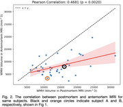 Figure 1
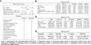 Figure 2
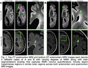 Figure 3
Peer Reviews
No public reviews on file for this paper yet. If you reviewed it on a platform where reviews are public (OpenReview, ICLR, NeurIPS, ICML), you can paste yours below so the community can read it here.
Videos
No videos yet. Explain this paper in a talk, walkthrough, or lecture? Add one.
Taxonomy
TopicsAutopsy Techniques and Outcomes · Advanced Neuroimaging Techniques and Applications · Forensic Anthropology and Bioarchaeology Studies