Evaluating Volumetric and White Matter Hyperintensity Lesion Measures for Low‐field MRI using WMH‐SynthSeg
Hope Shimony, Sarah J. Keefe, Kavon J Sharifi, Jude‐Patrick Nnamdi Okafor, Nelly Joseph‐Mathurin, Jessica N Banks, Jessica Hu, Stephen Jarman, Edmond Knopp, Madeline Paczynski, Joo Lim, Alexandra Venuto, Nupur Ghoshal, B. Joy Snider, Shaney Flores, Tammie L.S. Benzinger

TL;DR
This study evaluates a new method for analyzing brain scans from low-field MRI to detect white matter changes linked to Alzheimer's treatments.
Contribution
The paper introduces and evaluates the use of WMH-SynthSeg for low-field MRI to measure brain volumes and white matter lesions.
Findings
WMH-SynthSeg provides comparable volumetric measures to 3T MRI for large brain regions.
WMH lesion probability maps aligned with known ARIA but failed to capture the entire affected area.
WMH volumes in healthy controls were significantly lower than in patients on AAT therapy.
Abstract
White matter hyperintensities (WMH) are known predictors of amyloid‐related imaging abnormalities (ARIA) in patients undergoing anti‐amyloid immunotherapy (AAT) for Alzheimer disease. WMHs and brain volumetric changes can potentially be captured by low‐field magnetic resonance imaging (MRI) that imposes minimal safety risks to those with contraindicators to high‐field MRI. We investigate the recently released FreeSurfer WMH‐SynthSeg for volumetric and WMH lesion processing of low‐field MRI and its comparability to 3T MRI. Low‐field head MRI scans for 19 healthy controls and 23 patients undergoing AAT were acquired on a 0.064T Hyperfine SwoopÒ Portable MRI scanner. Treated patients also underwent a 3T MRI for ARIA screening by a clinical neuro‐radiologist per treatment protocol. Volumetric measures of cortical grey matter, white matter, ventricles, and hippocampus were extracted from…
Genes, proteins, chemicals, diseases, species, mutations and cell lines named across the full text — each resolved to its canonical identifier and authoritative record.
Click any figure to enlarge with its caption.
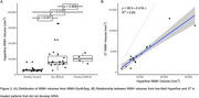 Figure 1
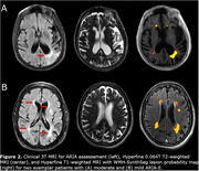 Figure 2
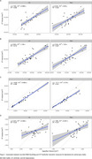 Figure 3
Peer Reviews
No public reviews on file for this paper yet. If you reviewed it on a platform where reviews are public (OpenReview, ICLR, NeurIPS, ICML), you can paste yours below so the community can read it here.
Videos
No videos yet. Explain this paper in a talk, walkthrough, or lecture? Add one.
Taxonomy
TopicsDementia and Cognitive Impairment Research · Intracerebral and Subarachnoid Hemorrhage Research · Cerebrospinal fluid and hydrocephalus