Cumulative environmental exposures adversely impact social behaviour and are associated with dysregulation of genes and proteins involved in epigenetic, ribosomal, and immune regulation in male mice
Morgan C. Bucknor, Brooke A. Keating, Velda X. Han, Brian S. Gloss, Pinki Dey, Nader Aryamanesh, Lee L. Marshall, Mark E. Graham, Ruwani Dissanayake, Xianzhong Lau, Shrujna Patel, Stela P. Petkova, Peter Valtchev, Anand Gururajan, Russell C. Dale, Markus J. Hofer

TL;DR
Cumulative environmental stressors in mice lead to social behavior issues and changes in genes and proteins linked to immune and epigenetic functions, especially in males.
Contribution
A novel 'triple-hit' mouse model was developed to study cumulative environmental effects on neurodevelopmental disorder risk and immune-related molecular changes.
Findings
Male mice exposed to combined stressors showed elevated NDD-related behavioral risk and social deficits.
Altered inflammatory and ribosomal pathways were observed in brain glia and peripheral immune cells.
Proteomic analysis revealed decreased proteins involved in inflammation, translation, and synaptic function in both brain and blood.
Abstract
This study investigated how cumulative environmental exposures influence offspring behaviour and inflammation-related molecular signatures in the brain and peripheral immune system. A novel "triple-hit" mouse model was developed using C57Bl/6JAusB mice (N = 70), combining preconceptual social stress, antenatal high-fat diet, and a postnatal immune challenge (poly(I:C), 10 mg/kg). At 12 weeks, offspring underwent behavioural tests relevant to neurodevelopmental disorders (NDDs), including the Elevated Plus Maze, 3-Chamber Social Preference, Self-Grooming, and Marble Burying. A composite NDD-risk index was calculated. Single-cell RNA sequencing (scRNA-seq) and bulk proteomics were performed on male triple-hit offspring to identify differentially expressed genes and proteins associated with inflammatory pathways. Male triple-hit offspring showed elevated NDD-related behavioural risk and…
Genes, proteins, chemicals, diseases, species, mutations and cell lines named across the full text — each resolved to its canonical identifier and authoritative record.
Click any figure to enlarge with its caption.
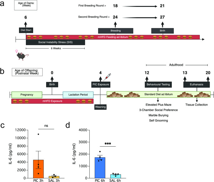 Figure 1
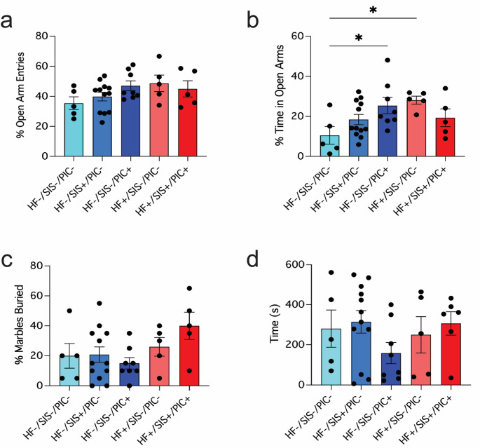 Figure 2
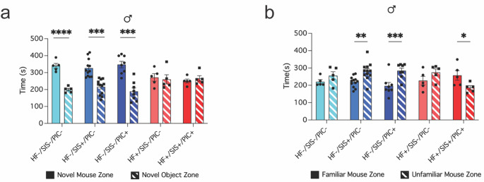 Figure 3
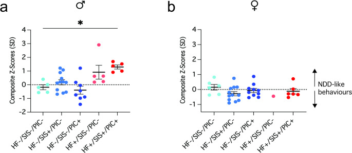 Figure 4
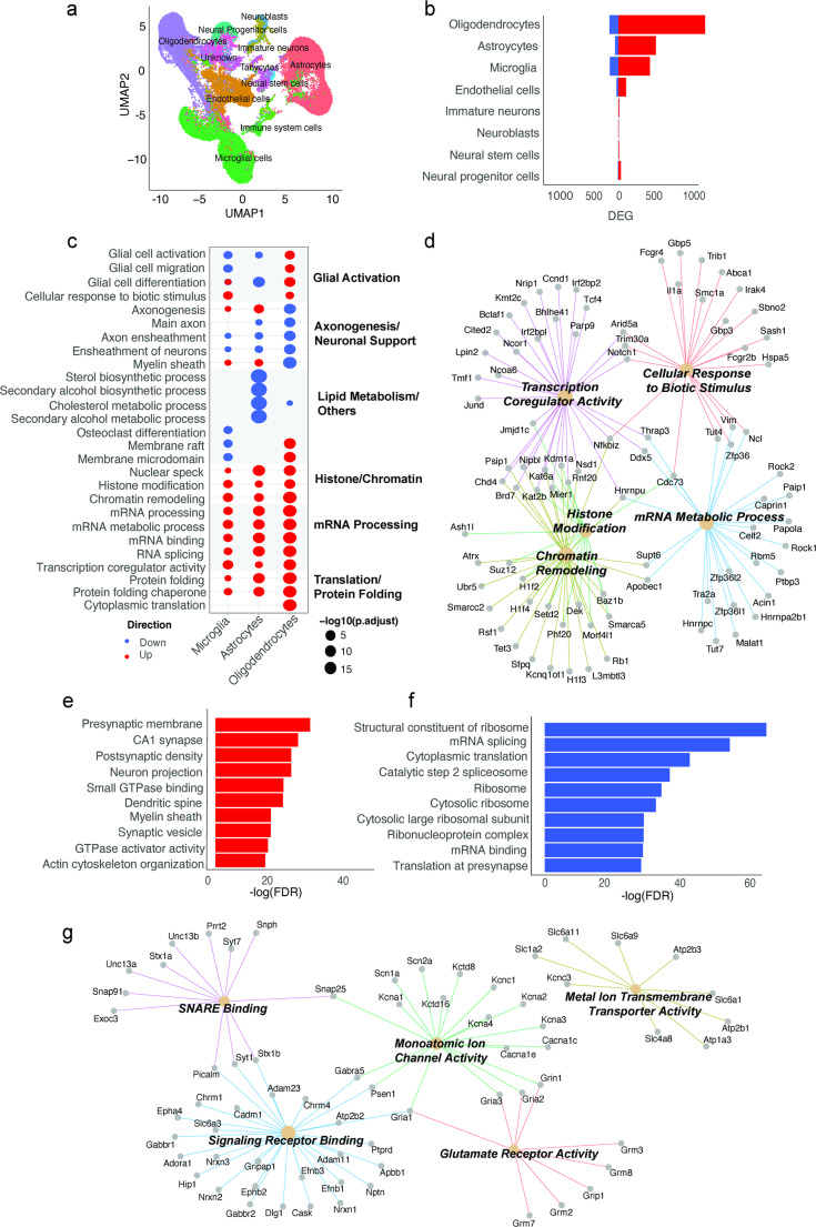 Figure 5
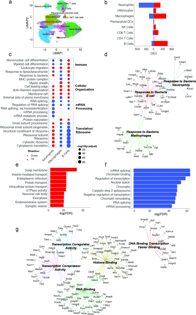 Figure 6
- —https://doi.org/10.13039/501100000925National Health and Medical Research Council
- —https://doi.org/10.13039/501100001774University of Sydney
Peer Reviews
No public reviews on file for this paper yet. If you reviewed it on a platform where reviews are public (OpenReview, ICLR, NeurIPS, ICML), you can paste yours below so the community can read it here.
Videos
No videos yet. Explain this paper in a talk, walkthrough, or lecture? Add one.
Taxonomy
TopicsTryptophan and brain disorders · Health, Environment, Cognitive Aging · Stress Responses and Cortisol
Introduction
First conceived in 1986, the neurodevelopmental hypothesis emerged as a primary explanation for the developmental origins of schizophrenia (SCZ) [1]. It broadly suggests that adverse gene-environment interactions during critical prenatal and postnatal periods can disrupt normal brain development, resulting in abnormal neuronal circuitry [2]. Since its inception, this hypothesis has been extended upon to capture the origins of additional neurodevelopmental disorders (NDDs) such as, intellectual disability, autism spectrum disorder (ASD), attention deficit/hyperactivity disorder (ADHD), motor disorders and tic disorders [3, 4]. These early childhood NDDs as well as adult-onset psychiatric disorders (e.g., SCZ and bipolar disorder), share clinical features, developmental delays, and common familial and environmental risk factors [4–8]. Accordingly, the definition of NDDs has broadened to include both early-onset conditions and adult psychiatric illnesses, given their shared neurodevelopmental basis [3, 6, 9].
NDDs are lifelong learning and behavioural conditions that are heterogeneous in presentation and lack effective disease-modifying therapies. Characteristic symptoms broadly include difficulties in social communication, impulsivity, cognitive flexibility, attention, language, sensory processing, learning, and motor control, which diverge from typical developmental trajectories and frequently co-occur [10–12]. There are profound sex and gender-related differences within NDDs as well, whereby males are more frequently diagnosed than females [12–14]. However, it is likely that females are underdiagnosed as opposed to being less susceptible or protected from disease [4, 12, 15–17]. The global prevalence of these conditions continues to rise annually [18, 19] and places a heavy burden on affected individuals and their families, public health care, and educational systems. Therefore, disentangling specific aetiological causes and pathological mechanisms underlying these conditions are a health priority.
It is now well-recognised that the aetiological landscape of NDDs is multifactorial and highly complex. These conditions are associated with a cocktail of different risk factors including genetic predisposition, the intrauterine experience (e.g., maternal infection or obesity) and postnatal environmental factors such as, childhood adversity, urban living, psychological trauma or infection [2, 4, 20–24]. While genetics are important risk determinants, they cannot fully explain the manifestation of disease. This is evidenced by low disease concordance rates observed in twin studies [25, 26], as well as many risk variants not consistently being expressed [27–29]. A growing body of clinical studies indicate that the cumulative effects of diverse environmental risk factors are more strongly linked to NDD risk [20, 30–34]. For example, one study developed a ‘polyenvironmic’ risk score (PERS) to predict the likelihood of psychosis in young relatives of individuals with SCZ based on cumulative environmental risk exposures (i.e. cannabis use, season of birth, paternal age, obstetric or perinatal complications and various types of childhood adversity) [35]. They observed a higher PERS was significantly associated with increased risk of psychosis. Another study reported that children exposed to multiple maternal exposures (i.e. autoimmunity, smoking, asthma, mood or anxiety disorders, hypertension, or diabetes) during pregnancy, faced a significantly higher risk of ADHD, with each added exposure increasing risk by ~ 50%, up to 3.3-fold for three or more [30]. Preclinical models have corroborated human aetiological evidence as well. For instance, animal models of maternal high-fat diet consumption during pregnancy observe a spectrum of NDD-associated behavioural changes in offspring [36–40] and structural brain alterations implicated in NDD pathophysiology [41]. They have also reliably confirmed that other established risk factors such as, prenatal infection and peripubertal stress exposure, are associated with abnormal brain development and behavioural alterations in offspring [22, 23, 42]. Consequently, exposure to a multifaceted range of developmental stressors in early life influences the onset and severity of NDDs.
However, it remains unclear how exactly cumulative stress exposures drive behavioural impairments and influence disease risk. Emerging evidence from animal studies point to epigenetic reprogramming or ‘priming’ of foetal immune and brain cells, promoting a disease-susceptible phenotype that compromises resilience to postnatal challenges and heightens the risk of future disease [29, 43–46]. Microglia, the brain’s resident immune cells, have been extensively implicated as highly susceptible to developmental epigenetic priming in response to developmental stress, due to their early embryonic colonisation and substantial cellular plasticity [47–55]. Neurodevelopmental impacts of other glial cells (i.e., astrocytes, oligodendrocytes) remain largely understudied. Moreover, peripheral immune priming is suggested to co-occur with microglial priming during foetal development, representing another crucial component of NDD pathogenesis [22, 49, 56, 57]. Peripheral immune dysregulation is common in NDD patients [56, 58], and with limited access to brain tissue in clinical settings, peripheral blood presents a compelling candidate as a surrogate biomarker for brain pathology. Yet, most neurodevelopmental animal models have derived mechanistic evidence by modelling isolated risk factors within one developmental window, limiting their ability to capture the multifaceted, lifelong nature of human exposures.
To our knowledge, this is the first study to integrate multiple sources of developmental stress within a single model spanning from preconception through the juvenile period. Here, we characterised a novel ‘triple-hit’ environmental stress model in mice to disentangle the complex human aetiological framework of NDDs. To do this, we integrated three diverse NDD risk factors in offspring, including, the maternal experience of psychosocial stress before conception, antenatal high-fat (HF) diet exposure and juvenile immune challenge (poly(I: C), PIC). We observed males exposed to all three stressors were more susceptible to developing NDD-associated behaviours compared to non-stressed controls and their female ‘triple-hit’ counterparts. Beyond behavioural phenotyping, we examined disease promoting peripheral-central nervous system (CNS) mechanisms by performing single-cell RNA transcriptomics and bulk proteomics in brain and peripheral blood cells. Our single-cell RNA transcriptomics and proteomic sequencing revealed alterations to peripheral immune functions, glial cell dysfunction, dysregulated ribosomal processes, synaptic homeostasis and chromatin remodelling in both the brain and blood of these offspring; all relevant pathways that share significant associations with the pathogenesis of various NDDs [29, 59–62]. Altogether, these findings emphasise the systemic impacts of early life adversity and offers novel insights into the potential of peripheral blood to serve as a reliable guide for future NDD therapeutic interventions.
Materials and methods
Animals and ethical approval
All animals were housed in a specific pathogen-free (SPF) environment at the Charles Perkins Centre (Sydney, Australia) animal facility (20–24℃, 40–70% humidity, 12 h light: dark cycle). All male and female offspring in this study were generated via on-site breeding from C57Bl/6Ausb dams as previously reported [63]. Male and female mice used for breeding were obtained from Australian BioResources (ABR; Moss Vale, NSW). All experiments were performed in compliance with the NSW Animal Research Act and the 8th edition of the NHMRC ‘Australian Code of Practice for the care and use of animals for scientific purposes.’ This study was reviewed and approved by the University of Sydney Animal Ethics Committee.
Maternal and offspring diets
Dams had ad libitum access to either a high-fat, no soy semi-pure diet (HF + diet, 43% kcal fat diet, SF21-208, Specialty Feeds, WA, Australia) or a low-fat, high-sugar, no soy semi-pure control diet formulation (HF- diet, 12.3% kcal fat diet, SF21-209, Specialty Feeds, WA, Australia) prior to, during gestation and during the lactation period. The detailed composition of maternal diets and time of exposure has been previously described [63]. F1 generation offspring were weaned at postnatal day (PND) 28 and transitioned to standard, grain-based laboratory chow (SF00-100, Specialty Feeds, WA Australia) for the remainder of the study.
Maternal stress exposure, onsite breeding and offspring poly(I: C) exposure
Refer to study timeline for experimental design (Fig. 1a–b). In brief, dams were randomised to either chronic high-fat diet consumption (HF + or HF-), six-weeks of social instability stress (SIS + or SIS-) or both in parallel as described previously [63]. This generated four distinct maternal stress groups: high-fat diet fed only (HF+/SIS-), high-fat diet fed and social instability stressed (HF+/SIS+), low-fat diet fed only (HF-/SIS-), and low-fat diet fed and social instability stressed (HF-/SIS+). The maternal SIS paradigm introduces chronic, unpredictable social stress by randomly changing the group cage composition multiple times per week and has been validated to cause stress in females [63–65].
At ≥16 weeks of age, females (N = 45 total) were paired for breeding and left undisturbed until birth of litters at our animal facility. However, due to significant maternal cannibalism [63], only (N = 19) total dams cared for offspring and each dam was bred only once. Given these reproductive outcomes, all surviving offspring from multiple litters were included and pooled for analyses described in this study (N = 70) (Table 1). Further breeding information is provided in Extended Methods (Table S1).
Following birth of a litter, offspring were left undisturbed until weaning. The day after weaning (PND 29; juvenile stage of development), offspring were randomised to receive either a single intraperitoneal injection of 10 mg/kg body weight poly(I: C) (high molecular weight, Cat# tlrl-pic-5, InvivoGen) or endotoxin-free 0.9% NaCl (Baxter, Australia). Following poly(I: C) or vehicle exposure. Poly(I: C)-induced immune activation (PIA) (10 mg/kg) was validated in an independent cohort of male C57bl/6 mice (n = 15). The dose was chosen based on previous literature [66–68]. The same batch of poly(I: C) was used between experimental animals and those in the independent cohort to avoid batch effects. PIA was defined by an acute increase in plasma IL-6 concentration at 3- and 6-hours post-injection (Fig. 1c–d). Plasma concentrations of IL-6 were determined using a commercial IL-6 ELISA kit (ab22503; sensitivity = 11.3 pg/mL; Abcam) according to manufacturer’s instructions. Plasma samples were diluted 1:8 and analysed in duplicate. Absorbance was measured at 450 nm using a Tecan infinite M1000 Pro plate reader. Concentrations of IL-6 were calculated using the AssayFit Pro 96 well curve fitting ELISA calculator (https://www.assayfit.com/home.html). Standard curve was determined by fitting the data to a 4-parameter logistic curve (4PL).
Next, adult offspring were behaviourally phenotyped and then euthanised for tissue collection. Further methodological details describing the maternal prenatal stress model can be obtained in Extended Methods. Offspring were group-housed with littermates and/or same-sex treatment group cage mates from other litters where possible for the remainder of the study.
Fig. 1. Experimental timeline and poly(I: C) validation. a Maternal high-fat diet and social instability stress paradigm to generate HF+/SIS + and/or HF+/SIS- females. Both dietary consumption and/or social instability stress (SIS) exposure began at 6 weeks of age. HF diet consumption continued into the lactation period. SIS exposure lasted for 6 weeks, prior to inducing pregnancy. HF + females were staggered for breeding 2 weeks apart, the first round of matings began at 18 weeks of age. Second round of matings paired females at 24-weeks of age. b Neonates were exposed to mHFD via maternal milk and left undisturbed until weaning. At 4 weeks of age, juvenile offspring were randomised to receive 10 mg/kg of poly(I: C) or NaCl vehicle solution via intraperitoneal injection. Behavioural testing began at 12 weeks of age, and immediately followed by euthanasia for tissue collection (animals aged 13 weeks, 15 weeks and one animal included for proteomics analyses was 20 weeks of age). c–d Results from poly(I: C:) validation in an independent cohort of male C7Bl/6 mice (N = 15). (c) Plasma IL-6 (pg/ml) concentration 3 h post- 10 mg/kg poly(I: C) intraperitoneal injection and/or saline vehicle. Mann-Whitney test (p = 0.0571); ns. (d) Plasma IL-6 (pg/ml) concentration 6 h post- 10 mg/kg poly(I: C) intraperitoneal injection and/or saline vehicle. Unpaired t-test (***p = 0.0002). Data shown as mean ± SEM. PIC = poly(I: C), SAL = saline, ns = not significant. Images created with biorender.com
Table 1. Developmental window of environmental exposures, sample group information and number of offspringDirect maternal exposure onlyDirect maternal and offspring exposure (antenatal-lactation)Direct offspring exposure only(PnD 29)PreconceptionAntenatalJuvenile♂offspring/group^c^♀offspring/group^d^Number of dams (litters) / groupmSIS^a^mHFD^b^Poly(I: C)(n)(n)(n)---564*+--121210-+-512--+8104*+++563N = 35N = 35N = 19
^a^mSIS = maternal social instability stress.
^b^mHFD = maternal high-fat diet.
^c^♂ = male.
^d^♀ = female.
^*^4 = indicates the same (HF-/SIS-) dams (n = 4) that were responsible for generating both (HF-/SIS-/PIC-) and HF-/SIS-/PIC+) offspring groups.
Behavioural testing
Behavioural testing began on both male and female offspring at 12-weeks of age and was carried out over a 2-week period. The same researcher conducted all tests and was blinded to offspring group conditions. All tests were performed during the light cycle between 0900 and 1700 h. To assess NDD-associated behaviours, we selected a battery of tests relevant to the symptomatology observed in NDDs [69]: elevated plus maze (anxiety-like behaviour), 3-chamber social preference (social engagement), marble burying and self-grooming (repetitive behaviours), with 2–4 days of rest between tests. Detailed description of all assay procedures is provided in brief below and expanded in Extended Methods. Raw data of elevated plus maze and 3-chamber social preference test was analysed using AnyMaze video tracking software (V7.20). Time spent grooming was manually scored using Behavioural Observation Research Interactive Software (Boris). The number of marbles buried was recorded by two independent researchers at the end of the testing period.
Elevated plus maze test
Elevated plus maze testing was performed as described previously [63]. Briefly, this test allows for assessment of anxiety-like conflict behaviour by allowing the mice to choose between entering the two open arms of the maze (natural exploratory drive) or entering and remaining in the safety of the two closed arms [70]. The parameters measured included total distance travelled, percentage of open arm entries and percentage of time spent in the open arms of the maze.
3-chamber social preference
Social impairments are one of the core symptoms of ASD and rodents regularly engage in high levels of social reciprocity [69, 71]. The 3-chamber social preference test assays for changes in sociability (time spent engaging with a novel mouse rather than an inanimate object) and social novelty (time spent engaging with a novel mouse rather than a familiar mouse) using a 3-chamber apparatus. This test includes three sequential 10-minute trials beginning with habituation, sociability and lastly, social novelty. The test mouse is placed in the centre chamber and allowed to freely explore both side chambers during the test. The habituation trial allows the mouse to freely explore the apparatus, with each side chamber consisting of only an empty wire cup. Following the habituation trial, the test mouse is confined to the centre chamber while a novel object (golf ball) is placed under one wire cup and a novel sex-matched mouse is placed under the other wire cup in the other side chamber. This trial assesses changes in sociability by measuring time spent in the chamber containing the novel object and novel mouse. For the last trial, the test mouse is confined to the centre chamber while the novel object is replaced with another novel sex-matched mouse. This trial assesses changes in social novelty or recognition by measuring the time spent in the chamber containing the unfamiliar mouse and familiar mouse.
Marble burying
Repetitive or ritualistic behaviours are clinically relevant symptoms of NDDs [69]. Therefore, to evaluate repetitive digging behaviours, we performed the marble burying test. The test mouse was placed in a standard cage with bedding 3–4 cm thick and overlaid with 20 black, glass marbles in a 4 × 5 arrangement. The number of marbles buried by test mice (defined as more than 2/3rds or more covered by the bedding) at the end of the 30 min test period was recorded.
Self-grooming
Spontaneous, unusually long repetitive bouts of grooming were scored over a period of 20 min in a novel, empty cage under dim lighting (< 50 lx) to assess repetitive, obsessive-compulsive like behaviour for each test mouse. The first 10 min period was unscored and considered the habituation phase, while the remaining 10 min were scored.
Integrated neurodevelopmental disorder (NDD) behavioural index
Integrated behavioural measures from complimentary tests can be z-normalised to reduce behavioural noise that is a common artifact of behavioural assays [72, 73]. A z-score is a standard deviation (SD) value that indicates how many standard deviations above or below a pooled sample is from the mean of a reference group (i.e. HF-/SIS-/PIC- offspring). Since these scores are all in the same unit (SD), the distribution has a mean of 0 and an SD of 1 [74]. Thus, a positive z-score (>0) indicates scores that are greater than the reference mean. Here, we have defined this as a translation to a greater propensity for NDD-associated behaviours. Whereas a negative z-score (< 0) translates to a reduced propensity for NDD-associated behaviours. This approach provides an overall measure of the animals’ emotionality dependent on the context of the behavioural tests integrated - which here includes anxiety-like behaviour (elevated plus maze), repetitive behaviours (marble burying and grooming), and social engagement (3-chamber social preference). Each behavioural test contributes equal weightage to the composite z-score with the exception of the 3-chamber social preference test. As this test comprises two independent trials, it is weighted twice in the composite z-score, aligning with its heightened relevance to social deficits characteristic of NDDs. Refer to Extended Methods for formula used and further contextual information.
Tissue collection for single-cell RNA sequencing and bulk proteomics
For single cell RNA (scRNA)-seq and bulk proteomic analyses, peripheral blood leukocytes and total forebrain tissue were analysed from male offspring for HF+/SIS+/PIC + and HF-/SIS-/PIC- for two reasons: (i) the male-biased prevalence of NDDs and (ii) it was evident from our behavioural analyses that the cumulative effects of pre- and postnatal stress had less impact on female offspring. The same 4 animals were used between scRNA-sequencing and proteomics experiments. scRNA-sequencing was performed using the HIVE™ Single Cell platform (Honeycomb Biotechnologies, Inc. USA). Bulk LC-MS/MS proteomics leveraged an untargeted approach and used the Dionex UltiMate 3000 HPLC system. Complete sample preparation information for scRNA-sequencing and proteomics is provided in Extended Methods.
For tissue collection, mice were deeply anesthetised with isoflurane (5% induction, 2.5% maintenance) (IsoFlo^®^, Abbott Laboratories, Botany, NSW, Australia). Up to 1 mL of peripheral whole blood was collected via cardiac puncture and placed into a 1.5 mL collection tube containing anticoagulant (10% blood volume) (Heparin, Sigma, H3393) on ice. The mouse was then perfused with PBS and the whole brain was dissected. The cerebellum and olfactory bulbs were excised along the anterior to posterior axis from approximately + 1.50 mm to -3.50 mm, leaving total forebrain tissue. Regions left intact included cerebral cortices, thalamus, hypothalamus, striatum and midbrain. The tissue was weighed and recorded before tissue homogenisation (gentleMACS Octo Dissociator with Heaters, Miltenyi Biotec, 130-096-427). A single-cell suspension was prepared using the Adult Brain Dissociation kit as per manufacturer’s instructions (Miltenyi Biotec, 130-107-677). To isolate peripheral leukocytes from whole blood, red blood cell lysis was performed using ammonium chloride solution (StemCell Technologies, 07850) at a volume: volume ratio of 9:1. The solution incubated at 4 °C for 10-minutes, then cells were washed in Dulbecco’s phosphate-buffered saline containing Ca^2+^, Mg^2+^, glucose and pyruvate (DPBS, ThermoFisher, 14287080) by centrifugation at 250 x g for 6-minutes at 4 °C.
scRNA-sequencing: HIVE CLX™ sample capture, library Preparation and sequencing
Once single-cell suspensions were generated for each sample condition, samples were loaded into the HIVE collectors according to the manufacturer’s instructions. Approximately 30,000 cells were loaded directly into a designated HIVE collector (Honeycomb Biotechnologies, Inc. USA) in 1% FBS in DPBS. Collectors were centrifuged at 30xG for 3 min to allow single-cells to settle into picowells of the HIVE collector, which contained mRNA-capture beads. Media was removed from the collector and 2 mL of CLX sample wash solution (provided by manufacturer) was added. After removing the wash solution, 1 mL of cell preservation solution (provided by manufacturer) was added (Honeycomb Biotechnologies, Inc. USA_Sample Capture protocol). HIVE collectors were stored at -80 °C until transferred to the Australian Genome Research Facility Ltd. (AGRF, Westmead, Australia) for transcriptome recovery and the library preparation.
All HIVE CLX™ devices were processed according to the manufacturer’s instructions (Honeycomb Biotechnologies, Inc. USA: Transcriptome Recovery and Library Preparation protocol). Briefly, HIVE collectors were sealed with a semi-permeable membrane, allowing for the addition of strong lysis solution and hybridization solution. Capture beads with transcripts were extracted from the collector by centrifugation. The remaining library preparation steps were performed in a 96-well format. The size profiles of the final libraries were determined on a TapeStation platform with a D5000 ScreenTape System (Agilent Technologies, Santa Clara, CA, USA), and the concentration of the final pooled library was determined by qPCR assay before sequencing on the Illumina NovaSeq X sequencing platform with custom primers (provided in the HIVE single-cell RNA seq processing kit) (AGRF, Melbourne, Australia).
scRNA-sequencing bioinformatics analysis
Raw base calls for brain and blood samples were processed using the Illumina BCL Convert 4.1.5 pipeline to generate FastQ files. FastQ files were analysed using BeeNet (v1.1.3) software, mapped to the reference Mus musculus genome (mm10.104) and imported into the R statistical environment (v4.3.1) using the Seurat,* tidyverse* and patchwork R packages. Preprocessing was performed according to beenet v1.3 workflow. Briefly, before expression data was merged, cells with a high mitochondrial transcript ratio (>0.20), less than 300 genes or 600 transcripts were excluded. Merged data were normalized and scaled using SCtransform before clustering and UMAP projection. ScType was used to assign cell types using the auto_detect_tissue_type workflow [75]. Data was split by cell type and then renormalized, markers between conditions were determined using FindMarkers with the parameters logfc.threshold = 0,test.use = “wilcox”,min.pct = 0.05. Pathway enrichment was determined via over representation analysis (ORA) to obtain Gene Ontology (GO) pathways for differentially expressed genes (adj p value < 0.05) using enrichGO in the CompareCluster function from ClusterProfiler [76].
Protein lysate sample Preparation for proteomics
Following brain and peripheral blood leukocyte cell isolation as described above, the remaining fraction of cells were lysed for protein for unbiased mass spectrometry analysis. Cell suspensions were incubated in 200 µL of 1X Lysis Buffer containing: 0.8% v/v Triton X-100, 50 mM HEPES (pH 7.4) with NaOH, EDTA free protease inhibitor, PhosSTOP and 2 µL of 2 mM phenylmethyl sulfonyl fluoride in ethanol (PMSF) at 85 °C for 10 min on a heating block. Samples were then stored at -80 °C until LC-MS analysis. Extended details are provided in Extended Methods concerning tissue lysis, protein digestion and peptide tagging for LC-MS/MS analysis.
LC-MS/MS analysis
LC-MS/MS was performed using a Dionex UltiMate 3000 RSLC nano system and Q Exactive Plus hybrid quadrupole-orbitrap mass spectrometer (ThermoFisher Scientific). Each HILIC fraction was loaded directly onto an in-house 300 mm long 0.075 mm inside diameter column packed with ReproSil Pur C18 AQ 1.9 μm resin (Dr Maisch, Germany). The column was heated to 50 °C using a column oven (PRSO-V1, Sonation lab solutions, Germany) integrated with the nano flex ion source with an electrospray operating at 2.3 kV. The S lens radio frequency level was 50 and capillary temperature was 250 °C.
All brain and blood sample fractions were analysed using data-dependent acquisition LC-MS/MS. MS scans were performed at 70,000 resolution with an automatic gain control target of 1,000,000 for a maximum ion time of 100 ms 375 to 1500). MS/MS scans were at 35,000 resolution with an automatic gain control target of 200,000 and maximum ion time of 100 ms. The loop count was 12, the isolation window was 1.1 m/z first mass was fixed at 120 m/z, and the normalized collision energy was 31. Singly charged ions and those > 8 + were excluded, with a 35 s dynamic exclusion.
Differential abundance protein and pathway enrichment analysis
For data cleaning, the raw ‘proteinGroups.txt’ LC-MS/MS output file was processed from MaxQuant v1.6.7.0. Each protein group must have had at least one unique peptide. Proteins were removed from further analysis if they matched contaminant or reverse sequence decoy entry prefixes (CON_ or REV_). Proteins with one or more missing values in any samples were also removed.Data was normalised by first identifying the UniProt accession for each protein group following rules from Engholm-Keller et al. (2019) [77]. Multi-mapped proteins were also excluded to avoid false enrichment due to alignment errors. The samples were first log (base 2) transformed, then between sample normalisation was performed using the ’scaled’ normalisation from the limma R package (v4.2.3). To remove batch effects from biological data, the remove unwanted variation RUV R package was used [78].Differential abundance analysis of proteins was performed using the adjusted abundance matrix. The results from this differential abundance analysis were then used for all analyses. For linear modelling, differential abundance analysis of proteins was performed using the limma R package. The linear model for comparing each pair of time points was fitted using the lmFit function and p-values calculated using the empirical Bayes method using the ‘eBayes’ function. Significant differentially expressed proteins were defined by (q-value < 0.05). Differentially expressed proteins were analysed for GO pathway enrichment analysis using the enrichGO function from the clusterProfiler R package.
Statistical analysis
No statistical methods were used to predetermine offspring sample sizes as this was limited by breeding outcomes from dams described previously [63]. All statistical testing was performed in the R statistical environment (v2023.12.1 + 402). GraphPad Prism software (v10.4.0) was used for graphical representation of physiological and behavioural phenotyping results. For physiological phenotyping, our analyses employed a mixed-effects model using the lme4 and car R Package to analyse the effect of age, group and their interaction on weight gained. The subject (offspring) was included as a random effect. Behavioural output from the elevated plus maze, marble burying, self-grooming and integrated z-score met assumptions of normality (Shapiro-Wilk test) and homogeneity of variance (Levene’s test) – therefore, one-way ANOVA was performed. Where a significant main effect of group was detected, Dunnett’s post-hoc test was performed. For social preference testing, we first fit a linear mixed-effects model using the (lme4) R Package and detected zero variance (standard deviation = 0) when including the random effect of offspring and litter identity. Therefore, a linear model (lm) was adopted which focused on fixed effects only (group and social zone). We then conducted Type III ANOVA on the model output to assess main effects (group) and interactions (group x social zone). Where significant main effects or interactions were detected, Tukey’s post-hoc test was performed.
Results: Behavioural Outcomes
Early life exposure to mHFD or poly(I: C) alone leads to increased time spent in the open arms of the plus maze
Mice underwent behavioural testing using NDD-associated assays at 12-weeks of age, starting with the elevated plus maze [69]. Description of male behaviours are provided below, while complementary results from female offspring are provided in supplementary material (Suppl. Fig. S2-3). Anxiety is not considered a core symptom of NDDs but is frequently reported as an associated symptom, depending on diagnostic criteria [79]. When we performed elevated plus maze testing, we found no statistically significant differences in the percentage of open arm entries between groups (Fig. 2a). However, one-way ANOVA revealed a main effect of group on percentage of time spent in the open arms (F(4, 30) = 2.913, p = 0.0379). Dunnett’s post-hoc test revealed HF-/SIS-/PIC + and HF+/SIS-/PIC- offspring spent more time in the open arms relative to HF-/SIS-/PIC- controls (14.9% (p = 0.030) and 17.6% (p = 0.020) mean increase in time, respectively (Fig. 2b). Repetitive behaviours were assayed using the marble burying test and observing grooming behaviours. Here, the marble burying test did not reveal any statistically significant differences between groups and nor did time spent grooming differ significantly (Fig. 2c-d). Overall, based on these tests, the most significant alterations in behaviour related to increases in exploratory drive in offspring exposed to only poly(I: C) or mHFD.
Fig. 2. Male offspring exposed to poly(I: C) or mHFD in isolation of other factors display increased time spent in open arms of plus maze. a–b Elevated plus maze, (a) percentage of open arm entries and (b) percentage of time spent in the open arms, (*p < 0.05); c marble burying test – percentage of marbles buried; d self-grooming test - time(s) spent grooming. (a–d) One-way ANOVA followed by Dunnett’s post-hoc test. Data shown as mean ± SEM; each individual data point represents one mouse
HF+/SIS+/PIC + offspring demonstrate deficits in social behaviours
A linear model was used to examine the effects of group and zone on time spent either with a novel mouse or novel inanimate object during the sociability trial. Type III ANOVA of this model revealed significant main effects of group (F(4, 60) = 4.80, p = 0.0019), zone (F(1, 60) = 22, p = p < 0.001) and interaction between group x zone (F(4, 60) = 7.84, p < 0.001). Tukey’s post-hoc test revealed significantly more time spent in the novel mouse zone versus novel object zone in HF-/SIS-/PIC- controls (+ 140.38s (p < 0.0001)), HF-/SIS+/PIC- and HF-/SIS-/PIC + offspring (+ 109.89s and + 158.66s (p < 0.001). This profound social engagement with the novel mouse is expected. Whereas, both HF + groups did not spend significantly more time with the novel mouse, instead they spent almost equal amounts of time in both zones (Fig. 3a). These changes in sociability suggest that exposure to mHFD alone, as well as in the context of additional mSIS and poly(I: C), result in social exploration deficits.
The same statistical approach was used for analysing the effects of group and social zone on time spent either with an unfamiliar or familiar mouse. Type III ANOVA revealed a significant interaction between group x zone (F(4, 60) = 5.139, p = 0.0013). Upon further investigation, Tukey’s post-hoc test revealed HF-/SIS+/PIC- and HF-/SIS-/PIC + spent significantly more time with the unfamiliar mouse than the familiar mouse [-61.5s (p = 0.0017) and − 88s (p = 0.0003)]. This distinction in time spent with familiar versus unfamiliar shows intact recognition and novelty interest. HF+/SIS+/PIC + offspring, in contrast, spent significantly more time in the familiar mouse zone compared to the novel (71.46s (p = 0.0165). These findings suggest that the cumulative effects of all stressors (HF+/SIS+/PIC+) lead to impairments in social memory or recognition (Fig. 3b).
Fig. 3. Male HF+/SIS+/PIC + offspring display deficits in social recognition and sociability. a–b 3-chamber social preference test, (a) total time (s) spent in novel mouse zone and novel object zone within group comparisons, (***p < 0.001, ****p < 0.0001); (b) total time (s) spent in familiar mouse zone and unfamiliar mouse zone within group comparisons, (*p < 0.05, **p < 0.01, ***p < 0.001) (a–b) Linear model followed by type III ANOVA and Tukey’s post-hoc test. Data shown as mean ± SEM; each individual data point represents one mouse
Integrated z-score indicates increased risk of neurodevelopmental disorder–like behaviours in adult male HF+/SIS+/PIC + offspring
To best interpret our behavioural findings across all tests, we performed an integrated z-score calculation. To do this we z-normalised each individual animal’s behavioural output, added them together and divided by the number of tests included in the integration to create a composite z-score (see Extended Methods). Since our behavioural phenotyping consisted of NDD-associated tests, we described this output as an integrated NDD behavioural index which broadly describes each groups’ propensity for displaying NDD-associated behaviours. Values > 0 are indicative of increased susceptibility to NDD-like behaviours, < 0 less susceptible, and 0 is no change. When we assessed the difference in z-score across male groups, one-way ANOVA detected a significant main effect of group (F(4, 30) = 5.73, p = 0.0015). Overall, HF+/SIS+/PIC + males exhibited a statistically significant increase in z-score compared to HF-/SIS-/PIC- controls, and Dunnett’s post-hoc test confirmed this (+ 1.5 standard deviation difference (p = 0.0101) (Fig. 4a). When we performed the same analysis in female counterparts, we found the absence of any increased risk for neurobehavioural impairments. Instead, all female groups clustered closely around zero (Fig. 4b). Lastly, to see if sex may influence NDD-like behavioural susceptibility, we fit a linear model followed by type III ANOVA which revealed a significant interaction between sex x group (F(4, 60) = 4.99, p = 0.001). Tukey’s post-hoc test revealed female HF+/SIS+/PIC + offspring displayed a significantly lower average z-score compared to male HF+/SIS+/PIC + offspring (-1.4 standard deviation difference (p = 0.0003) (Fig. 4a-b). These results indicate firstly, that mHFD, mSIS and postnatal poly(I: C) exposure differentially impact males and females; and male HF+/SIS+/PIC + offspring are most vulnerable to NDD-associated behavioural impairments compared to HF-/SIS-/PIC- controls.
Fig. 4. Male HF+/SIS+/PIC + offspring exhibit increased susceptibility to neurodevelopmental abnormalities compared to HF-/SIS-/PIC- controls, with no similar effect observed in females. a–b Integrated NDD-behavioural z-score incorporating elevated plus maze, marble burying, self-grooming and social preference testing output. (a) (*p < 0.05); (b) no significant difference. (a–b) One-way ANOVA followed by Dunnett’s post-hoc test. Data shown as mean ± SEM; each individual data point represents one mouse. Males (♂), females (♀)
Transcriptome and Proteome Changes in the Brain and Periphery
scRNA sequencing in brain glial cells reveals dysregulation in glial function, mRNA processing and epigenetic regulation pathways
Following our behavioural phenotyping results that revealed adult male HF+/SIS+/PIC + offspring to be especially vulnerable to NDD-associated behavioural deficits, we sought to investigate what biological pathways in specific cell types could be mediating this phenotype relative to HF-/SIS-/PIC- controls (n = 2/group). To do this, we characterised the transcriptomic profile of the forebrain at single-cell resolution. Relevant quality control information is provided in (Suppl. Fig. S6a–c). Further information regarding sample preparation is provided in Extended Methods.
Cell clustering and uniform manifold approximation and projection (UMAP) analysis of samples revealed 11 distinct cell type clusters (Fig. 5a). Statistically significant (FDR < 0.05) differentially expressed genes (DEGs) ranged from 12 to 1333 across captured cell types in HF+/SIS+/PIC + versus HF-/SIS-/PIC- offspring, with the highest proportions of DEGs found in oligodendrocytes, astrocytes and microglia (Fig. 5b). Therefore, we restricted our functional gene ontology (GO) pathway enrichment using Over-representation Analysis (ORA) from these DEGs to these 3 primary glial cells.
The top five up- and downregulated pathways in microglia, astrocytes and oligodendrocytes showed dysregulation in glial functions, axonogenesis and neuronal support, lipid metabolism, histone/chromatin, mRNA processing and translation/protein folding pathways (Fig. 5c). We observed a variable direction of change across all three cell types in glial cell activation, differentiation, axonogenesis and myelin sheath. Histone modification, chromatin remodelling, transcription coregulator activity, and protein folding were exclusively upregulated in all glial cells. Microglia in particular showed significant upregulation in pathways relating to inflammation, histone and chromatin, and mRNA processing. The top five upregulated ORA GO pathways in microglia were subclustered as a network connecting enriched pathway terms with genes annotated with each specific pathway term: cellular response to biotic stimulus, mRNA metabolic process, histone modification, chromatin remodelling, and transcription coregulator activity (Fig. 5d).
Bulk proteomics in brain reveals downregulation in translation and upregulation in synaptic pathways
To complement our scRNA sequencing findings in this model, we performed untargeted bulk proteomics in the same setting. Principal component analysis (PCA) of differential protein abundance revealed distinct separation between HF+/SIS+/PIC + and HF-/SIS-/PIC- controls (n = 3/group) (Suppl. Fig. S7a). Of 7859 statistically significant (FDR < 0.05) proteins, 2842 were downregulated and 3216 were upregulated in HF+/SIS+/PIC + offspring relative to HF-/SIS-/PIC- controls (Suppl. Fig. S7b). We performed ORA GO analysis based on these differentially expressed proteins (DEPs). Top 10 upregulated pathways included synaptic function and plasticity terms such as presynaptic membrane, CA1 synapse, postsynaptic density and synaptic vesicle (Fig. 5e). While top 10 downregulated pathways were predominantly related to ribosome biogenesis and mRNA translation (Fig. 5f).
We explored the most enriched upregulated ‘presynaptic membrane’ pathway further by clustering the genes in this pathway into themes based on GO molecular function (Fig. 5g). There were five themes that emerged in the genes enriching this pathway including SNARE binding (Snap25, Stx1a, Unc13a, Snph), Signalling receptor binding (Nrxn1, Nrxn2, Slc6a3), Monoatomic ion channel activity (Cacna1c, Kcna1, Kcna2, Kcna3, Psen1), Glutamate receptor activity (Grin1, Gria2, Gria3) and Metal ion transmembrane transporter activity (Slc6a1, Atp1a3, Atp2b1).
Fig. 5. Brain transcriptome and proteome in male HF+/SIS+/PIC + offspring versus controls. a UMAP (uniform manifold approximation of projection) of single-cell populations captured for sequencing: astrocytes, endothelial cells, immature neurons, immune system cells, microglial cells, neural progenitor cells, neural stem cells, neuroblasts, oligodendrocytes, tanycytes, unknown b proportions of significantly differentially expressed genes (DEGs) (FDR < 0.05) across brain cell types. Oligodendrocytes (1333), astrocytes (567) and microglia (560) display largest proportions of upregulated DEGs. Colours indicate DEG direction of change (red: up; blue: down). c Dot plot displaying GO analysis of DEGs (FDR < 0.05) HF+/SIS+/PIC + versus HF-/SIS-/PIC- controls in select cell types (microglia, astrocytes, oligodendrocytes) presented on x-axis. Enriched pathway terms are displayed on left y-axis and clustered biological themes of like terms presented on right y-axis. Size of each coloured dot is indicative of the negative log10 of enriched pathways adjusted p-value. The colour of each dot is indicative of the direction of change. d Connectivity network (CNET) plot of top five upregulated GO pathways and corresponding genes that enrich for each pathway in microglia: transcription coregulator activity, cellular response to biotic stimulus, mRNA metabolic process, chromatin remodelling, histone modification. Each enriched pathway is represented by the respective colours and corresponding genes’ adjusted p-value. e Bar plot of bulk proteome depicting top 10 enriched GO pathways amongst proteins with increased abundance. Statistical significance depicted by negative log(FDR). f Bar plot of bulk proteome depicting top 10 enriched GO pathways amongst proteins with decreased abundance. Statistical significance depicted by negative log(FDR). g Subclustered CNET plot of the upregulated GO pathway ‘presynaptic membrane’ in proteome. Subclusters are based on molecular function and include: SNARE binding, signalling receptor binding, glutamate receptor activity, monoatomic ion channel activity and metal ion transmembrane transporter activity. Each enriched subclustered pathway is depicted by the respective colours and corresponding genes’ adjusted p-value
ScRNA sequencing in peripheral blood leukocytes reveals dysregulation in immune, mRNA processing and translation pathways
UMAP analysis of these samples revealed 12 distinct cell type clusters (Fig. 6a). Statistically significant (FDR < 0.05) DEGs ranged from 13 to 873 across cell types in HF+/SIS+/PIC + versus HF-/SIS-/PIC- offspring. Here, we observed the largest proportion of DEGs in neutrophils, macrophages and naïve B Cells (Fig. 6b). Next, we performed ORA GO enrichment analysis and we restricted our focus to these 3 cell types based on these DEGs.
Top five up- and downregulated pathways in neutrophils, macrophages and naïve B cells showed dysregulation in immune, cellular organization, mRNA processing, and ribosomal/translational pathways (Fig. 6c). We observed upregulated cellular organization including myelin sheath, actin filament organization, and downregulated mRNA processes including RNA splicing, mRNA metabolic process, across all three cell types. However, immune pathways including mononuclear cell differentiation, response to lipopolysaccharide, response to bacteria, and translational pathways showed variable directions. Neutrophils exhibited mixed regulation in immune pathways while showing upregulation in ribosomal and translational pathways. In contrast, naïve B cells predominantly revealed upregulated immune pathways but downregulated ribosomal and translational pathways.
The response to bacteria pathway was upregulated in neutrophils and naïve B cells, but downregulated in macrophages, with different genes enriching this pathway (Fig. 6d). Genes which enriched the response to bacteria pathway included the following in neutrophils (Cd14,* Casp4*,* S100a8*,* S100a9), macrophages (Irak2, Nfkbia*,* Cx3cr1*) and naïve B cells (Il1b,* S100a8*,* S100a9*,* Irak4*,* Irf8).*
Peripheral blood proteomics reveals downregulated chromatin and mRNA processing pathways
We also performed bulk proteomics in peripheral blood leukocytes in the same setting. PCA analysis of differential protein abundance revealed distinct separation between HF+/SIS+/PIC + and HF-/SIS-/PIC- controls (Suppl. Fig. S7c). Of 4464 statistically significant (FDR < 0.05) proteins, 2720 were downregulated and 3134 were upregulated in stressed offspring relative to controls (Suppl. Fig. S7d). We performed ORA GO analysis based on these DEPs. Top 10 upregulated pathways included intra- and extracellular transport functions such as Golgi membrane, vesicle-mediated transport, synaptic vesicle, protein transport, and GTPase activity (Fig. 6e). Top 10 downregulated pathways were predominantly related to splicing and epigenetic modifications such as, chromatin binding/remodelling, and mRNA processing (Fig. 6f).
Fig. 6. Peripheral blood transcriptome and proteome in male HF+/SIS+/PIC + offspring versus controls. a UMAP (uniform manifold approximation of projection) of single-cell populations captured for sequencing: basophils, CD4 + NKT-like cells, classical monocytes, macrophages, naïve B cells, naïve CD8 + T cells, natural killer cells, neutrophils, plasmacytoid dendritic cells, platelets, unknown, ɣ-δ-T cells b Proportions of significantly differentially expressed genes (DEGs) (FDR < 0.05) across leukocyte cell types. Neutrophils (873) display largely downregulated DEGs, macrophages (682) and naïve B cells (647) display largely upregulated DEGs. All three cell types display largest proportions of DEGs. Colours indicate DEG direction of change (red: up; blue: down). c Dot plot displaying GO analysis of DEGs (FDR < 0.05) HF+/SIS+/PIC + versus HF-/SIS-/PIC- controls in select cell types (neutrophils, macrophages, B cells) presented on x-axis. Enriched pathway terms are displayed on left y-axis and clustered biological themes of like terms presented on right y-axis. Size of each coloured dot is indicative of the negative log10 of enriched pathways adjusted p-value. The colour of each dot is indicative of the direction of change. d CNET plot of linked genes from enriched ‘response to bacteria’ GO pathway in the transcriptome in macrophages (downregulated), naïve B cells (upregulated) and neutrophils (upregulated). e Bar plot of bulk proteome depicting top 10 enriched GO pathways amongst proteins with increased abundance. Statistical significance depicted by negative log(FDR). f Bar plot of bulk proteome depicting top 10 enriched GO pathways amongst proteins with decreased abundance. Statistical significance depicted by negative log(FDR). g Subclustered CNET plot of the downregulated GO pathway ‘chromatin binding’ in proteome. Subclusters are based on molecular function and include: transcription coregulator activity, transcription corepressor activity, RNA binding, histone binding and DNA binding transcription factor binding. Each enriched subclustered pathway is depicted by the respective colours and corresponding genes’ adjusted p-value
We explored this highly downregulated chromatin binding pathway further by clustering the genes in this pathway into themes based on GO molecular function (Fig. 6g). There were five themes that emerged and the genes enriching these pathways included histone binding (Hdac2,* Kmt2a*,* Kdm5a*,* Chd1*,* Chd2*,* Chd3*,* Chd7*,* Brd2*,* Brd3*,* Brd4*), transcription coregulator activity (Ep300,* Rela*), transcription corepressor activity (Hdac1,* Hdac3*,* Hdac7*), DNA binding transcription factor binding (Kdm1a,* Kat6a*), and RNA binding (Fmr1,* Dnmt1*,* Hmgn2*,* Hmgn5*).
Discussion
In this study, we explored the behavioural and molecular impacts of cumulative environmental stress exposures on foetal neurodevelopment into adulthood. Furthermore, we aimed to identify transcriptomic and proteomic alterations in the brain and periphery to uncover overlapping pathway interactions implicated in the pathogenesis of NDDs.
We demonstrated that cumulative environmental exposures impact social behaviours in male HF+/SIS+/PIC + offspring, a phenotype which aligns with one of the core symptoms of autism and multifactorial aetiology of NDDs. In addition, triple-hit males displayed the highest NDD-behavioural risk profile compared to non-stressed controls. While we did not perform analyses between single-hit versus triple-hit groups due to small sample sizes and statistical limitations, we hypothesise that larger sample numbers may capture significant effects in single-hit or double-hit models. Nonetheless, we propose that these results are translationally meaningful, as epidemiological evidence demonstrate that cumulative maternal and postnatal environmental influences markedly increase the risk of autism and ADHD [20, 30, 52, 80, 81]. In contrast, we did not observe any social deficits in males exposed to only mSIS or postnatal poly(I: C). Interestingly, HF+/SIS-/PIC- offspring displayed normal social recognition but displayed sociability deficits comparable to our triple-hit group. This suggests that chronic mHFD exposure alone may drive sociability deficits. These results also align with a recent study showing mHFD-induced MIA impaired sociability in male offspring via dysregulated tryptophan metabolism [82]. Here, we presume that chronic mHFD exposure poses as a significant developmental modulator on its own and in the face of additional environmental exposures in early life. Furthermore, these results emphasise the clinical significance of poor maternal diet during pregnancy and the neurodevelopmental consequences of chronic exposure.
While not the main focus of this study, there appeared to be a sex-specific vulnerability to these behavioural deficits. Female HF-/SIS+/PIC- and HF+/SIS+/PIC + offspring showed a reduced number of open arm entries relative to controls and there were no robust differences in time spent in the open arms of the EPM or repetitive behaviours (Suppl. Fig. S2). These results (entries versus time) might indicate arousal or risk-assessment differences rather than conventional ‘anxiety’ shifts, which require further study. Furthermore, we did not detect any alterations to social behaviours in female HF+/SIS+/PIC + offspring (Suppl. Fig. S3) compared to sex-matched controls or an abnormal NDD-behavioural risk profile compared to male HF+/SIS+/PIC + offspring. The sexual dimorphism underlying NDDs is highly nuanced; in the context of autism, there are two contrasting theories concerning the underdiagnosis of females. The ‘female protective effect’ suggests that females require a greater etiological load to exhibit the same behavioural symptoms as males, while the ‘female autism phenotype’ posits that female symptom presentation does not align well with current diagnostic criteria, leading to masking or ‘camouflaging’ [15, 83]. While we could not address these complexities here due to the nature of our study design, they warrant further exploration in a context of cumulative environmental exposures.
Having established a robust male-specific behavioural phenotype in triple-hit offspring, we next sought to explore underlying molecular changes in the brain and periphery that may mediate these effects and support future hypothesis-driven validation studies. In the transcriptome of microglia, astrocytes and oligodendrocytes, we observed a consistent theme of immune-related dysregulation, particularly in microglia. Of the top five upregulated pathways in these cells, genes that enriched for the cellular response to biotic stimulus pathway are all implicated in regulating inflammation, neurodevelopment and microglial activity (e.g., Il1a,* Irak4*,* Notch1*) [84–87]. Our pathway enrichment analysis in the transcriptomes of neutrophils, macrophages and naïve B cells revealed immune pathways to be dysregulated, with both up- and downregulation. Moreover, the direction of change varied between innate and adaptive immune cells. Peripheral macrophages displayed a distinct set of genes that enriched for the downregulated expression of the response to bacteria pathway in HF+/SIS+/PIC + male offspring. The CX3C chemokine receptor 1 (Cx3cr1) gene particularly stands out, as the significance of this G protein-coupled receptor is primarily restricted to microglial functions. Appropriate CX3CR1 activation is required for microglia-mediated synaptic pruning during early development [56] and animal models deficient in this receptor display autism-like behaviours [88]. Its dysregulated expression in human peripheral blood and post mortem brain tissue has also been shown to be highly associated with NDDs such as schizophrenia [89] and ASD [90–92]. Thus, its dysregulation in peripheral macrophages here highlights neuro-immune interactions that are implicated in the pathogenesis of NDDs. Moreover, these findings complement existing evidence suggesting chronic immune dysregulation between the periphery and the brain may be a key pathological mechanism associated with NDDs [56, 93–95]. Human cases frequently report alterations in circulating levels of cytokines, immunoglobulins, immune cell composition and abnormal innate or adaptive immune responses [56, 58, 94–96]. Collectively, these transcriptomic alterations identified in our preclinical model strengthen the premise that peripheral blood cells could serve as reliable biomarkers for NDDs.
Beyond immune dysregulation, ribosomal biogenesis, translation and mRNA processing were additional functional pathways we found to be disturbed across the periphery and CNS. The transcriptome of peripheral blood macrophages and naïve B cells revealed cytoplasmic translation and ribosome associated functions to be significantly downregulated, whereas neutrophils showed upregulation. In all brain glia cells, pathways such as mRNA processing and translation were exclusively upregulated. Dysregulated translational control and disrupted proteostasis have emerged as further disease promoting mechanisms associated with NDD risk. In the context of ASD, specific mutations in autism-risk genes have been linked to either the amplification or suppression of translation [60]. Similarly, translation pathway dysregulation has been observed in peripheral blood monocytes from children with ASD [97]. Furthermore, mouse models demonstrate pharmacological or genetic interventions of dysregulated translation lead to either amelioration or complete rescue of NDD-like behavioural deficits [98–100]. Altogether, our findings align with both human and animal data which support a role for aberrant translational control in NDD pathogenesis—warranting further, more rigorous investigation.
Building on these transcriptomic findings, we profiled the brain and peripheral blood proteome to determine whether these pathway alterations extend to the protein level and overlap between the CNS and periphery. Synaptic impairments are a common pathological hallmark of NDDs [101–104], and we were intrigued to find evidence of synaptic protein alterations in the proteome of male HF+/SIS+/PIC + offspring. The regulation of proteostasis is important for maintaining synaptic homeostasis, as balanced synaptic protein turnover is crucial for the remodelling of synapses [60, 99, 105]. In the brain, proteins with significantly increased abundance showed enriched GO pathways involved in synaptic structure and function. Further, we observed translation at presynapse to be a pathway enriched in proteins with significantly reduced abundance. This was consistent with our findings of dysregulated translation and ribosome functions in the transcriptome and thus, emphasises the significance of appropriate regulation of synaptic protein turnover in the context of normal neurodevelopment. Moreover, further analysis of the presynaptic membrane pathway in proteins with increased abundance in the brain revealed several genes linked to autism and other NDDs, including: Grin1,* Nrxn1*,* Gria1-3*, and Cacna1c. These genes have functional relevance to synaptogenesis, glutamate receptor activity, ion channel activity or receptor binding – processes often dysregulated in NDDs [90, 101, 106, 107]. While we did anticipate finding synaptic protein alterations in the brain, we did not expect this theme to be common to peripheral immune cells as well. Specifically, pathways such as neuronal cell body,* endomembrane system* and synaptic vesicle were enriched in proteins of increased abundance in the blood, indicating changes in synapse-related protein expression in peripheral immune cells. Collectively, these findings capture impaired synaptic stability and regulation that is evident in the brain and mirrored in the periphery. These findings may hold clinical relevance for blood-based biomarker development.
Lastly, it is well-established that various maternal factors during pregnancy—including lifestyle, metabolic conditions, and infections—can markedly influence the offspring’s epigenome and related molecular machinery, substantially increasing the risk of NDDs [108]. Additionally, in a small, but important percentage of cases, mutations in chromatin remodelling genes and histone demethylases appear to be overrepresented in children with NDDs [59, 109, 110]. Here, we observed alterations to epigenetic modifiers and changes to transcriptional regulation in both the transcriptome and proteome of male triple-hit offspring. The transcriptome of brain glia displayed pronounced histone modification and chromatin remodelling pathway upregulation; while that of the peripheral blood displayed downregulation in pathways associated with post-transcriptional regulation. Proteomic analyses of peripheral immune cells revealed reduced protein expression in pathways strongly associated with chromatin. Interestingly, we observed both the microglial transcriptome, and the peripheral blood proteome shared gene families enriched in histone-associated functions, particularly those within the chromatin remodelling protein (e.g. CHDs) or histone lysine demethylase enzyme (e.g. KDMs) families. Both have influence on various aspects of neuronal development, such as neural progenitor generation, cell-specific differentiation and expansion, migration and circuit integration [111]. Animal studies have further examined how prenatal stress-induced epigenetic reprogramming affects foetal brain development. Longitudinal poly(I: C)-based models have revealed transgenerational inheritance of DNA methylation and histone modifications in offspring brains following in utero exposure, coinciding with NDD-associated behaviours [107, 112]. Similarly, prenatal exposure to non-immunogenic stressors such as mHFD has been shown to reprogram offspring epigenetic machinery in a region- and sex-specific manner, associated with increased anxiety-like behaviour [113]. Although epigenome-based drug discovery is still in its infancy, emerging preclinical evidence indicates that targeting histone deacetylase inhibitors (HDAC), euchromatic histone methyltransferases (EHMT) and lysine-specific histone demethylase 1 A (LSD1) holds therapeutic promise—restoring synaptic function and ameliorating behavioural impairments [114]. In our model, we observe notable post-transcriptional alterations in both the CNS and peripheral blood as a consequence of cumulative environmental stressors. Future studies should build on our preliminary hypotheses by directly measuring DNA methylation and histone modifications, both to confirm epigenetic priming between the brain and blood and to identify potential therapeutic targets.
We acknowledge several limitations in this study. First, we recognise that our behavioural analyses were limited in statistical power due to low sample size in our mHFD exposed offspring. While translationally relevant, exposing dams to multiple stressors and maintaining them on a high-fat diet for prolonged periods significantly affects reproductive outcomes [63]. For future longitudinal studies modelling multiple environmental stressors in mice, it is important to note a large number of females and resources are necessary to generate sufficient numbers of offspring of both sexes. Controlling for litter effects is an important consideration in NDD models, as rodents from the same litter are more phenotypically similar than those from different litters due to shared genetics and maternal care [115]. We do not dismiss that pooling litters in our study may have influenced variance and group differences we observed. In addition, differences in maternal care could be another potential mediator that we did not quantify directly due to significant cannibalism in HF + dams [63]. To reduce these sources of confounding, future studies should refine methods to evenly distribute cumulative stress across preconception and gestation, and consider cross-fostering to both increase litter numbers and disentangle prenatal from postnatal influences.
In regard to the sex-specificity of our findings – we were unable to determine if the absence of behavioural deficits in females at 12 weeks is a reflection of true resilience or delayed expression of stress effects. Several factors may account for this finding: (1) the behavioural tests used in this study were not sensitive to a female-specific phenotype, suggesting that expanding the behavioural battery—by adding additional time points and NDD-relevant assays [for review, see: [69]—could better capture female behavioural changes; (2) the ‘female protective effect’ described above; and (3) a possible delay in the onset of detectable behavioural alterations in females. Furthermore, without molecular profiling in females, we cannot discern whether our findings reflect male-specific vulnerability or female-specific resilience, such as compensatory molecular mechanisms that preserve behaviour. Literature investigating sex-specific responses to prenatal or postnatal stress demonstrate that females do exhibit unique behavioural alterations [71, 116] and transcriptional reprogramming in the brain following early developmental adversity [99, 113]. Therefore, it is clear there are nuanced sex-specific responses underlying developmental stress exposure we could not capture here but should be prioritised in future cumulative stress-based investigations.
In addition, our mechanistic findings are exploratory and are not positioned to either confirm nor establish biological causality. We propose future work validate our scRNA-sequencing results by either testing several NDD-associated genes by RT-qPCR in an independent, sex-balanced cohort or bulk RNA-sequencing to capture broader expression changes and confirm pathway-level effects. In line with this, our proteomics analyses were limited in sensitivity due to a small sample size (n = 3). To validate these protein level changes, in-vitro functional validation experiments could be performed as well as western blot, or targeted mass spectroscopy. Lastly, while our results support translational inferences, they are limited to a preclinical context and cannot be fully extrapolated to humans. Nevertheless, key strengths of our model include its ability to closely examine multiple developmental exposures across the life course, interrogate different tissues, and inform future preclinical and clinical studies focused on NDD pathophysiology.
Overall, we conclude that the cumulative effects of multiple early-life environmental exposures persist in adulthood and in a sex-specific manner. In males, we identified overlapping peripheral-CNS immune, ribosomal and epigenetic alterations that are associated with autism-like social impairments. We speculate that these behavioural changes may be a consequence of maternal stress-induced epigenetic priming in peripheral immune cells and glia in utero, which significantly increase disease susceptibility following postnatal poly(I: C) exposure. These convergent molecular impairments we observed between the periphery and the brain should be characterised and independently validated further in a cumulative context and open the potential of peripheral blood to serve as a reliable prognostic and diagnostic biomarker for NDDs.
Supplementary Information
Below is the link to the electronic supplementary material.
Supplementary Material 1
Supplementary Material 2
The reference list from the paper itself. Each links out to its DOI / PubMed record.
- 1Schmitt A, Falkai P, Papiol S. Neurodevelopmental disturbances in schizophrenia: evidence from genetic and environmental factors. J Neural Transm [Internet]. 2023;130:195–205. Available from: 10.1007/s 00702-022-02567-510.1007/s 00702-022-02567-5PMC 966013636370183 · doi ↗ · pubmed ↗
- 2Doherty JL, Owen MJ. Genomic insights into the overlap between psychiatric disorders: implications for research and clinical practice. Genome Med. 2014;6.10.1186/gm 546PMC 406206324944580 · doi ↗ · pubmed ↗
- 3Thapar A, Cooper M, Rutter M. Neurodevelopmental disorders. The Lancet Psychiatry [Internet]. 2017;4:339–46. Available from: 10.1016/S 2215-0366(16)30376-510.1016/S 2215-0366(16)30376-527979720 · doi ↗ · pubmed ↗
- 4GBD 2019 Mental Disorders Collaborators. Global, regional, and national burden of 12 mental disorders in 204 countries and territories, 1990–2019: a systematic analysis for the Global Burden of Disease Study 2019. The Lancet Psychiatry [Internet]. 2022;9:137–50. Available from: 10.1016/S 2215-0366(21)00395-310.1016/S 2215-0366(21)00395-3PMC 877656335026139 · doi ↗ · pubmed ↗
- 5Han VX, Patel S, Jones HF, Nielsen TC, Mohammad SS, Hofer MJ et al. Maternal acute and chronic inflammation in pregnancy is associated with common neurodevelopmental disorders: a systematic review. Transl Psychiatry [Internet]. 2021;11. Available from: 10.1038/s 41398-021-01198-w 10.1038/s 41398-021-01198-w PMC 782047433479207 · doi ↗ · pubmed ↗
- 6Bergdolt L, Dunaevsky A. Brain changes in a maternal immune activation model of neurodevelopmental brain disorders. Prog Neurobiol [Internet]. 2019;175:1–19. Available from: 10.1016/j.pneurobio.2018.12.00210.1016/j.pneurobio.2018.12.002PMC 641350330590095 · doi ↗ · pubmed ↗
- 7Hilker R, Helenius D, Fagerlund B, Skytthe A, Christensen K, Werge TM et al. Heritability of Schizophrenia and Schizophrenia Spectrum Based on the Nationwide Danish Twin Register. Biol Psychiatry [Internet]. 2018;83:492–8. Available from: 10.1016/j.biopsych.2017.08.01710.1016/j.biopsych.2017.08.01728987712 · doi ↗ · pubmed ↗
- 8Smoller JW, Andreassen OA, Edenberg HJ, Faraone SV, Glatt SJ, Kendler KS. Psychiatric genetics and the structure of psychopathology. Mol Psychiatry [Internet]. 2019;24:409–20. Available from: 10.1038/s 41380-017-0010-410.1038/s 41380-017-0010-4PMC 668435229317742 · doi ↗ · pubmed ↗