In vivo detectability of tau‐PET tracers in Alzheimer's disease
Cécile Tissot, Nesrine Rahmouni, Hsin‐Yeh Tsai, Joseph Therriault, Arthur C. Macedo, Stijn Servaes, Jenna Stevenson, Firoza Z Lussier, Jacob Ziontz, Peiwei Liu, Lydia Trudel, Brian A. Gordon, Belen Pascual, Val J Lowe, David N. Soleimani‐Meigooni, Hwamee Oh, William E Klunk

TL;DR
This study compares the effectiveness of different tau-PET tracers in detecting tau deposits in Alzheimer's disease, finding that [18F]MK6240 is the most reliable tracer.
Contribution
The study introduces a novel comparison of tau-PET tracer detectability using noise to dynamic range ratio (NRR) across multiple brain regions and over time.
Findings
[18F]MK6240 consistently showed the lowest NRR, indicating better detectability of tau pathology in Alzheimer's disease.
Longitudinal analyses confirmed [18F]MK6240's superior detectability across the entire brain, especially in AD-related regions.
Differences in NRR among tracers were most pronounced in the frontal medial temporal regions.
Abstract
Tau‐PET tracers are essential for visualizing pathology in Alzheimer's (AD). High detectability to tau is crucial for early detection and monitoring of tau deposition. This study compares the noise to dynamic range ratio (NRR) of [18F]FTP, [18F]MK6240, [18F]PI2620, and [18F]RO948, cross‐sectionally and longitudinally. 460 individuals from the HEAD study (23 cognitively unimpaired (CU) young, 249 CU old and 188 cognitively impaired) underwent [18F]FTP and [18F]MK6240 tau‐PET scans; 94 additionally received [18F]PI2620 and [18F]RO948. A subset of 28 individuals (15 CU and 13 CI) underwent [18F]FTP and [18F]MK6240 follow‐up scans (1.5 ± 0.1 years later). Annual change was measured as [(followup‐baseline)/time between scans]. Noise was calculated as the standard deviation (SD) of CU Aβ‐ participants aged ≤65 (SDCUAβ‐≤65). The dynamic range was calculated as the SD across all subjects…
Genes, proteins, chemicals, diseases, species, mutations and cell lines named across the full text — each resolved to its canonical identifier and authoritative record.
Click any figure to enlarge with its caption.
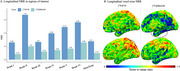 Figure 1
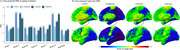 Figure 2
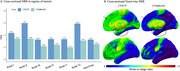 Figure 3
Peer Reviews
No public reviews on file for this paper yet. If you reviewed it on a platform where reviews are public (OpenReview, ICLR, NeurIPS, ICML), you can paste yours below so the community can read it here.
Videos
No videos yet. Explain this paper in a talk, walkthrough, or lecture? Add one.
Taxonomy
TopicsMedical Imaging Techniques and Applications · Dementia and Cognitive Impairment Research · Functional Brain Connectivity Studies