A Case of a Huge Granulomatous Epulis in the Mandibular Alveolar Region
Yu Hara, Kunio Yoshizawa, Junya Furukawa, Atsuya Ishiyama, Riku Kohara, Karen Gomi, Akinori Moroi, Koichiro Ueki

TL;DR
A 60-year-old man had a large benign tumor in his lower gum, which was successfully removed and did not return after six months.
Contribution
This paper presents a rare case of a large granulomatous epulis in the mandible, emphasizing its clinical and histopathological features.
Findings
The lesion was a 55 × 35 × 20 mm mass in the mandibular gingival region.
Histopathological analysis confirmed the diagnosis of granulomatous epulis.
The patient showed no recurrence after six months of follow-up.
Abstract
Epulis is a general diagnostic term for benign localized tumors that occur in the gingiva. Clinical diagnosis is usually straightforward based on the location and morphology of the lesion; however, large lesions may mimic malignancy. We report a case of a large granulomatous epulis in the mandibular alveolar region. A male in his 60s presented with swelling in the right mandible. Examination revealed a well‐defined, pedunculated soft tissue mass in the right mandibular gingival region. Then, 1 month after the initial visit, the lesion (55 × 35 × 20 mm) was resected under general anesthesia. Histopathological examination confirmed granulomatous epulis, and the patient has remained free of recurrence after 6 months of follow‐up.
Genes, proteins, chemicals, diseases, species, mutations and cell lines named across the full text — each resolved to its canonical identifier and authoritative record.
Click any figure to enlarge with its caption.
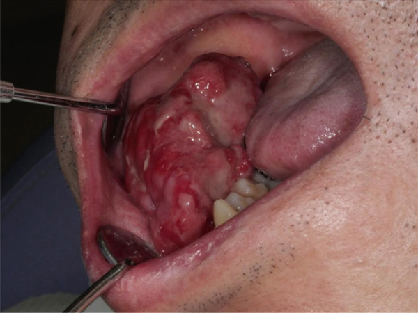 Figure 1
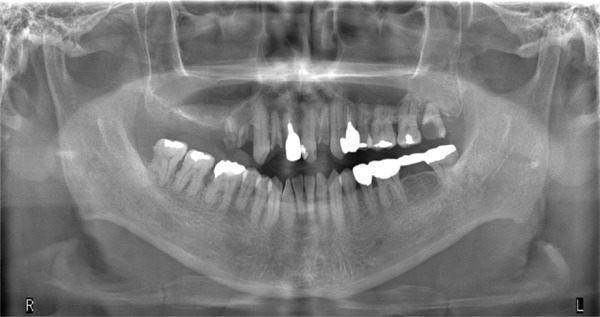 Figure 2
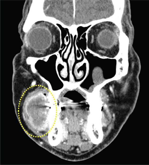 Figure 3
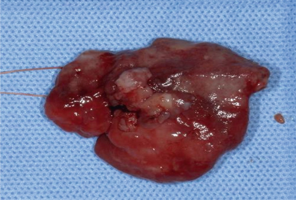 Figure 4
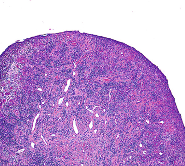 Figure 5
Peer Reviews
No public reviews on file for this paper yet. If you reviewed it on a platform where reviews are public (OpenReview, ICLR, NeurIPS, ICML), you can paste yours below so the community can read it here.
Videos
No videos yet. Explain this paper in a talk, walkthrough, or lecture? Add one.
Taxonomy
TopicsOral and Maxillofacial Pathology · Oral and Craniofacial Lesions · Oral and gingival health research
1. Introduction
Epulis is a general term describing benign localized growths of the gingiva, often resulting from chronic irritation such as sharp tooth edges, poor prostheses, or neglected dental treatment. Histopathologically, granulomatous epulis consists of proliferative granulation tissue covered by mucosal epithelium with inflammatory cell infiltration and edema.
We report a case of a large granulomatous epulis arising from the mandibular alveolar region. Although not rare, this case was initially suspected to be malignant due to its size and surface irregularity. The diagnostic process and final histopathological confirmation are discussed.
2. Case Presentation
A male in his 60s presented in October 2024 with swelling of the right mandibular gingiva. His medical history included hypertension and diabetes mellitus, with no relevant family or medication history. The patient had first noticed the swelling approximately 8 months earlier but did not seek treatment because it was painless.
Extraoral examination revealed preserved facial symmetry. Intraorally, a pedunculated, reddish mass with an irregular surface was noted in the right mandibular gingiva, raising suspicion of malignancy (Figure 1).
Intraoral photograph at the initial examination showing gingival swelling of the right mandible with an irregular surface.
Panoramic radiography showed no evidence of bone resorption (Figure 2). Contrast‐enhanced CT imaging revealed a soft tissue mass without calcification or bony invasion (Figure 3). Based on these findings, a provisional diagnosis of a right mandibular tumor was made.
Panoramic radiograph showing no bone resorption.
Contrast‐enhanced CT image showing no calcification or bone invasion.
In November 2024, the lesion was completely excised under general anesthesia using a scalpel and electrocautery (Figure 4). The surgical wound was sutured, and healing was uneventful. Histopathological analysis revealed squamous epithelial tissue with inflammatory cell infiltrates, including lymphocytes, plasma cells, and neutrophils, with fibrosis and granulation tissue formation in the stroma (Figure 5).
Excised specimen measuring 55 × 35 × 20 mm with a smooth surface.
Histopathological image (H&E stain, × 40) showing squamous epithelium with inflammatory infiltration and granulation tissue formation.
A final diagnosis of granulomatous epulis was made. At 6 months postoperatively, the patient showed good healing with no evidence of recurrence.
3. Discussion
Epulis is not a true neoplasm but a reactive benign lesion. Among its variants, granulomatous epulis frequently presents with ulcers, erythema, or an irregular surface that can mimic malignant tumors, making it commonly encountered. [1] In this case, the lesion′s large size (55 × 35 × 20 mm) and irregular surface initially raised clinical suspicion of malignancy. While epulis is relatively easy to diagnose clinically based on location and characteristics, large lesions or those with unusual surface features can sometimes clinically mimic malignancy at first glance. Indeed, cases have been reported where lesions suggestive of epulis were histopathologically found to be metastatic carcinoma to the gingiva [2], malignant salivary gland tumors [3], squamous cell carcinoma, or Burkitt lymphoma [4]. Therefore, when a neoplastic lesion cannot be ruled out, performing a preoperative biopsy is crucial. Findings suggestive of a tumor include the following: (1) large lesion size, (2) rapid growth, (3) broad base, (4) alveolar bone resorption, (5) multinodular appearance, (6) granular texture, (7) ulceration, and (8) easy bleeding. In our case, the large lesion size meant a neoplastic lesion could not be excluded. However, imaging studies showed no evidence of infiltration or destruction. A preoperative biopsy was performed, and histopathology confirmed a benign inflammatory lesion, leading to excision. Performing a preoperative biopsy is crucial [5].
While this case is not particularly rare, the reasoning process in the diagnostic pathway holds educational value. The absence of pain and the patient′s poor oral hygiene likely contributed to the delayed diagnosis and enlargement of the lesion. Therefore, regular follow‐up and oral hygiene management are necessary. Clinicians encountering large gingival masses should consider a broad differential diagnosis including pyogenic granuloma, marginal osteoclastoma, marginal giant cell granuloma, and squamous cell carcinoma.
Accurate diagnosis requires correlating clinical findings, imaging findings, and histopathological findings, followed by removal of the causative stimulus. Reporting detailed diagnostic reasoning enhances the educational value of such cases and helps clinicians distinguish benign reactive lesions from malignant tumors.
4. Conclusion
Large granulomatous epulis lesions may mimic malignancy due to their size and irregular appearance. This case emphasizes the importance of systematic diagnostic assessment combining clinical, imaging, and histopathological findings to ensure correct diagnosis and management.
Ethics Statement
This case report adheres to the Declaration of Helsinki, and written informed consent was obtained from the patient.
Conflicts of Interest
The authors declare no conflicts of interest.
Funding
No funding was received for this manuscript.
The reference list from the paper itself. Each links out to its DOI / PubMed record.
- 1Matsusue Y. , Yamamoto K. , Nakagami K. , and Kirita T. , Two Cases of Relatively Large Epulis Suspected of a Tumor from Clinical Findings, Journal of the Japanese Society of Stomatology. (2017) 23, no. 1, 46–53, 10.6014/jjsom.23.46. · doi ↗
- 2Yoko A. , Yoshinori K. , Tadahide N. , Ito H. , Matsumoto K. , and Numao A. , Clinical Study of tumors metastasizing to the Oral Cavity, Journal of the Japanese Society of Oral and Maxillofacial Surgeons. (2001) 47, no. 9, 559–562, 10.5794/jjoms.47.559. · doi ↗
- 3Miyauchi M. , Igaki K. , Ito Y. , Harada N. , Sugiyama M. , and Ishikawa T. , An uncommon Case of acinic cell carcinoma Presenting with a caulescent lump at the retromolar region, Journal of the Japanese Society of Oral and Maxillofacial Surgeons. (2001) 47, no. 10, 607–610, 10.5794/jjoms.47.607. · doi ↗
- 4Sekiwa T. , Uehara T. , Uehara H. , Honda M. , Terakado M. , and Mugishima H. , A Case of Burkitt Lymphoma involving the Right Maxillary Gingiva in a 2-year-7-month-old Infant, Japanese Journal of Oral and Maxillofacial Surgery. (2001) 47, no. 3, 174–177, 10.5794/jjoms.47.174. · doi ↗
- 5Baba N. , Matsuo N. , Kawasaki G. , Mizuno A. , Fujita S. , and Okabe H. , A Case of Giant Osteoplastic Epulis Causing Facial Deformity, Journal of the Japanese Stomatological Association. (1999) 48, no. 1, 92–96, 10.11277/stomatology 1952.48.92. · doi ↗