A fishy situation – expanding the differential diagnosis of the fish-mouth papilla
Omar El Ouarzadi, Reda Goudrar, Marc-André Smith, Marcel Tomaszewski

Abstract
Genes, proteins, chemicals, diseases, species, mutations and cell lines named across the full text — each resolved to its canonical identifier and authoritative record.
Click any figure to enlarge with its caption.
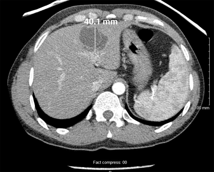 Fig. 1
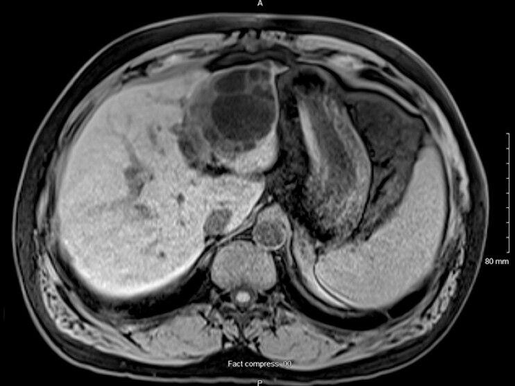 Fig. 2
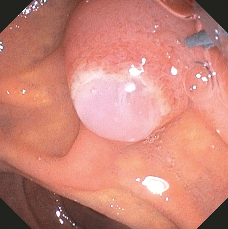 Fig. 3
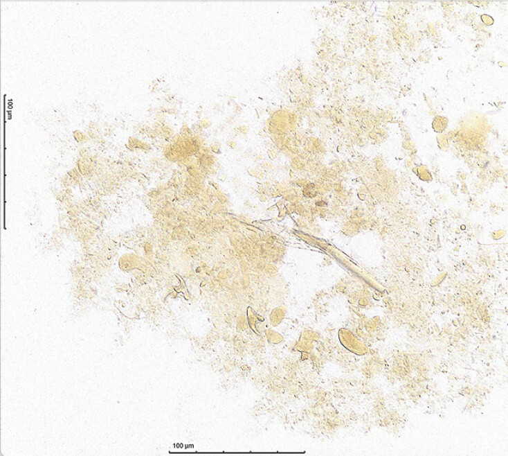 Fig. 4
Peer Reviews
No public reviews on file for this paper yet. If you reviewed it on a platform where reviews are public (OpenReview, ICLR, NeurIPS, ICML), you can paste yours below so the community can read it here.
Videos
No videos yet. Explain this paper in a talk, walkthrough, or lecture? Add one.
Taxonomy
TopicsCongenital Ear and Nasal Anomalies · Sinusitis and nasal conditions · Oral and Maxillofacial Pathology
Hydatid disease (echinococcosis) is a parasitic infection that presents with hepatic cysts and pulmonary involvement. Hepatic hydatid disease can lead to an obstruction of the biliary tree in the case of cyst rupture 1 . This case report provides endoscopic images of a hepatic hydatid cyst mimicking the fish-mouth appearance of papilla, usually pathognomonic for main duct intraductal papillary mucinous neoplasm (MD-IPMN 2 ).
A 35-year-old man, from North Africa, presented with acute on chronic epigastric pain. Initial work-up revealed increased alanine aminotransferase (648 IU/L) and total bilirubin (99 μmol/L) levels. A computed tomographic scan showed a multilobulated, septated, calcified lesion in the inferior left liver, suggestive of a hepatic cyst ( Fig. 1 ). He was later lost to follow-up.
A CT scan showed a multi-lobulated, septated, calcified lesion in the inferior left liver, suggestive of a hepatic cyst. CT, computed tomography.
Nine years later, he returned to the emergency room with work-up confirming cholangitis. MRCP showed poorly defined obstructive filling defects within a slightly dilated bile of 8 mm in diameter ( Fig. 2 ). Interval growth of the hepatic cyst and left intrahepatic biliary ductal dilatation was noted.
MRI revealed interval growth of the hepatic cyst and left intra-hepatic biliary ductal dilatation. MRI, magnetic resonance imaging.
ERCP demonstrated a white, soft, mucinous substance protruding from the ampulla ( Video 1 ). The endoscopic images were suggestive of a fish-mouth papilla ( Fig. 3 ). After biliary sphincterotomy, the spontaneous discharge of thick, white membranes occurred.
Fish-mouth papilla appearance of the ampulla, and discharge of thick, white membranes after sphincterotomy.Video 1
ERCP: a white, soft, mucinous substance protruding from the ampulla, suggestive of a fish-mouth papilla.
Evaluation of the biliary aspirate with wet mount iodine microscopy identified numerous hooklets ( Fig. 4 ) and confirmed the diagnosis of a compressive echinococcal cyst. He was first treated with albendazole and then referred to hepatobiliary surgery for resection. Both ERCP and left hepatectomy conferred a significant clinical and biochemical improvement.
Wet mount iodine microscopy: biliary aspirate with numerous hooklets, confirming echinococcosis.
A ruptured hydatid cyst can lead to the fish-mouth papilla, which is typically pathognomonic for MD-IPMN. ERCP was effective in the treatment of cholangitis in the context of biliary obstruction from hydatid cyst rupture and biliary aspirate confirmed the diagnosis.
Endoscopy_UCTN_Code_CCL_1AB_2AG_3AD
The reference list from the paper itself. Each links out to its DOI / PubMed record.