Combined digestive endoscopy and laryngoscopy for successful management of an early-stage epiglottic carcinoma
Xun-Mei Duan, Yu Bao, Ye-Han Zhou, Zhen-Ming Zhang

Abstract
Genes, proteins, chemicals, diseases, species, mutations and cell lines named across the full text — each resolved to its canonical identifier and authoritative record.
Click any figure to enlarge with its caption.
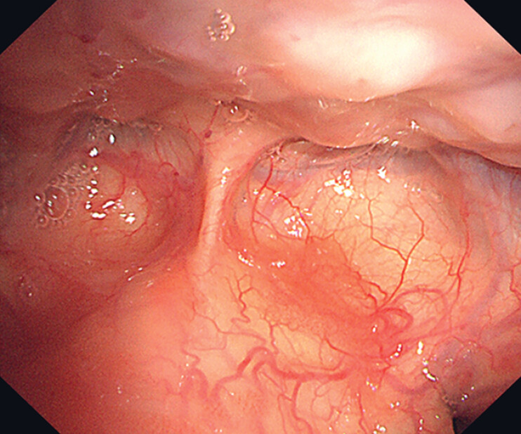 Fig. 1
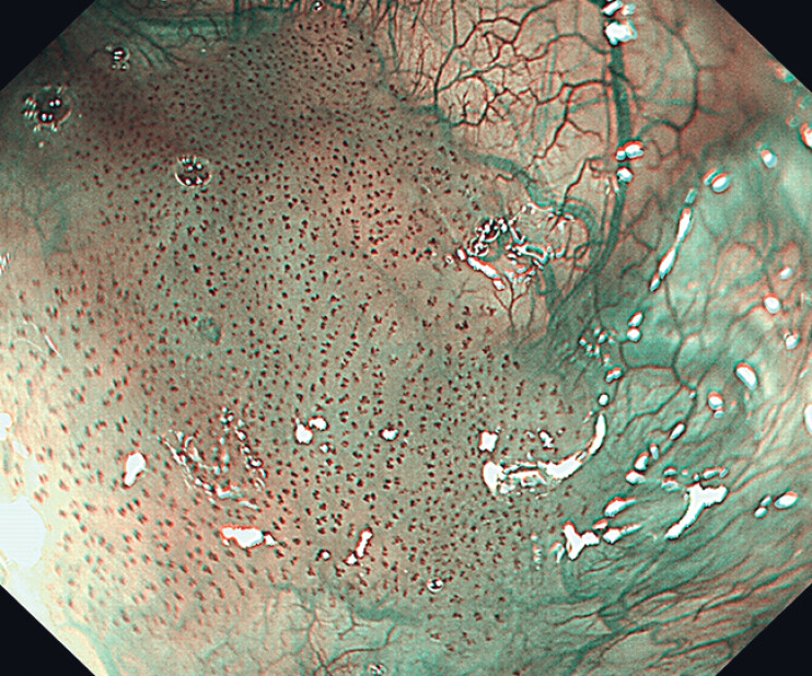 Fig. 2
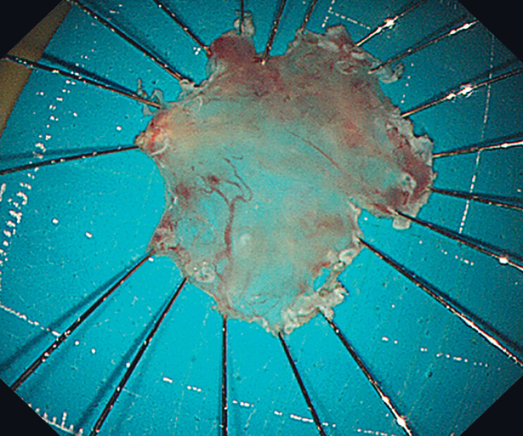 Fig. 3
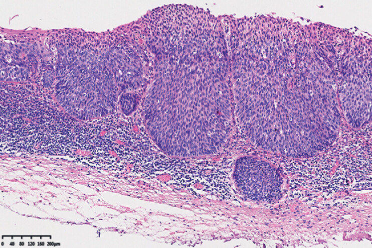 Fig. 4
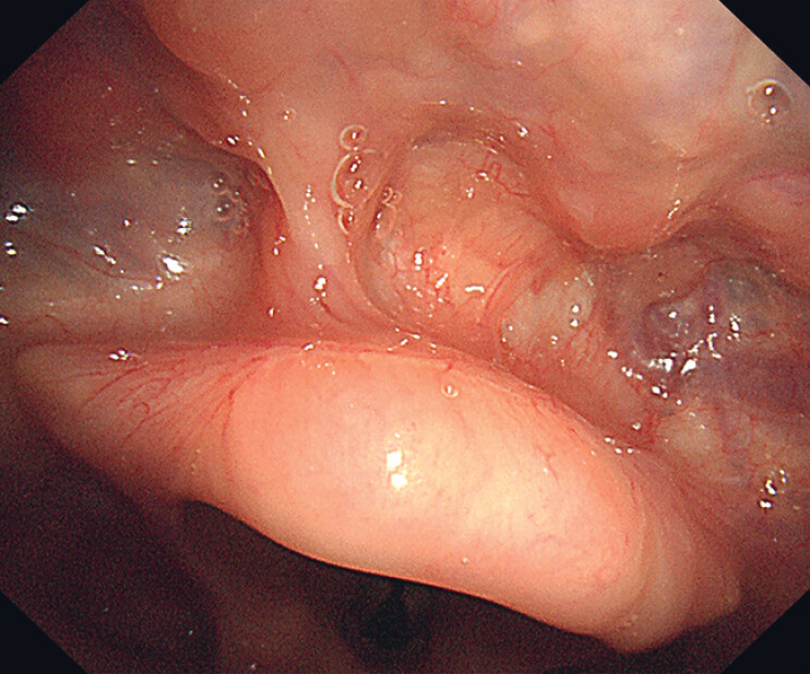 Fig. 5
- —The Early Cancer Standardization Capacity Building Fund Program
- —Sichuan Province Medical Young Innovative Research Project Program
Peer Reviews
No public reviews on file for this paper yet. If you reviewed it on a platform where reviews are public (OpenReview, ICLR, NeurIPS, ICML), you can paste yours below so the community can read it here.
Videos
No videos yet. Explain this paper in a talk, walkthrough, or lecture? Add one.
Taxonomy
TopicsHead and Neck Cancer Studies · Dysphagia Assessment and Management · Esophageal Cancer Research and Treatment
A 70-year-old man with a history of resected esophageal carcinoma was hospitalized due to the discovery of several lesions on the surface of the epiglottis during follow-up laryngoscopy. Laryngoscopy revealed multifocal and irregular lesions scattered on the lingual surface of the epiglottis. These flat (0-IIb), reddish lesions displayed B1-type intraepithelial papillary capillary loops under narrow-band imaging ( Fig. 1 , Fig. 2 ). A tumor biopsy revealed in situ squamous cell carcinoma. Meanwhile, enhanced computed tomography indicated an absence of lymph node involvement.
The lesions in the epiglottis were flat (0-IIb) and slightly reddish under white light laryngoscopy.
The appearance of the neoplastic lesions under narrow-band imaging.
Endoscopic submucosal dissection (ESD) was performed under general anesthesia induced via nasal intubation ( Video 1 ). A therapeutic gastroscope was used for the procedure. The constrained anatomical space of the epiglottic vallecula necessitated the use of a snare traction approach to facilitate submucosal dissection in challenging areas. Subsequently, en bloc resection of the lesions was achieved ( Fig. 3 ). No adverse events were observed during or after the procedure. Histopathological analysis confirmed the local invasion of squamous cell carcinoma into the lamina propria and showed that R0 resection was achieved ( Fig. 4 ). A follow-up laryngoscopy conducted 3 months post-ESD revealed complete healing of the surgical site ( Fig. 5 ), and subsequent routine follow-ups over 46 months detected neither local recurrence nor lymphadenopathy.
Combined digestive endoscopy and laryngoscopy for the successful management of an early-stage epiglottic carcinoma.Video 1
The resected specimen of the lesions.
Histopathological analysis confirmed the presence of squamous cell carcinoma that locally invaded the lamina propria.
Follow-up laryngoscopy showing that the surgical wound has completely healed with scar formation.
In this case, the epiglottic mucosal lesions were initially overlooked on gastroscopy; however, they were subsequently detected by laryngoscopy. This finding highlights the complementary diagnostic value of laryngoscopy alongside gastroscopy in screening high-risk populations for pharyngeal cancer 1 . Complete endoscopic resection via digestive endoscopy was then performed, demonstrating its essential role in the treatment of early-stage pharyngeal cancer 2 3 . Post–ESD follow-up was conducted via laryngoscopy under local anesthesia, providing a well-tolerated and patient-friendly approach. This case highlights the clinical significance of an integrated strategy combining digestive endoscopy and laryngoscopy for the comprehensive management of early pharyngeal cancer, supporting its broader adoption in clinical practice.
Endoscopy_UCTN_Code_TTT_1AO_2AG_3AD
The reference list from the paper itself. Each links out to its DOI / PubMed record.
- 1Lu G Zhang Q Kang S Evaluating hypopharyngeal carcinoma using narrow band imaging and oxygen-injected laryngoscope: New technique Am J Otolaryngol 20234410373310.1016/j.amjoto.2022.10373336527815 · doi ↗ · pubmed ↗
- 2Okada K Tsuchida T Ishiyama A Endoscopic mucosal resection and endoscopic submucosal dissection for en bloc resection of superficial pharyngeal carcinomas Endoscopy 20124455656410.1055/s-0032-130972022638778 · doi ↗ · pubmed ↗
- 3Matsuura N Kato M Iwata K Efficacy and safety of the water pressure method for endoscopic submucosal dissection in superficial pharyngeal cancer Endosc Int Open 202412 E 621E 62810.1055/a-2284-918438681148 PMC 11052648 · doi ↗ · pubmed ↗