Cortical freewater increases in areas of tau tangle aggregation, independent of cortical thinning
Brandon J Hall, Etienne Aumont, Arnaud Boré, Joseph Therriault, Seyyed Ali Hosseini, Nesrine Rahmouni, Arthur C. Macedo, Stijn Servaes, Gleb Bezgin, Jaime Fernandez Arias, Yi‐Ting Wang, Tevy Chan, Lydia Trudel, Jenna Stevenson, Andrea Benedet, Gallen Triana‐Baltzer

TL;DR
The study finds that tau tangle accumulation in Alzheimer's disease is linked to increased freewater in specific brain regions, independent of other factors like cortical thinning.
Contribution
The study identifies a novel link between tauopathy and microstructural changes in the brain, independent of amyloid and cortical thinning.
Findings
Tau PET correlates with freewater in gray matter of temporal and occipital regions.
p-tau217 correlates with freewater in periventricular white matter in tau-positive individuals.
Astrogliosis may contribute to microstructural changes linked to tau tangles.
Abstract
It is unclear how cortical microstructure changes across biological stages of Alzheimer's disease (AD), and how this occurs in response to amyloid plaques, tauopathy, and inflammation. We hypothesize that tauopathy is chiefly responsible. To test this, we investigated how different measures of tauopathy, including plasma and PET, influence the microstructure in the whole brain, temporal meta‐ROI, and hippocampus. We sampled 301 participants (59% female, age 67±10) from the TRIAD cohort with T1 MRI, diffusion‐weighted MRI, amyloid PET (F18‐NAV4694), tau PET (F18‐MK6240), and plasma biomarkers. We used the NODDI‐flow pipeline to estimate the diffusion characters of the TRIAD cohort (Dparallel, Dorthogonal,Disotropic) and run the NODDI‐Bingham algorithm to calculate whole‐brain isotropic volume fraction images (“freewater”). FreeSurfer 7 was used to derive hippocampal and temporal…
Genes, proteins, chemicals, diseases, species, mutations and cell lines named across the full text — each resolved to its canonical identifier and authoritative record.
Click any figure to enlarge with its caption.
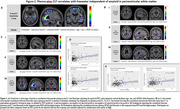 Figure 1
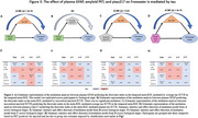 Figure 2
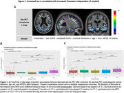 Figure 3
Peer Reviews
No public reviews on file for this paper yet. If you reviewed it on a platform where reviews are public (OpenReview, ICLR, NeurIPS, ICML), you can paste yours below so the community can read it here.
Videos
No videos yet. Explain this paper in a talk, walkthrough, or lecture? Add one.
Taxonomy
TopicsDementia and Cognitive Impairment Research · Advanced Neuroimaging Techniques and Applications · Cerebrospinal fluid and hydrocephalus