Silent Propagation of Classical Scrapie Prions in Homozygous K222 Transgenic Mice
Natalia Fernández-Borges, Alba Marín-Moreno, Juan Carlos Espinosa, Sara Canoyra, Olivier Andréoletti, Juan María Torres

TL;DR
This study shows that homozygous K222 transgenic mice can silently carry classical scrapie prions without showing symptoms, which could impact breeding programs.
Contribution
The study reveals silent prion propagation in K222 homozygous mice, challenging assumptions about resistance to classical scrapie.
Findings
Homozygous K222-Tg516 mice did not show clinical signs of scrapie but accumulated prion proteins.
Few isolated scrapie PrP plaques were observed in mice inoculated with highly adapted isolates.
Silent carriers could emerge in goat populations if K222 is included in breeding programs.
Abstract
Classical scrapie affects sheep and goats. To control prevalence in sheep, the European Union initiated breeding programs targeting resilient genotypes. Although certain goat polymorphisms, such as Q222K, are linked to resistance, specific breeding programs have not been implemented. Hemizygous transgenic mice carrying the goat K222 cellular prion protein (PrP) allele (K222-Tg516) exhibited resistance to several classical scrapie isolates. We inoculated homozygous K222-Tg516 and Q222-Tg501 mice with various scrapie isolates. Homozygous K222-Tg516 mice reached the end of their lifespan without exhibiting clinical signs; we observed brain proteinase K–resistant PrP accumulation in those mice that was lower than in Q222-Tg501 mice. Histologically, K222-Tg516 brains lacked prion-related lesions, except for the presence of few isolated scrapie PrP plaques in cases of isolates highly adapted…
Genes, proteins, chemicals, diseases, species, mutations and cell lines named across the full text — each resolved to its canonical identifier and authoritative record.
Click any figure to enlarge with its caption.
 Figure 1
 Figure 2
 Figure 3
 Figure 4
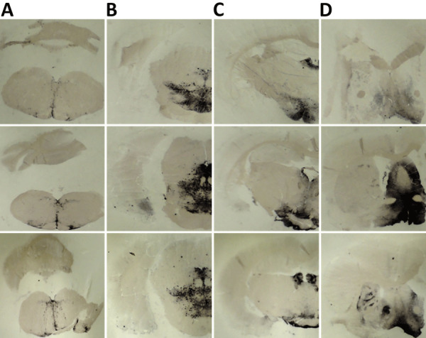 Figure 5
 Figure 6
 Figure 7
| Category | Isolate | Species | Origin | Goat PrP genotype† | Description | Supplier |
|---|---|---|---|---|---|---|
| I | 198/9 | Sheep | Italy | wt; S240S | Classical scrapie isolate from a naturally infected sheep | ISS |
| II | S2 | Goat | Spain | wt; S240P | Classical scrapie isolate from a naturally infected goat | UNIZAR |
| Goat | France | wt | Classical scrapie isolate from an experimentally infected goat | ENVT | ||
|
| Goat | France | K222 | Classical scrapie isolate from an experimentally infected goat | ENVT | |
| II + III | UKA2 | Goat | United Kingdom | wt; S240P | Classical scrapie isolate from a naturally infected goat | APHA |
|
| F14 | Goat | France | wt; I142M, S240P | Classical scrapie isolate from a naturally infected goat | INRA |
| IV | F10 | Goat | France | wt; S240P | Classical scrapie isolate from a naturally infected goat | INRA |
|
| C1 | Goat | Cyprus | wt | Classical scrapie isolate from a naturally infected goat | VS |
| Negative control | Healthy goat brain | Goat | France | wt | Brain from a noninfected goat | INRA |
| Category | Isolate | 1st passage | 2nd passage | |||
|---|---|---|---|---|---|---|
| Mean survival time ± SD, d | No. diseased and PrPres-positive/no. inoculated | Mean survival time ± SD, d | No. diseased and PrPres-positive/no. inoculated | |||
| I | 198/9 | 592 ± 13 | 6/6 |
| 536 ± 46 | 5/5 |
| II | S2 | 228 ± 15 | 6/6 | 233 ± 4 | 6/6 | |
| 379 ± 31 | 5/5 | ND | NA | |||
|
| 415 ± 40 | 6/6 |
| ND | NA | |
| II + III | UKA2 | 245 ± 36 | 5/5 | 252 ± 8 | 6/6 | |
|
| F14 | 526 ± 46 | 4/4 |
| 241 ± 22 | 4/4 |
| IV | F10 | 449 ± 19 | 5/5 | 372 ± 14 | 6/6 | |
| F10/K222-Tg516 | 495 ± 26 | 3/3 | ND | NA | ||
|
| C1 | 483 ± 15 | 4/4 |
| 301 ± 10 | 4/4 |
| Negative control | Healthy goat brain | >650 | 0/6† | >650 | 0/6† | |
| Category | Isolate | 1st passage | 2nd passage | ||||
|---|---|---|---|---|---|---|---|
| Mean survival time ± SD, d | No. diseased and PrPres-positive/no. inoculated | Mean survival time ± SD, d | No. diseased and PrPres-positive/no. inoculated | ||||
| I | 198/9 | >650 | 1/6† |
| ND | NA | |
| II | S2 | >650 | 3/4† | >650 | 7/7† | ||
| >650 | 5/5† | >650 | 5/5† | ||||
|
| >650 | 4/4† |
| >650 | 6/6† | ||
| II + III | UKA2 | >650 | 4/4† | >650 | 5/5† | ||
|
| F14 | >650 | 4/4† |
| >650 | 5/5† | |
| IV | F10 | >650 | 6/6† | >650 | 5/5† | ||
| F10/K222-Tg516 | >650 | 5/5† | >650 | 6/6† | |||
|
| C1 | >650 | 7/7† |
| ND | NA | |
| Negative control | Healthy goat brain | >650 | 0/6† | >650 | 0/6† | ||
Peer Reviews
No public reviews on file for this paper yet. If you reviewed it on a platform where reviews are public (OpenReview, ICLR, NeurIPS, ICML), you can paste yours below so the community can read it here.
Videos
No videos yet. Explain this paper in a talk, walkthrough, or lecture? Add one.
Taxonomy
TopicsPrion Diseases and Protein Misfolding · Animal Genetics and Reproduction · Neurological diseases and metabolism
Scrapie is a fatal infectious neurodegenerative disease inherent to sheep and goats that falls within the spectrum of transmissible spongiform encephalopathies (TSEs) or prion diseases. Of note, various mammals, including cattle with bovine spongiform encephalopathy (BSE), mink with transmissible mink encephalopathy, cervids with chronic wasting disease, and humans with Creutzfeldt-Jakob disease, can also succumb to TSEs. The hallmark of those diseases is posttranslational conversion of the host cellular prion protein (PrP), PrP^C^, into a misfolded pathologic isoform causing scrapie, PrP^Sc^, which accumulates within the central nervous system of affected individuals (1).
Infection with TSEs in an organism is influenced by 2 main factors: the similarity between the primary PrP sequence of the host (recipient) and the donor (inoculum), and the prion strain (2). Together, those factors define the concept of the transmission barrier. Sheep and goats share the same PrP primary sequence, although polymorphisms differ between the animals. In sheep, high susceptibility to classical scrapie is associated with the V_136_R_154_Q_171_ and A_136_R_154_Q_171_ alleles, whereas the A_136_R_154_R_171_ genotype is linked to resistance (3–8). To control and decrease classical scrapie in sheep, European Union member states have established breeding programs on the basis of the selection of the resistant A_136_R_154_R_171_ allele, although the variant does not confer resistance against the atypical/Nor98 scrapie strain (9). In goats, some polymorphisms, such as I_142_M (10–13) and N_146_S (14), have been associated with resistance to scrapie infection.
The most promising results of studies were in regard to goat-resistant polymorphisms for the goat Q_222_K polymorphism. The lysine allele (K_222_) was first reported to confer resistance in Italy (15,16), and similar results were later found in France (10) and Greece (17,18). Cell-free conversion assays also indicated that K_222_ provides protection against the ME7 scrapie strain (19). Experimental studies in goats found that heterozygous Q/K_222_ and homozygous K_222_ goats either showed resistance to classical scrapie or exhibited clear delays in incubation times after intracerebral or oral inoculation (20–23) and reduced contribution of K_222_ to proteinase K–resistant PrP (PrP^res^) formation in Q/K_222_ heterozygous goats infected with scrapie (24). In addition, Q/K_222_ heterozygous goats were found to harbor a relative abundance of the natural α-cleaved PrP^C^ fragment C1, which has also been detected in classical scrapie-resistant R_171_ sheep (25). Furthermore, Q/K_222_ heterozygous goats inoculated with goat BSE showed neither evidence of clinical prion disease nor PrP^Sc^ accumulation in the brain or peripheral tissues (26,27), but low infectivity was detected after long postinoculation times (26). Finally, 1 goat harboring the K_222_-PrP^C^ variant tested positive for atypical/Nor98 scrapie, indicating that the genotype may still be susceptible to this scrapie strain (28). All those results were replicated using a hemizygous transgenic mouse line expressing the K_222_-PrP^C^ allele, which was found to be resistant to several classical scrapie isolates and cattle BSE, while susceptible to goat or sheep BSE and atypical scrapie (29,30).
We conducted our study on the transgenic homozygous mouse line, along with its control counterpart harboring the wild-type glutamine allele (Q_222_). We intracranially inoculated the mice with several isolates representative of different categories of classical scrapie strains to test whether animals still remained uninfected, as previously reported (29), or if they mimicked the results found in homozygous goats (22).
Methods
Ethics Considerations
We performed animal experiments in strict accordance with the recommendations included in the guidelines of European Community Council 2010/63/UE and made all efforts to minimize animal suffering. The Committee on the Ethics of Animal Experiments of the Instituto Nacional de Investigación y Tecnología Agraria y Alimentaria and the General Directorate of the Madrid Community Government approved the study (permit nos. CEEA 2011–050, PROEX 263/15).
Prion Transmission Studies
We intracranially inoculated 20 μL of 10% (wt/vol) brain homogenate from previously characterized classical scrapie isolates (Table 1) into the right parietal lobe of 5–7 transgenic mice (6–7 weeks old), which expressed either the wild-type goat PrP^C^ (Q_222_-Tg501) or the K_222_-PrP^C^ variant (K_222_-Tg516) (29,30) in homozygosity. PrP^C^ expression levels of both mice lines were 2- to 4-fold the physiologic levels found in goat brain (29). We used a 25-gauge disposable hypodermic needle to inoculate animals while they were anesthetized with isoflurane.
After inoculation, we monitored mice daily and assessed their neurologic status twice a week. We euthanized animals when the progression of prion disease was evident, at the end of their lifespan (around 650 days postinoculation), or at previously established endpoints as part of a kinetic study. We harvested mouse brains and sliced them sagittally. We fixed half of each brain in 10% buffered formalin for histopathologic analysis and homogenized the remaining portion as 10% (wt/vol) in 5% glucose to detect PrP^res^ by Western blot.
We calculated survival time as the mean number days postinoculation for all mice that tested positive for PrP^res^ in the brain, with the SD included. We expressed attack rate as the proportion of PrP^res^-positive mice among all the inoculated mice.
Western Blotting
We homogenized mouse brain tissue in 5% glucose solution in distilled water using grinding tubes (Bio-Rad Laboratories, https://www.bio-rad.com) and adjusted to 10% (wt/vol) using a TeSeE Precess 48TM homogenizer (Bio-Rad) according to the manufacturer’s instructions. We determined PrP^res^ presence in transgenic mouse brains by Western blot analysis of 10–100 µL of 10% (wt/vol) brain homogenate, as previously described (32). We incubated membranes with the Sha31 monoclonal antibody (mAb) (33), which recognizes the 148_YEDRYYRE_155 epitope of the goat PrP sequence. We detected immunocomplexes with horseradish peroxidase-conjugated mouse IgG (GE HealthCare, https://www.gehealthcare.com) after 1 hour of incubation. We visualized immunoreactivity by chemiluminescence with ECL Select (GE HealthCare). We captured images using ChemiDoc XRS + System (Bio-Rad) and processed them using Image Lab 5.2.1 software (Bio-Rad).
Histologic Analysis
To analyze brain tissue, we trimmed and dehydrated formalin-fixed brains, embedded them in paraffin wax, and cut 4-μm slices. We dewaxed and rehydrated the specimens by standard procedures. We established the vacuolar lesion profile of the brains in accordance with published standard methods and semiquantitatively scored vacuolation on a scale of 0–5 in different brain areas (34,35).
For immunohistochemical (IHC) demonstration of PrP^Sc^ accumulation, tissue sections underwent antigen retrieval and hydrogen peroxide quenching as previously described (36). We incubated the sections with 2A11 mAb (37), which recognizes the 163_QVYYRPVDQ_171 epitope of the goat PrP sequence. Subsequently, we subjected the sections to antigen retrieval and inactivation of endogenous peroxidase activity before incubating them with the 2A11 mAb. We used a commercial immunoperoxidase technique (VECTASTAIN Elite ABC Kit; Vector Laboratories, https://vectorlabs.com), according to the manufacturer’s instructions. Finally, we counterstained the sections with Mayer’s hematoxylin. We used the Sha31 mAb (33) for paraffin-embedded tissue blotting, as previously described (38,39).
Results
Homozygous K222-Tg516 Mice and Resistance to Classical Scrapie PrPSc
We intracranially inoculated homozygous K_222_-Tg516 with classical scrapie isolates (Table 1) previously characterized as representative of different prion strains circulating in Europe (31*, *40). Although all mice expressing the wild-type goat PrP (Q_222_-Tg501) developed recognizable prion disease, K_222_-Tg516 mice reached the end of their lifespan without showing clinical signs indicative of prion disease (Table 2). After second passage, survival times were still prolonged, even reaching the end of the mice’s lifespan again (Table 2). However, in both first and second passages, Western blot analysis showed the presence of PrP^res^ in the brains of K_222_-Tg516 animals inoculated with the different classical scrapie isolates (Figure 1, panel A). For the 198/9 and S2 isolate, the percentage of PrP^res^-positive animals in the first passage was not 100% of the inoculated animals (Tables 2, 3). At least for the S2 isolate, 100% of the inoculated mice were PrP^res^-positive by the completion of the second passage (Tables 2, 3). Comparison between the PrP^res^ signature of the original inoculum and the PrP^res^ obtained molecular mass for the nonglycosylated band, depending on the individual (Figure 1, panel A). In addition, brain PrP^res^ accumulation in K_222_-Tg516 mice was remarkably reduced compared with that in Q_222_-Tg501 mice for most of the inoculated isolates, with the exception of F14 and F10 (Figure 1, panel A).
Proteinase K–resistant PrP (PrPres) accumulation in brains of K222-Tg516 and Q222-Tg501 homozygous mice in study of propagation of classical scrapie prions. A) Comparison of the biochemical profile of brain PrPres from classical scrapie isolates in K222-Tg516 mice with that in Q222-Tg501 mice using Sha31 monoclonal antibody. Exposure time and dilution factor are specified. B) Comparison of the biochemical profile of brain PrPres of CP060146 and F10 isolates of classical scrapie, before (left) and after (right) adaptation to the K222-PrPC context, in Q222-Tg501 mice, using the Sha31 monoclonal antibody. Molecular weight markers are indicated on the right side of each band.
K_222_-Tg516 PrP^res^-positive animals exhibited only a few vacuolations that were difficult to distinguish from those resulting from the physiologic aging process (Figure 2). Immunohistochemistry of K_222_-Tg516 mice inoculated with CP060146/K_222_ goat and F10/K_222_-Tg516 inocula revealed only a few large and focalized PrP^Sc^ plaques and lacked any other type of deposits affecting neurons or microglia cells (Figures 3, 4). Those PrP^Sc^ deposits were restricted to the mesencephalon, thalamus, and hypothalamus areas (Figures 3, 4). We detected no deposits for the remaining inoculations (data not shown). Consistent with our findings, paraffin-embedded tissue blotting showed clear PrP^res^ deposition only in K_222_-Tg516 mice inoculated with CP060146/K_222_ goat (Figure 5) and F10/K_222_-Tg516 inocula (data not shown), with deposition to the exact same brain areas affected by IHC (Figures 3, 4). We detected no deposits for the remaining inoculations (data not shown).
Histologic analysis of brain tissue from K222-Tg516 homozygous mice inoculated with classical scrapie in study of propagation of classical scrapie prions. Comparative analysis shows the vacuolar lesion profile in homozygous K222-Tg516 mice inoculated with different scrapie isolates compared with noninoculated mice. G, gray matter; W, white matter.
Immunohistochemistry results of brain tissue in study of propagation of classical scrapie prions. Images are of tissue specimens from K222-Tg516 mice inoculated with F10 goat scrapie isolate at second passage. Results are visualized using the Sha31 monoclonal antibody. A) Thalamus specimen. B) Hippocampus specimen. C) Midbrain specimen. Original magnification ×40.
Immunohistochemistry results of brain tissues in study of propagation of classical scrapie prions. Images are of tissue specimens from K222-Tg516 mice inoculated with CP060146/K222 goat isolate. Results are visualized using the Sha31 monoclonal antibody. A) Hippocampus specimen tested at first passage. B) Midbrain specimen tested at first passage. C) Hippocampus specimen tested at second passage. D) Midbrain specimen tested at second passage. Original magnification ×40.
Paraffin-embedded tissue blotting results of brain tissues in study of propagation of classical scrapie prions. Images are of brain specimens from 3 distinct K222-Tg516 mice inoculated with CP060146/K222 goat isolate. Results are visualized with the Sha31 monoclonal antibody. A) Cerebellum specimens. B) Thalamus specimens. C) Hippocampus specimens. D) Cerebral cortex specimens. Proteinase K–resistant prion protein is visible as dark staining in similar brain regions in the 3 mice. Original magnification ×20.
Proteinase K Studies in K222-Tg516 Mice
The differential brain PrP^res^ accumulation observed between K_222_-Tg516 and Q_222_-Tg501 mice (Figure 1, panel A) can be attributed to 2 alternative hypotheses. There could be a genuine reduction in PrP^res^ accumulation for these classical scrapie isolates in K_222_-Tg516 mice. Alternatively, the produced PrP^res^ might be more susceptible to proteinase K treatment, resulting in a weaker Western-blotting signal. To distinguish between those 2 possibilities, we performed proteinase K resistance analyses using different enzyme concentrations in both Q_222_-Tg501 and K_222_-Tg516 mice inoculated with F10 (which exhibited similar PrP^res^ accumulation between K_222_-Tg516 and Q_222_-Tg501 mice) and CP060146 (which showed reduced PrP^res^ accumulation in K_222_-Tg516 mice compared with Q_222_-Tg501 mice) isolates. In all cases, proteinase K consistently acted at a concentration of 50 µg/mL (which falls within the normal proteinase K concentration range for routine Western blotting); we observed the same pattern and signal intensity at a concentration of 500 µg/mL (Figure 6). However, protease action did not achieve proper PrP^res^ resolution at concentrations of 1 µg/mL and 0.1 µg/mL (Figure 6). Those results suggest that both isolates, when replicating in either Q_222_-PrP^C^ or K_222_-PrP^C^ contexts, retain the same proteinase K sensitivity. Thus, the differences in Western blotting signals detected previously (Figure 1, panel A) truly account for reduced brain PrP^res^ accumulation in K_222_-Tg516 mice.
Proteinase K digestion studies conducted as part of study of propagation of classical scrapie prions. K222-Tg516 and Q222-Tg501 homozygous mice were inoculated with classical scrapie. A) Proteinase K–resistant prion protein (PrPres) sensitivity in the brains of Q222-Tg501 and K222-Tg516 mice initially inoculated with F10 scrapie isolate and subsequently reinoculated into both the original model and its counterpart. B) PrPres sensitivity in the brains of Q222-Tg501 and K222-Tg516 mice initially inoculated with CP060146 scrapie isolate and subsequently reinoculated into both the original model and its counterpart. In both cases, proteinase K concentrations of 500, 50, 1 and 0.1 µg/mL were tested. Western blot visualizations were done using the Sha31 monoclonal antibody. Molecular weight markers are indicated on the right side of each band.
Transmission in K222-Tg516 Mice and Host-Induced Reversible Strain Adaptations
After the second passage in K_222_-Tg516 mice or adaptation in a K_222_ homozygous goat, F10 and CP060146 isolates were transmitted back into Q_222_-Tg501 mice (Table 3). The purpose of those inoculations was to determine whether replication in the K_222_ context resulted in host-induced reversible adaptations of the strain, including changes in prion strain characteristics such as biologic properties (mean survival time and proportion of PrP^res^-positive animals) and biochemical properties (brain PrP^res^ accumulation and PrP^res^ glycosylation pattern). In both cases, the survival times were comparable to those observed for the primary transmission of the same inocula in Q222-Tg501 mice. Specifically, survival time was 449 + 19 days (5/5) for the original F10 inoculum from a wild-type goat versus 495 + 26 days (3/3) after adaptation in K_222_-Tg516 mice and 379 ± 31 days (5/5) for the original CP060146 inoculum from a wild-type goat versus 415 + 40 days (6/6) after adaptation in a K_222_ goat (Table 2). The PrP^res^ signatures obtained were identical to those observed after the primary transmission of these isolates in Q_222_-Tg501 mice (Figure 1, panel B).
Differences between K222 and Q222 PrPres Formation Kinetics
Once we confirmed the lower brain PrP^res^ accumulation in K_222_-Tg516 mice compared with the Q_222_-Tg501 control counterparts, we conducted kinetic studies on PrP^res^ formation in both transgenic lines using the goat isolates F10 and CP060146, which had been previously adapted for propagation in a K_222_-PrP^C^ context. Of interest, both K_222_ and Q_222_-PrP^res^ appeared at equal levels by 300 days postinoculation (Figure 7). However, Q_222_-PrP^res^ accumulation continued to increase steadily until the time of death, whereas K_222_-PrP^res^ remained at low levels throughout the lifespan of the mice (Figure 7).
Kinetic studies of proteinase K–resistant prion protein (PrPres) detection in K222-Tg516 and Q222-Tg501 homozygous mice inoculated with classical scrapie in study of propagation of classical scrapie prions. Brain PrPres from mice euthanized at various time points postinoculation were analyzed by Western blotting and visualized using the Sha31 monoclonal antibody. A) Q222-Tg501 and K222-Tg516 mice inoculated with the CP060146 classical scrapie isolate adapted to the K222 cellular prion protein (PrPC) context (CP060146/K222-goat). B) Q222-Tg501 and K222-Tg516 mice inoculated with the F10 classical scrapie isolate adapted to the K222-PrPC context (F10/K222-Tg516). Molecular weight markers are indicated on the right side of each band.
Discussion
Previous studies conducted in heterozygous Q/K_222_ and homozygous K_222_ goats (20–23), as well as in hemizygous K_222_-Tg516 mice (29), have highlighted the Q_222_K polymorphism as one of the most promising candidates for reducing prion disease transmission in goats. Although the K_222_ allele has been consistently reported in certain countries in Europe, such as Italy (15,16), France (10), and Greece (17,42), in other countries, such as the United Kingdom, the polymorphism has been reported as infrequent (43). However, once the supposed protective effect against prion diseases is confirmed, the frequency of the K_222_ allele could increase across different countries through selective breeding programs.
Transgenic mice expressing K_222_-PrP^C^ in homozygosity emerge as the optimal tool for definitively testing the susceptibility or resistance that allele confers to prions. Our model enables the testing of multiple prion strains more rapidly and cost-effectively than the model using goats. In our study, classical scrapie isolates representing different classical scrapie strains circulating within Europe (40–42) were selected and used to challenge homozygous K_222_-Tg516 mice.
Once the expression level is increased, homozygous K_222_-Tg516 mice become susceptible to all tested classical scrapie isolates (Table 3). The K_222_-PrP^C^ variant is capable of sustaining PrP^Sc^ replication even in the absence of the Q_222_-PrP^C^ variant, which was identified as responsible for most accumulated brain PrP^res^ in Q_222_K heterozygous goats (24). Furthermore, K_222_-Tg516 mice exhibit consistently lower brain PrP^res^ accumulation than Q_222_-Tg501 mice (Figure 1). The explanation that K_222_-PrP^res^ is more sensitive to proteinase K treatment and so reduced detection of brain PrP^res^ accumulation has been ruled out (Figure 6). Therefore, we recommend careful analysis of the general features and behavior of classical scrapie K_222_-PrP^res^.
K_222_-Tg516 mice inoculated with classical scrapie did not develop typical prion pathology and showed no clinical signs of prion disease, which suggests that classical scrapie K_222_-PrP^res^ might not be toxic or might not induce the signaling pathways leading to neuronal death. Those conclusions are not only caused by insufficient time for the onset of neuronal death pathways within the animal lifespan; second passages in K_222_-Tg516 yielded identical results to the first ones. However, we noted that the lower brain PrP^res^ accumulation in K_222_-Tg516 animals could lead to a misinterpretation of those results. The reduced accumulation might reflect insufficient replication within the animal’s lifespan, possibly caused by consistently low replication rates, as suggested by our kinetic experiments, or by more efficient clearance of PrP^res^ aggregates. Those factors could explain why transmission does not necessarily result in prion disease, highlighting a dissociation between infectivity and toxicity of classical scrapie K_222_-PrP^res^.
All circulating prion strains must be considered in the design of breeding selection programs. Programs aimed at controlling and reducing classical scrapie in sheep, implemented by EU member states, have identified sheep herds that are more susceptible to atypical/Nor98 scrapie (44). In our study, K_222_-Tg516 mice died without exhibiting overt clinical signs after inoculation with different classical scrapie isolates; we found that PrP^res^ accumulated in their brains (Table 1). Of note, K_222_-derived PrP^res^ retained infectivity when transmitted back to Q_222_-Tg501 mice, recovering the strain characteristics observed in the original inocula. Our findings suggest that, under the experimental conditions we established, the K_222_ allele does not confer full resistance to classical scrapie agents.
Of interest, the reversibility of strain features observed upon reinoculation of K_222_-derived PrP^res^ into Q_222_-Tg501 mice is reminiscent of the phenomenon of nonadaptive prion amplification as described previously (45). In that model, PrP^Sc^ can replicate transiently in a nonpermissive host without inducing a permanent adaptation of the strain. Our data are consistent with that concept; the classical scrapie agents replicated in K_222_-Tg516 mice but reverted to their original biochemical and biologic properties upon passage back into a permissive Q_222_ context. That interpretation reinforces the view that the K_222_ allele may enable subclinical or low-efficiency replication of classical scrapie agents without supporting stable strain selection or adaptation.
It is important to note that the use of transgenic models with PrP overexpression may enhance prion replication efficiency, potentially uncovering low-level or subclinical conversion events that might not occur under physiologic PrP expression in goats. In addition, all animals were inoculated intracerebrally; that route does not mimic natural exposure and bypasses key peripheral barriers such as the gut and associated lymphoid tissues, which play a critical role in determining prion susceptibility and pathogenesis under field conditions. Therefore, although our results highlight the potential for silent propagation of classical scrapie strains in the context of the K_222_ variant, extrapolation to the natural host should be made with caution.
Interest has grown for in-depth characterization of the strains of Q/K_222_ heterozygous goats affected with scrapie, which are abundant in various regions of Greece. The interest lies in determining whether prions propagated under the K_222_ allele can act as potential silent carriers of the disease, as shown in previous studies. Furthermore, understanding whether the presence of the K_222_ allele induces a change in the biologic properties of the strains and their potential transmission to other animal species is crucial.
Overall, our results underscore the need for further in vivo studies using physiologically relevant models or natural hosts to fully evaluate the protective efficacy of the K_222_ allele. Until such evidence becomes available, the inclusion of the K_222_ polymorphism in breeding selection programs should be critically considered, especially in regions where classical scrapie strains with known zoonotic potential remain present. Furthermore, experiments conducted in classical BSE-inoculated Q/K_222_ heterozygous goats have shown at least low infectivity in goat tissues after long postinoculation periods (26), whereas heterozygous K_222_-Tg516 mice were already fully susceptible to goat BSE (29). In addition, at least 1 Q/K_222_ heterozygous goat tested positive for atypical/Nor98 scrapie (28), and homozygous K_222_-Tg516 mice were found to be completely susceptible to atypical/Nor98 scrapie (30). Taken together, those data suggest that the protective effect of the Q_222_K polymorphism may be limited, and its use in breeding programs should be carefully evaluated.
The reference list from the paper itself. Each links out to its DOI / PubMed record.
- 1Prusiner SB. Molecular biology of prion diseases. Science. 1991;252:1515–22. 10.1126/science.16754871675487 · doi ↗ · pubmed ↗
- 2Marín-Moreno A, Fernández-Borges N, Espinosa JC, Andréoletti O, Torres JM. Transmission and replication of prions. Prog Mol Biol Transl Sci. 2017;150:181–201. 10.1016/bs.pmbts.2017.06.01428838661 · doi ↗ · pubmed ↗
- 3Baylis M, Chihota C, Stevenson E, Goldmann W, Smith A, Sivam K, et al. Risk of scrapie in British sheep of different prion protein genotype. J Gen Virol. 2004;85:2735–40. 10.1099/vir.0.79876-015302967 · doi ↗ · pubmed ↗
- 4Houston F, Goldmann W, Foster J, González L, Jeffrey M, Hunter N. Comparative susceptibility of sheep of different origins, breeds and PRNP genotypes to challenge with bovine spongiform encephalopathy and scrapie. P Lo S One. 2015;10:e 0143251. 10.1371/journal.pone.014325126587837 PMC 4654545 · doi ↗ · pubmed ↗
- 5Belt PB, Muileman IH, Schreuder BE, Bos-de Ruijter J, Gielkens AL, Smits MA. Identification of five allelic variants of the sheep Pr P gene and their association with natural scrapie. J Gen Virol. 1995;76:509–17. 10.1099/0022-1317-76-3-5097897344 · doi ↗ · pubmed ↗
- 6Bossers A, Schreuder BE, Muileman IH, Belt PB, Smits MA. Pr P genotype contributes to determining survival times of sheep with natural scrapie. J Gen Virol. 1996;77:2669–73. 10.1099/0022-1317-77-10-26698887505 · doi ↗ · pubmed ↗
- 7Hunter N, Foster JD, Goldmann W, Stear MJ, Hope J, Bostock C. Natural scrapie in a closed flock of Cheviot sheep occurs only in specific Pr P genotypes. Arch Virol. 1996;141:809–24. 10.1007/BF 017181578678828 · doi ↗ · pubmed ↗
- 8Hunter N, Moore L, Hosie BD, Dingwall WS, Greig A. Association between natural scrapie and Pr P genotype in a flock of Suffolk sheep in Scotland. Vet Rec. 1997;140:59–63. 10.1136/vr.140.3.599023905 · doi ↗ · pubmed ↗