The Vogt Collection: reactivating a treasure facilitating brain research, neurology and psychiatry
Katrin Amunts

TL;DR
The Vogt Collection, a historic brain histology archive, is being digitized to enhance modern neuroscience and interdisciplinary research.
Contribution
Digitizing the Vogt Collection to bridge historical brain data with contemporary neuroscience and related fields.
Findings
Digitization will make the Vogt Collection accessible for modern neuroscience research.
The collection will support interdisciplinary studies in medicine, history, and ethics.
The project connects historical data with current scientific and medical advancements.
Abstract
Over 125 years ago, Cécile and Oskar Vogt began assembling an extensive collection of brain histological sections and related documents. Katrin Amunts explains how digitizing these materials will connect them with modern neuroscience, creating resources for research spanning basic science, medicine, history and ethics.
Genes, proteins, chemicals, diseases, species, mutations and cell lines named across the full text — each resolved to its canonical identifier and authoritative record.
Click any figure to enlarge with its caption.
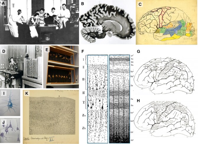 Figure 1
 Figure 2
- —Gesellschaft von Freunden und Förderern
- —Heinrich-Heine University Düsseldorf10.13039/501100003484
- —Medical Faculty of Heinrich-Heine Universität Düsseldorf10.13039/501100009400
- —European Union’s Horizon Europe Programme
Peer Reviews
No public reviews on file for this paper yet. If you reviewed it on a platform where reviews are public (OpenReview, ICLR, NeurIPS, ICML), you can paste yours below so the community can read it here.
Videos
No videos yet. Explain this paper in a talk, walkthrough, or lecture? Add one.
Taxonomy
TopicsNeurology and Historical Studies
** This year marks the 150th anniversary of Cécile Vogt's and the 155th anniversary of Oskar Vogt's births. These two remarkable researchers created a most comprehensive and unique collection of histological sections and other objects—what is now called ‘the Vogt Collection’.**
To realize their ambitious research goals and to better understand the background against which neurological and psychiatric conditions emerge, the Vogts collected brains and studied them. During their professional lives, which spanned over 60 years, they processed large series of histological sections—altogether about 850 000 sections from different species (Fig. 1).
Cécile Vogt studied medicine in Paris and became internationally renowned for her research into the extrapyramidal system of the brain, neurostimulation and myelinization, among other things. Oskar Vogt's work laid the foundations for an interdisciplinary approach to brain research and the mapping of the brain with focus on myeloarchitecture.^1^ In the first decade of the 20th century, Korbinian Brodmann—who was assistant of the Vogts in Berlin—published seminal papers about the cytoarchitecture of various brain regions, which he then integrated and expanded in his 1909 monograph that includes his famous cytoarchitectonic map with 43 areas (for an overview see Zilles^3^). While Brodmann's map is widely cited and dominates his legacy, Vogt's myeloarchitectonic approach was less well known for many years and is only currently experiencing something of a renaissance.^4,5^
At the turn of the 19th to the 20th century, new ideas and approaches emerged in different places and laboratories, changing the fields of neurology, psychiatry and neurosurgery and leading to progress in basic brain research. Oskar Vogt worked in the laboratory of Paul Flechsig in Leipzig, who studied the basis of myelinization during development, with Joseph Jules Dejerine in Paris, who published one of the most comprehensive textbooks on connectivity and anatomy and with Otto Binswanger in Jena, one of the founders of neuropsychiatry. Cécile Vogt studied in Pierre Marie's lab in Paris, a famous neurologist and neuropathologist, where she met Oskar, whom she married 1899.
The interest of the Vogts in the brain was driven by questions coming from neurology and psychiatry. Against this background, they developed their own ambitious research program and brought together a strong team of researchers and technicians in Berlin (Fig. 1). Moreover, the Vogts collaborated with Otfried Foerster, a distinguished neurologist and neurosurgeon, to explore the relationship of neurostimulation with the underlying brain anatomy. Such comparisons were facilitated by stimulation experiments in monkey brains, which were later studied histologically (Fig. 1).
Interestingly, the research programme of the Vogts combined different disciplines, including neuroanatomy, architectonics, histology, histopathology and electrophysiology, with neurochemistry and pharmacology, experimental and human genetics, physiology, psychology and phonetics. Accordingly, Oskar Vogt conceived and founded the Kaiser Wilhelm Institute for Brain Research in Berlin with such departments, where he was until 1937.
The development of this program was tightly linked with an increasing number of histological sections from brains of healthy human subjects, patients, non-human primates and other species. In total, the Vogts generated sections from about 1600 human brains and more than 100 species of non-human primates and other animals including rats, bats, deer, cats, ravens, dolphins and horses. Some sections result from cutting blocks of hemispheres or slabs of hemispheres, while others were processed as entire organs. Most of the sections have been stained either for cell bodies or myelin. To be able to process large sections, a new microtome was designed and manufactured. Embedding and staining techniques were improved, novel photographic techniques developed, and processes carefully monitored and documented. Altogether, the histological collection was stored on approximately 28 000 shelves in 70 collection cabinets, each with up to 400 shelves. Sometimes shelves were occupied by several layers of sections. These sections are well documented in protocols and patient folders. In addition, the Vogts collected about 11 000 glass plates, slides and photos, 20 000 reprints and various other objects.
The enormous quantity of histological sections is certainly much more than can be analysed in a single researcher's lifetime, and it would be interesting to know what the Vogts thought about this. The answer lies perhaps in the 12 000 sheets in more than 1200 bound volumes—the basis for the Vogt Archive—which also have been preserved.
After the Vogts’ death, the Collection with its various components was moved to Düsseldorf, Germany, where it is now hosted by the Cécile and Oskar Vogt Institute for Brain Research at the University Hospital Düsseldorf (https://www.uniklinik-duesseldorf.de/patienten-besucher/klinikeninstitutezentren/c-u-o-vogt-institut-fuer-hirnforschung/sammlungen-1). The Collection was maintained and expanded by the successors, Adolf Hopf (director 1960–88) and later Karl Zilles (until 2012). The author of this essay took over in 2013. Adolf Hopf and Karl Zilles also took care of the written legacy: the Vogt Archive was developed by Ursula Grell from the 1990s. It allowed her to respond to requests from researchers worldwide to use material from the archive. The resulting publications include books, monographs and journal articles that shed light on the Vogts, their collections and contemporary history from various perspectives (for an overview of the literature see the Supplementary material). However, the spatial conditions in which the collection and the archive have been stored, and their physical and non-digital presence do not allow the Vogt Collection to be made accessible to a broader scientific public.
The histological collection was studied rather little, except for one brain, which was used as a control or reference brain in several publications, for example, Mai et al.^5^ The patient records and protocols were stored in folders and were almost untouched since the 1950s. After the death of the two Vogts, several studies of the Vogt school were published (e.g. by Gerhard, Beheim-Schwarzbach and later Hopf; Supplementary material), which contributed detailed, albeit difficult to reproduce, descriptions of various brain regions. For a long time, more research was done on the collection than with the collection. As is often the case with anatomical collections, large parts were stored in basement rooms that were difficult to access. In this case, however, they were kept dry, clean and safe. Thus, they are in good shape and can now be ‘reactivated’. The conceptual, methodical, technical and informatics basis to handle such a large collection is available nowadays. Digitalization of the histological sections and documentations seems to be straight forward. But what are the arguments to start such a gigantic project? What are the historical and ethical preconditions? These must guide neuroscientific research, and they must be in place from the very beginning. An increase of scientific use is therefore only possible with deep historical research.
One argument is that in the past, histological collections have contributed many times to progress in brain research. For example, the Yakovlev collection was instrumental in better understanding brain development and pushed quantitative studies.^6^ Many sections are invaluable, and without the preservation of this collection there would be no access to them anymore. While this has been true for a long time, interesting new lines of research have also emerged, that shed new light on the historical sections of the Vogts and similar collections:
Such lines of research seem to align well with Vogt's vision of bringing together different disciplines, including genetics, surgery and pharmacology, through innovative methods, to improve our understanding of the pathogenesis of neurological diseases.
Although many of the Vogts’ views and research results have been overturned by new findings over the years, the Vogt Collection remains an invaluable part of our cultural heritage, offering a unique reflection of science and its history. It was assembled over a period of six decades, a period marked by major social upheavals. Importantly, this included the era of National Socialism. Although the Vogts were persecuted by the Nazis and were not involved in their euthanasia programmes, it has been known since 1988 at the latest that some of the existing sections were resulting from such programmes after the murder of patients and later were included in the collection through the Vogts’ personal relationships, e.g. with Bernhard Patzig [see Bogerts (1988) in the Supplementary material]. In response, my predecessor has decided to remove such sections from research. A project for in-depth investigation is now planned to address this issue.
When the six decades of the Vogt's research is evaluated in relation to today, new neuroscientific and ethical concepts have emerged, and changes in the organization of research itself can be observed. Whether for the history of neurology, psychiatry and psychology, for sociological, ethical and political aspects of 20th century sciences, or for research into the brain in all its facets, the Vogt Collection and Archive is a treasure trove for research in a wide range of areas from basic science to medicine, history and ethics. Building on the archive and the digitization of all histological sections and documents, this unique collection is now to be fully catalogued and made accessible to the broad scientific public in order to make it available as a source and inspiration for research.
Supplementary Material
awaf365_Supplementary_Data
The reference list from the paper itself. Each links out to its DOI / PubMed record.
- 1Vogt C, Vogt O. Allgemeinere Ergebnisse unserer Hirnforschung (English translation: Results of our brain research in a broader context). J Psychol Neurol. 1919;25:292–398.
- 2Vogt C, Vogt O. Die vergleichend-architektonische und die vergleichend-reizphysiologische Felderung der Großhirnrinde unter besonderer Berücksichtigung der menschlichen. Naturwissenschaften. 1926;14:1190–1194.
- 3Zilles K . Brodmann: A pioneer of human brain mapping—His impact on concepts of cortical organization. Brain. 2018;141:3262–3278.30358817 10.1093/brain/awy 273PMC 6202576 · doi ↗ · pubmed ↗
- 4Nieuwenhuys R, Broere CAJ. A new 3D myeloarchitectonic map of the human neocortex based on data from the Vogt-Vogt school. Brain Struct Funct. 2023;228:1549–1559.37378856 10.1007/s 00429-023-02671-6PMC 10751253 · doi ↗ · pubmed ↗
- 5Mai JK, Majtanik M, Paxinos G. Atlas of the human brain. 4th ed. Academic Press; 2015.
- 6Kretschmann HJ, Kammradt G, Cowart EC, et al The Yakovlev collection. A unique resource for brain research and the basis for a multinational data bank. J Hirnforsch. 1982;23:647–656.7169526 · pubmed ↗
- 7Amunts K, Lepage C, Borgeat L, et al Big Brain: An ultrahigh-resolution 3D human brain model. Science. 2013;340:1472–1475.23788795 10.1126/science.1235381 · doi ↗ · pubmed ↗
- 8Axer M, Amunts K. Scale matters: The nested human connectome. Science. 2022;378:500–504.36378967 10.1126/science.abq 2599 · doi ↗ · pubmed ↗