Individual functional connectivity constraints on spatial progression of tau pathology in Alzheimer's disease
Harry H Behjat, Jacob W. Vogel, Olof Strandberg, Nicola Spotorno, Jonathan Rittmo, Lyduine E. Collij, Alexa Pichet Binette, Yu Xiao, Danielle van Westen, Erik Stomrud, Sebastian Palmqvist, Niklas Mattsson‐Carlgren, Dimitri Van De Ville, Ruben Smith, Oskar Hansson

TL;DR
This study shows that individual brain connectivity patterns better explain the spread of tau protein in Alzheimer's disease, supporting the idea that tau spreads through neural connections.
Contribution
The study provides individual-level evidence that tau pathology progression is constrained by functional brain connectivity.
Findings
Hybrid functional connectivity (FC) models explained tau PET patterns better than group-average FC across Alzheimer's disease stages.
Individual FC was more predictive of future tau PET changes compared to canonical patterns in MCI and AD dementia.
Aβ PET patterns were not better explained by individual FC compared to canonical patterns, suggesting different spread mechanisms.
Abstract
Tau PET patterns show notable spatial heterogeneity across subjects in Alzheimer's disease (AD). In vitro findings suggest that tau may spread ‘prion‐like’ across neuronal connections in an activity‐dependent manner, a hypothesis strengthened by group‐level studies showing association between resting‐state functional connectivity (FC) networks and tau deposition patterns. This hypothesis would be better supported by evidence at the individual level, an investigation that is the focus of this study. Structural MRI, resting‐state fMRI, tau‐PET, and amyloid‐β (Aβ)‐PET data from 733 participants aged 50+ from the BioFINDER‐2 study were used: 402 cognitively unimpaired (CU, CSF Aβ+=89), 157 with mild cognitive impairment (MCI), and 174 with AD dementia, with 523 follow‐up tau‐PET scans (323 CU, 109 MCI, 91 AD); individuals with MCI or AD dementia were all CSF Aβ+. fMRI data were…
Genes, proteins, chemicals, diseases, species, mutations and cell lines named across the full text — each resolved to its canonical identifier and authoritative record.
Click any figure to enlarge with its caption.
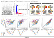 Figure 1
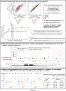 Figure 2
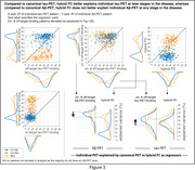 Figure 3
Peer Reviews
No public reviews on file for this paper yet. If you reviewed it on a platform where reviews are public (OpenReview, ICLR, NeurIPS, ICML), you can paste yours below so the community can read it here.
Videos
No videos yet. Explain this paper in a talk, walkthrough, or lecture? Add one.
Taxonomy
TopicsFunctional Brain Connectivity Studies · Dementia and Cognitive Impairment Research · Alzheimer's disease research and treatments