Automatic optimization of flat-field corrections by evaluation and enhancement (EVEN) in multimodal optical microscopy
Elena Corbetta, Matteo Calvarese, Patrick Then, Hyeonsoo Bae, Tobias Meyer-Zedler, Bernhard Messerschmidt, Orlando Guntinas-Lichius, Michael Schmitt, Christian Eggeling, Juergen Popp, Thomas Bocklitz

TL;DR
The EVEN method uses machine learning to automatically improve image quality in optical microscopy by correcting uneven illumination.
Contribution
EVEN introduces a machine learning workflow for automatic image correction and quality assessment in multimodal optical microscopy.
Findings
EVEN integrates image metrics into a Linear Discriminant Analysis model to detect and predict image quality.
The method was successfully applied to multimodal nonlinear imaging and multichannel fluorescence measurements.
EVEN simplifies further processing by automatically optimizing corrections in single-channel images.
Abstract
Uneven illumination affects all images acquired by optical microscopes, especially large, multicolour and nonlinear measurements. Although removal is possible with various algorithms, evaluating raw and processed images is challenging due to the lack of established workflows for image quality assessment. This manuscript describes a machine learning-based method, EVEN (Evaluation and Enhancement), to assess and optimise corrections in optical microscopy. EVEN integrates quantitative image metrics into a Linear Discriminant Analysis model to detect and predict image quality, automatically optimising corrections. The method can be integrated into the optical microscopy pipeline to simplify further processing and analysis. Here, we show the implementation and application of EVEN in different processing scenarios, including multimodal nonlinear imaging of human and neck tissue slices and…
Genes, proteins, chemicals, diseases, species, mutations and cell lines named across the full text — each resolved to its canonical identifier and authoritative record.
Click any figure to enlarge with its caption.
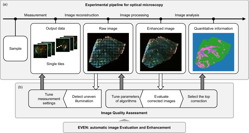 Figure 1
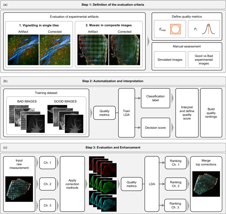 Figure 2
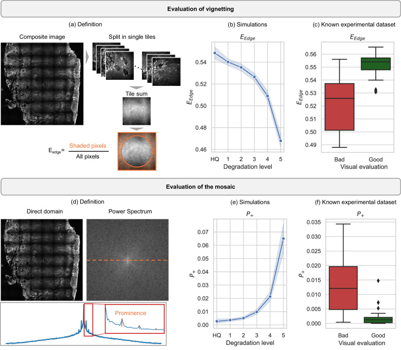 Figure 3
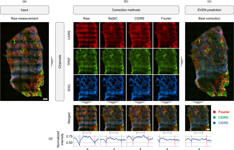 Figure 4
 Figure 5
Peer Reviews
No public reviews on file for this paper yet. If you reviewed it on a platform where reviews are public (OpenReview, ICLR, NeurIPS, ICML), you can paste yours below so the community can read it here.
Videos
No videos yet. Explain this paper in a talk, walkthrough, or lecture? Add one.
Taxonomy
TopicsCell Image Analysis Techniques · Advanced Fluorescence Microscopy Techniques · Digital Holography and Microscopy