Fixel-based analysis reveals detailed white matter changes in semantic dementia
Maria Luisa Mandelli, Yann Cobigo, Ilaria Perretti, Dana Leichter, Celina Alba, Rian Bogley, Nick Wellman, Siddarth Ramkrishnan, Zachary A. Miller, Bruce L. Miller, William W. Seeley, Howard J. Rosen, Maria Luisa Gorno-Tempini

TL;DR
This study shows that fixel-based analysis provides more detailed insights into white matter changes in semantic dementia compared to traditional methods.
Contribution
The study introduces fixel-based analysis as a more sensitive method for detecting white matter alterations in semantic dementia.
Findings
FBA detected additional white matter pathways affected in semantic dementia, such as the tapetum and anterior commissure.
Both FBA and TBSS identified damage to anterior temporal lobe connected tracts in semantic dementia patients.
FBA offers greater anatomical precision and sensitivity to structural changes in white matter compared to TBSS.
Abstract
Accurately characterizing white matter (WM) microstructure is critical for understanding neurodegenerative diseases such as semantic dementia (SD). Regionally constrained techniques like tract-based spatial statistics (TBSS) rely on diffusion-tensor imaging (DTI) and assume a single fiber population per voxel, limiting their sensitivity to complex architecture. Fixel-based analysis (FBA) overcomes these constraints by resolving multiple fiber populations (fixels) within a single voxel, enabling more anatomically specific assessment of WM organization. Multi-shell diffusion MRI from 16 semantic-variant PPA (svPPA) and 15 semantic-behavioral fronto-temporal dementia (sbvFTD) cases—with imaging-confirmed left- and right-predominant temporal atrophy, respectively—and 44 neurologically healthy controls were analyzed using both TBSS-DTI and whole-brain FBA. Fiber-specific metrics of fiber…
Genes, proteins, chemicals, diseases, species, mutations and cell lines named across the full text — each resolved to its canonical identifier and authoritative record.
Click any figure to enlarge with its caption.
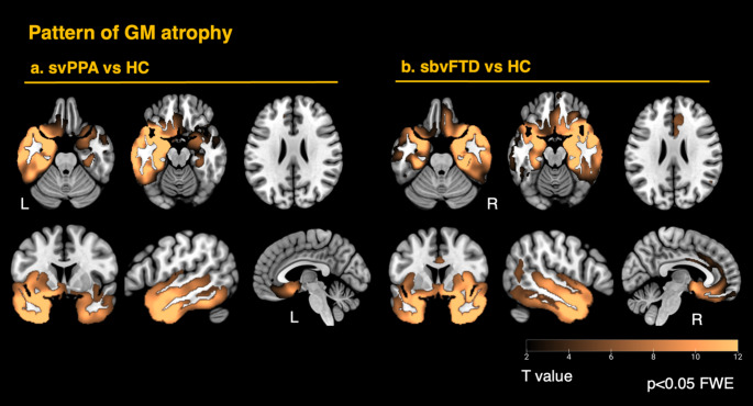 Figure 1
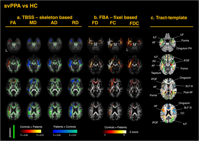 Figure 2
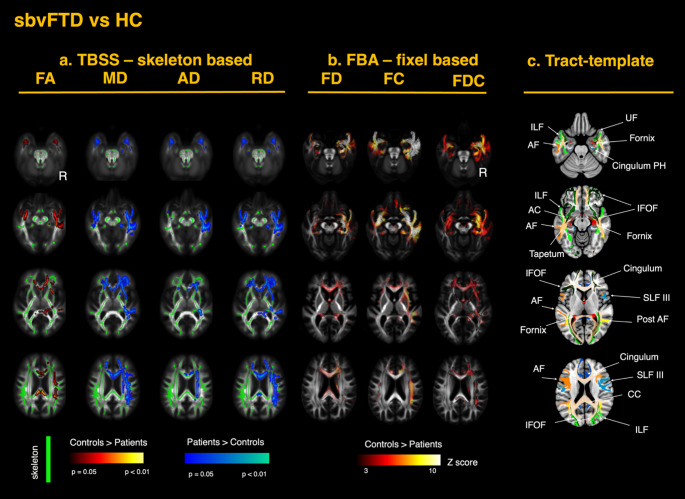 Figure 3
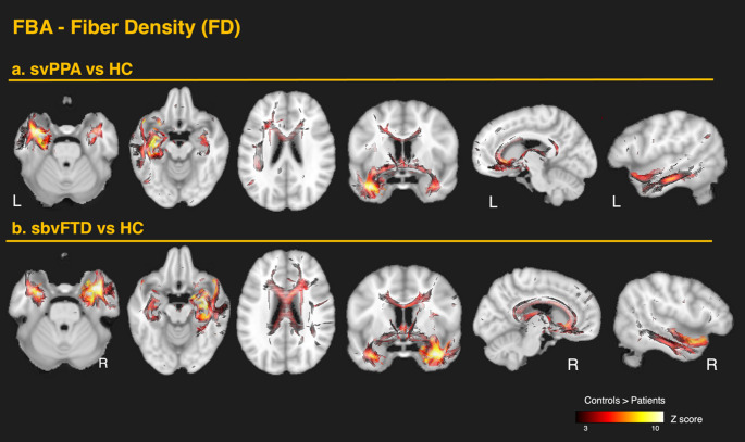 Figure 4
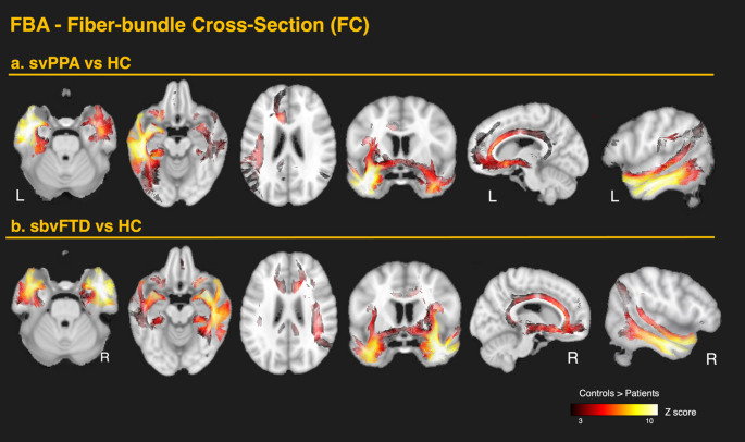 Figure 5
Peer Reviews
No public reviews on file for this paper yet. If you reviewed it on a platform where reviews are public (OpenReview, ICLR, NeurIPS, ICML), you can paste yours below so the community can read it here.
Videos
No videos yet. Explain this paper in a talk, walkthrough, or lecture? Add one.
Taxonomy
TopicsAdvanced Neuroimaging Techniques and Applications · Dementia and Cognitive Impairment Research · Epilepsy research and treatment