Differences in growth rates and spatial patterns of white matter hyperintensities between beta‐amyloid burden and vascular risk
Jeremy F. Strain, Maryam Rahmani, Chia‐Ling Phuah, Donna Dierker, Jingqin Luo, Christopher J. Owen, Andrei G. Vlassenko, Hussain Jafri, Pierrick Bourgeat, Jurgen Fripp, Liang Jin, Krista L. Moulder, Tammie L.S. Benzinger, Chengjie Xiong, Jin‐Moo Lee, Michael W Weiner

TL;DR
This study finds that white matter hyperintensities (WMH) in the brain grow faster in people with higher beta-amyloid levels, especially in the parietal region, and are less linked to vascular risk factors over time.
Contribution
The study reveals distinct longitudinal associations between beta-amyloid burden and vascular risk with WMH progression in specific brain regions.
Findings
Higher baseline beta-amyloid burden is independently associated with increased growth rates of WMH.
The parietal region shows a unique and persistent longitudinal association with beta-amyloid burden.
Vascular risk factors influence WMH volume but not their progression in specific brain regions.
Abstract
There is increasing evidence for an association between white matter hyperintensities (WMH) and brain beta‐amyloid deposition. WMH are not exclusive correlates of a single etiology, and the spatial topography can associate with different pathologic markers of vascular or neurodegenerative disease. How WMH are longitudinally associated with brain beta‐amyloid burden requires further investigation, particularly with respect to co‐existent vascular risk factors and differences across brain regions. We retrospectively measured WMH on MRI and vascular risk factors in a combined neuroimaging data set comprised of the ADNI, AIBL and OASIS3 studies, which included harmonized centiloid estimates of beta‐amyloid burden from PET imaging. WMH were measured using the TrUE‐Net algorithm. Vascular risk factors were extracted from provided clinical data and used to calculate individual revised…
Genes, proteins, chemicals, diseases, species, mutations and cell lines named across the full text — each resolved to its canonical identifier and authoritative record.
Click any figure to enlarge with its caption.
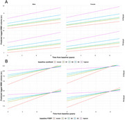 Figure 1
Peer Reviews
No public reviews on file for this paper yet. If you reviewed it on a platform where reviews are public (OpenReview, ICLR, NeurIPS, ICML), you can paste yours below so the community can read it here.
Videos
No videos yet. Explain this paper in a talk, walkthrough, or lecture? Add one.
Taxonomy
TopicsDementia and Cognitive Impairment Research · Intracerebral and Subarachnoid Hemorrhage Research · Acute Ischemic Stroke Management