Predictive Value of Serum Autotaxin for Hepatocellular Carcinoma Recurrence After Curative Radiofrequency Ablation
Takanobu Iwadare, Hiroyuki Kobayashi, Takefumi Kimura, Taiki Okumura, Taro Nakajima, Shun‐ichi Wakabayashi, Yuki Yamashita, Naoyuki Fujimori, Hideo Kunimoto, Satoshi Shimamoto, Koji Igarashi, Takuro Uchida, Takeji Umemura, Naoki Tanaka

TL;DR
This study shows that high levels of a protein called autotaxin in the blood can predict if liver cancer will return after a specific treatment called radiofrequency ablation.
Contribution
The study identifies autotaxin as a novel and robust biomarker for predicting hepatocellular carcinoma recurrence after curative radiofrequency ablation.
Findings
Serum autotaxin (ATX) levels were a strong independent predictor of hepatocellular carcinoma recurrence after radiofrequency ablation.
ATX demonstrated superior predictive performance with an area under the curve of 0.729, sensitivity of 0.857, and specificity of 0.629.
High ATX levels were associated with significantly higher recurrence rates compared to low ATX levels (p = 0.0006).
Abstract
Despite recent treatment advancements, the high recurrence rate of hepatocellular carcinoma (HCC) following curative therapy remains a significant challenge. Autotaxin (ATX) is a key biomarker in chronic liver disease that has a yet unclarified role in predicting HCC recurrence. This study examined whether precurative radiofrequency ablation (RFA) serum ATX level could serve as a predictor of HCC recurrence after treatment. Fifty‐six HCC patients (37 [66%] male; median age: 74 years) treated by curative RFA were retrospectively analyzed. Twenty‐one patients experienced HCC recurrence during follow‐up. ATX demonstrated superior predictive performance for HCC recurrence after RFA, with an area under the receiver operating characteristic curve of 0.729, sensitivity of 0.857, and specificity of 0.629. Kaplan–Meier analysis revealed that patients with high ATX had a significantly higher…
Genes, proteins, chemicals, diseases, species, mutations and cell lines named across the full text — each resolved to its canonical identifier and authoritative record.
Click any figure to enlarge with its caption.
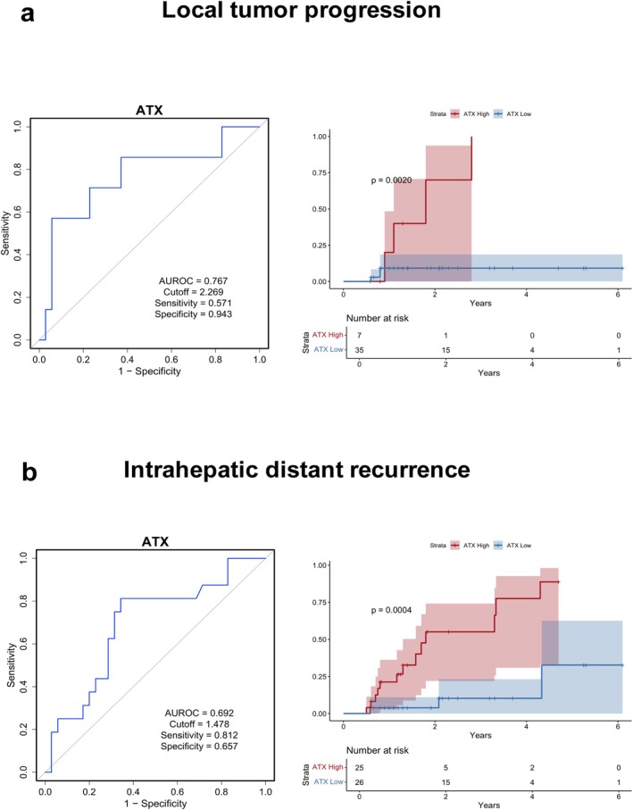 Figure 1
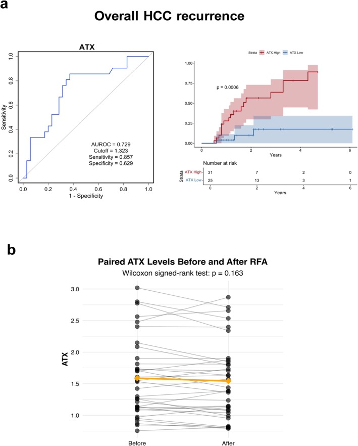 Figure 2
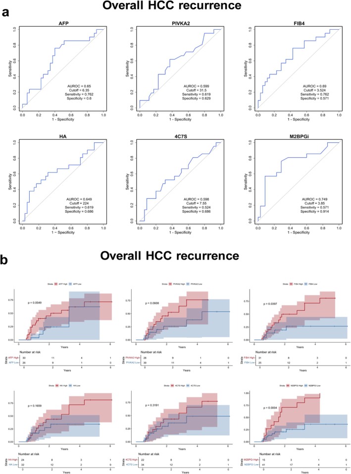 Figure 3
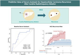 Figure 4
Peer Reviews
No public reviews on file for this paper yet. If you reviewed it on a platform where reviews are public (OpenReview, ICLR, NeurIPS, ICML), you can paste yours below so the community can read it here.
Videos
No videos yet. Explain this paper in a talk, walkthrough, or lecture? Add one.
Taxonomy
TopicsSphingolipid Metabolism and Signaling · Caveolin-1 and cellular processes · Intracerebral and Subarachnoid Hemorrhage Research