Metal artifact reduction in spectral computed tomography for intracavity brachytherapy in cervical cancer patients: a prospective study
Yuliang Sun, Yining Chen, Zheng Zeng, Bing Zhou, Haoran Xu, Junfang Yan, Ke Hu, Fuquan Zhang

TL;DR
This study shows that combining high-energy virtual monochromatic images with metal artifact reduction improves imaging quality in cervical cancer brachytherapy.
Contribution
The study demonstrates the clinical benefits of using 140 keV VMI with MAR for optimal artifact reduction and contouring accuracy in cervical cancer patients.
Findings
VMI + MAR significantly reduced artifact index in the cervix and rectum compared to no-MAR across all energies.
140 keV + MAR achieved the highest SNR, CNR, and best artifact reduction with improved contouring consistency.
Higher VMI energy reduced geometric distortion and improved delineation reproducibility for target and organs at risk.
Abstract
This study aims to evaluate the effectiveness of combining virtual monochromatic images (VMI) with metal artifact reduction (MAR) algorithms in reducing metal artifacts in cervical cancer patients undergoing intracavitary brachytherapy. Fifty cervical cancer patients scheduled for intracavitary brachytherapy underwent spectral computed tomography (CT) images. VMI + MAR/no-MAR were reconstructed at energies ranging from 40 to 140 keV. CT attenuation and image noise (standard deviation) were measured to calculate the signal-to-noise ratio (SNR), contrast-to-noise ratio (CNR) for the most prominent artifacts, and the artifact index (AI) for the cervix, bladder, and rectum. Objective metrics were compared between the VMI + MAR and VMI + no-MAR groups. Subjective image quality was evaluated using a 5-point Likert scale, focusing on artifact reduction, soft tissue contrast, and contouring…
Genes, proteins, chemicals, diseases, species, mutations and cell lines named across the full text — each resolved to its canonical identifier and authoritative record.
Click any figure to enlarge with its caption.
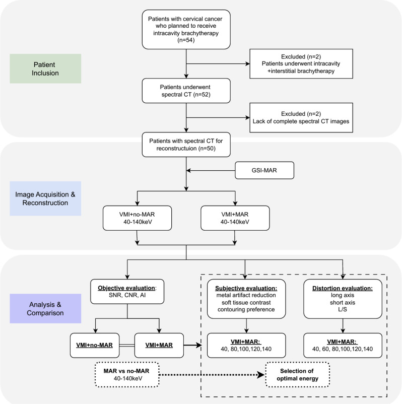 Figure 1
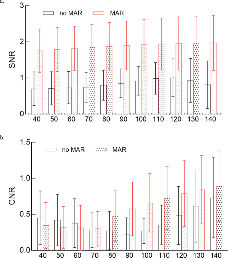 Figure 2
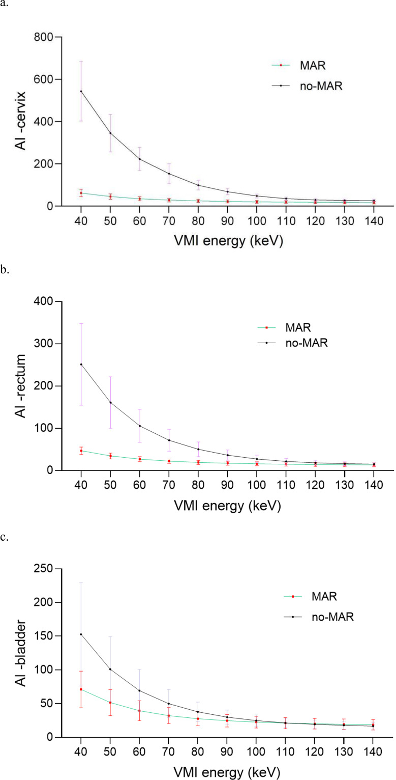 Figure 3
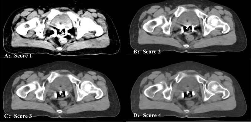 Figure 4
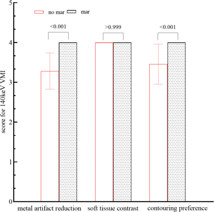 Figure 5
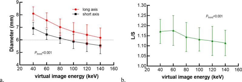 Figure 6
Peer Reviews
No public reviews on file for this paper yet. If you reviewed it on a platform where reviews are public (OpenReview, ICLR, NeurIPS, ICML), you can paste yours below so the community can read it here.
Videos
No videos yet. Explain this paper in a talk, walkthrough, or lecture? Add one.
Taxonomy
TopicsAdvanced X-ray and CT Imaging · Dental Radiography and Imaging · Advanced X-ray Imaging Techniques