Altered Functional Specialization and Interhemispheric Coordination in Rhegmatogenous Retinal Detachment: Associations With Gene Expression, Neurotransmitter Receptor Distribution, and SVM–SHAP Classification: A Multimodal Neuroimaging–Transcriptomics Study Integrating Functional Metrics and Interpretable Machine Learning
Yu Ji, Yuan‐Yuan Wang, Xiao‐Rong Wu

TL;DR
This study explores how retinal detachment affects brain function, linking changes to gene activity and neurotransmitter systems using advanced imaging and machine learning.
Contribution
The study introduces a novel multimodal approach combining neuroimaging, transcriptomics, and interpretable machine learning to explore brain changes in retinal detachment.
Findings
RRD patients showed altered brain function in the frontal, occipital lobes, and thalamus.
Genes linked to these changes are involved in synaptic signaling and neuroplasticity.
The SVM–SHAP model identified thalamic connectivity as a key biomarker for RRD.
Abstract
Previous studies have reported functional alterations in the brains of patients with rhegmatogenous retinal detachment (RRD). However, it remains largely unclear whether RRD affects hemispheric specialization and interhemispheric coordination, and how these alterations relate to underlying gene expression patterns and neurotransmitter receptor distributions. We employed the Autonomy Index (AI) and Connectivity between Functionally Homotopic Voxels (CFH) to quantify alterations in hemispheric specialization and interhemispheric cooperation in patients with RRD. Transcriptome–neuroimaging spatial correlation analysis was performed by integrating gene expression data from the Allen Human Brain Atlas (AHBA) to identify genes associated with AI and CFH alterations. Enrichment and protein–protein interaction analyses were conducted to characterize the biological processes and molecular…
Genes, proteins, chemicals, diseases, species, mutations and cell lines named across the full text — each resolved to its canonical identifier and authoritative record.
Click any figure to enlarge with its caption.
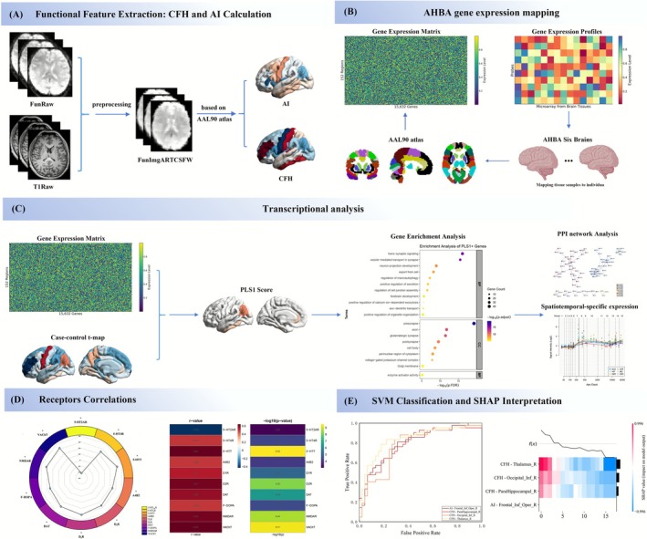 Figure 1
 Figure 2
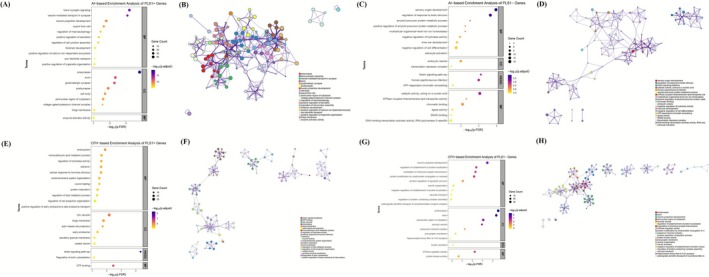 Figure 3
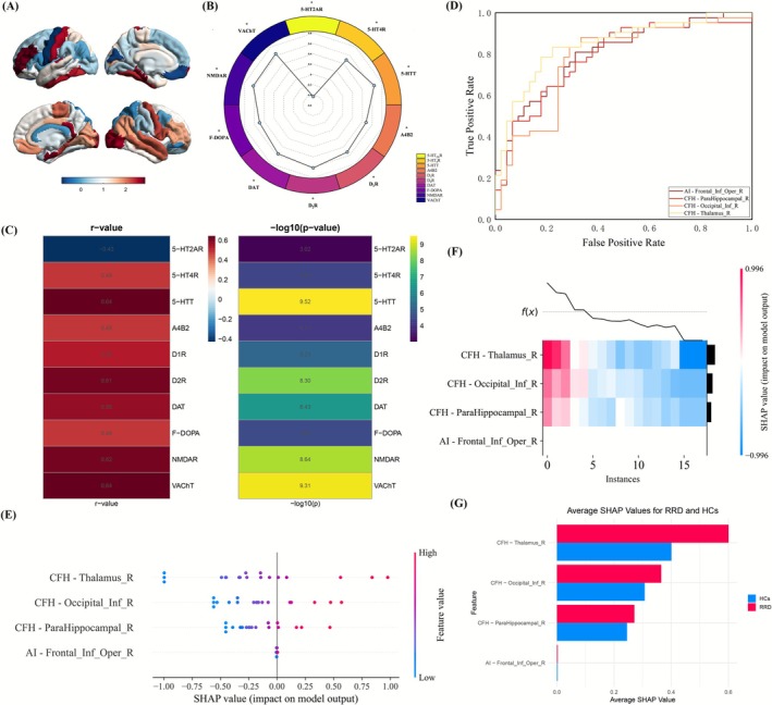 Figure 4
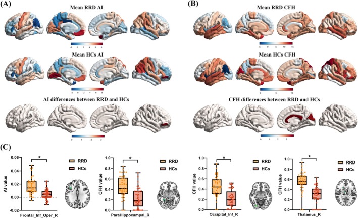 Figure 5
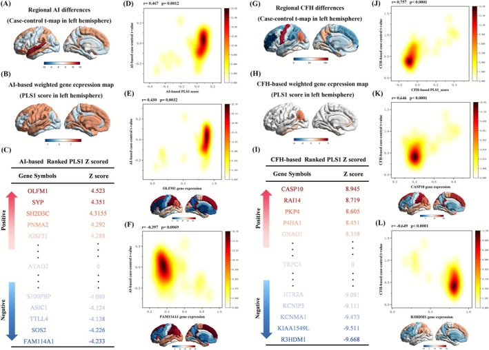 Figure 6
Peer Reviews
No public reviews on file for this paper yet. If you reviewed it on a platform where reviews are public (OpenReview, ICLR, NeurIPS, ICML), you can paste yours below so the community can read it here.
Videos
No videos yet. Explain this paper in a talk, walkthrough, or lecture? Add one.
Taxonomy
TopicsRetinal and Macular Surgery · Retinal Development and Disorders · Retinopathy of Prematurity Studies