Noninvasive in vivo deoxycytidine kinase (dCK)-PET identifies tumor-draining lymph nodes upon immune checkpoint inhibitor therapy
Cécile Philippe, Jonathan Cotton, Gregory D. Bowden, Simone Pöschel, Philipp Knopf, Barbara Schörg, Irene Gonzalez-Menendez, Dominik Sonanini, Lukas Flatz, Martin Allen-Auerbach, Caius G. Radu, Johannes Czernin, Leticia Quintanilla-Martinez, Marcus Hacker, Bernd J. Pichler

TL;DR
This study shows that PET imaging with [18F]FAC and [18F]CFA can detect immune cell activation in tumor-draining lymph nodes during immunotherapy, offering a noninvasive way to monitor treatment response.
Contribution
The study introduces [18F]FAC and [18F]CFA as novel radiotracers for noninvasive monitoring of immune activation in tumor-draining lymph nodes during immunotherapy.
Findings
Activated T cells and macrophages show significantly higher [18F]FAC uptake in vitro.
CIT induces increased [18F]FAC uptake in tumor-draining lymph nodes in preclinical and clinical settings.
Ex vivo analysis confirms elevated [18F]FAC uptake in T cells from CIT-treated tumor-draining lymph nodes.
Abstract
Efficient application of immunotherapy necessitates advanced whole-body imaging techniques to monitor sites of immune cell activation. Deoxycytidine kinase (dCK), a key enzyme in the deoxynucleotide salvage pathway, is upregulated in proliferating immune cells and can be targeted by the radiotracers [18F]FAC (preclinical) and [18F]CFA (clinical), allowing for noninvasive monitoring of immune activation in lymphatic organs via positron emission tomography (PET). In this study, we aimed to assess the efficacy of [18F]FAC in detecting immune activation upon immune checkpoint inhibitor therapy (CIT). In vitro, activated T cells and macrophages exhibited significantly higher [18F]FAC uptake compared to their naïve counterparts. In vivo, preclinical [18F]FAC-PET/MRI revealed a CIT-induced significant increase in [18F]FAC uptake in tumor-draining lymph nodes (TDLNs) compared to contralateral…
Genes, proteins, chemicals, diseases, species, mutations and cell lines named across the full text — each resolved to its canonical identifier and authoritative record.
Click any figure to enlarge with its caption.
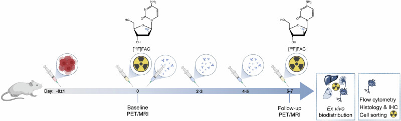 Figure 1
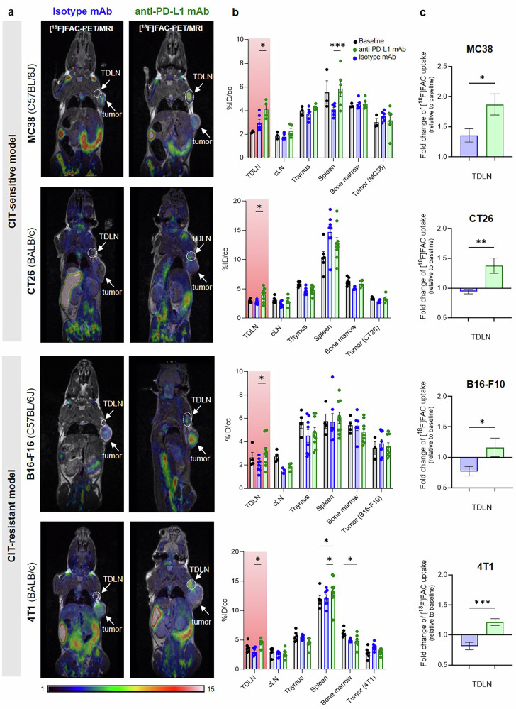 Figure 2
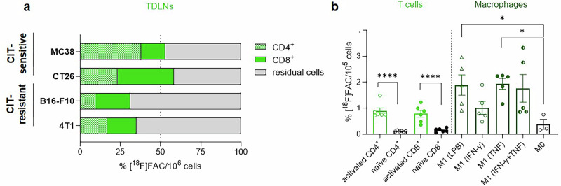 Figure 3
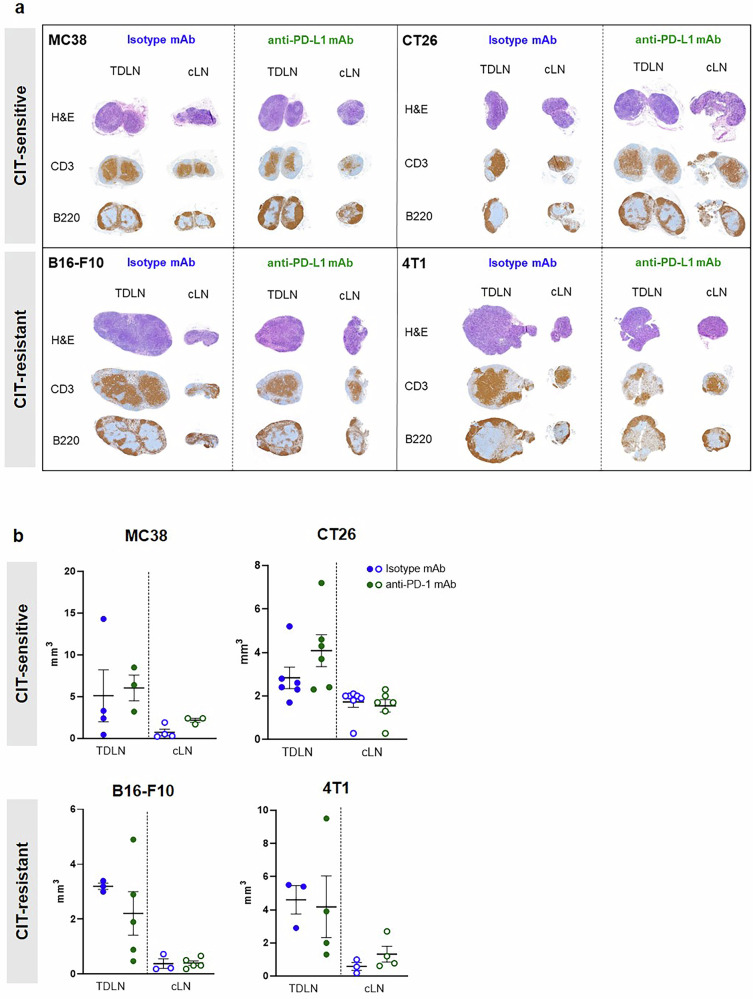 Figure 4
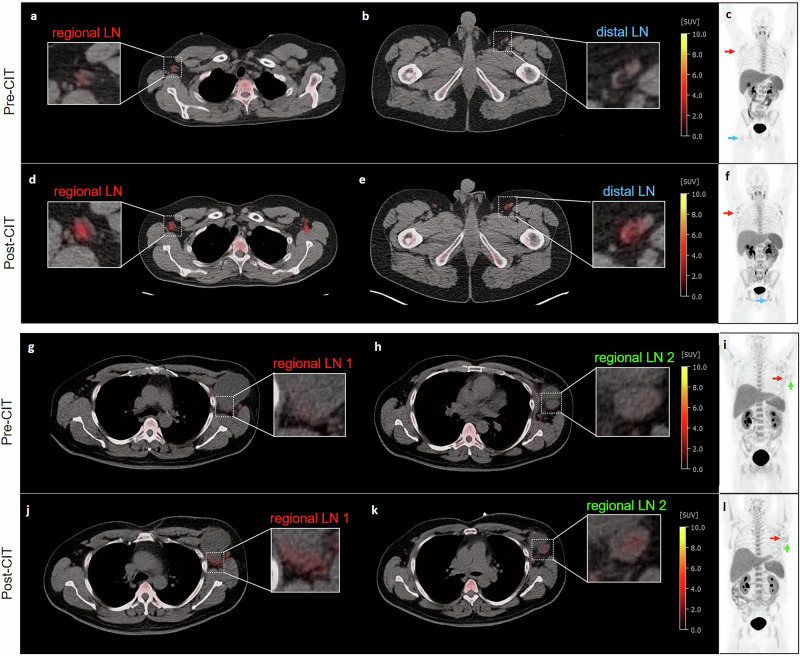 Figure 5
Peer Reviews
No public reviews on file for this paper yet. If you reviewed it on a platform where reviews are public (OpenReview, ICLR, NeurIPS, ICML), you can paste yours below so the community can read it here.
Videos
No videos yet. Explain this paper in a talk, walkthrough, or lecture? Add one.
Taxonomy
TopicsCancer Immunotherapy and Biomarkers · Immune cells in cancer · Immunotherapy and Immune Responses