Prevalence of Oral Alterations and Correlation Between Oral and Cutaneous Neurofibromas in Neurofibromatosis Type 1: A Retrospective Case–Control Study
Pâmella de Pinho Montovani, Gabriela Pizão Werneck Moreira da Costa, Rafaela Elvira Rozza‐de‐Menezes, Karin Soares Cunha

TL;DR
This study found that people with Neurofibromatosis Type 1 have more oral changes and a link between mouth and skin tumors.
Contribution
The study identifies specific oral alterations and their correlation with cutaneous neurofibromas in NF1 patients.
Findings
Oral mucosal alterations and exostoses were significantly more common in NF1 individuals.
Enlarged fungiform papillae and oral neurofibromas were the most frequent oral changes in NF1.
Oral and cutaneous neurofibromas counts were correlated in NF1 patients.
Abstract
The aim of this study was to determine the prevalence of oral alterations detectable through physical examination in NF1 individuals. Additionally, we assessed the correlation between the number of oral and cutaneous neurofibromas. This retrospective study evaluated oral alterations in individuals with and without NF1. In the NF1 group, associations between oral and cutaneous neurofibromas, age, sex, pregnancy, and family history of NF1 were assessed. A total of 327 participants were evaluated (81 with NF1 and 246 controls). Oral mucosal alterations (92.6% vs. 79.3%) and exostoses (12.3% vs. 4.5%) were significantly more prevalent in the NF1 group. The most frequent oral alterations were enlarged fungiform papillae (46.9% vs. 8.1%), coated tongue (45.7% vs. 29.3%), neurofibromas (38.3% versus none), physiological melanin pigmentation (30.9% vs. 10.6%), and exostoses (12.3% vs. 4.5%).…
Genes, proteins, chemicals, diseases, species, mutations and cell lines named across the full text — each resolved to its canonical identifier and authoritative record.
Click any figure to enlarge with its caption.
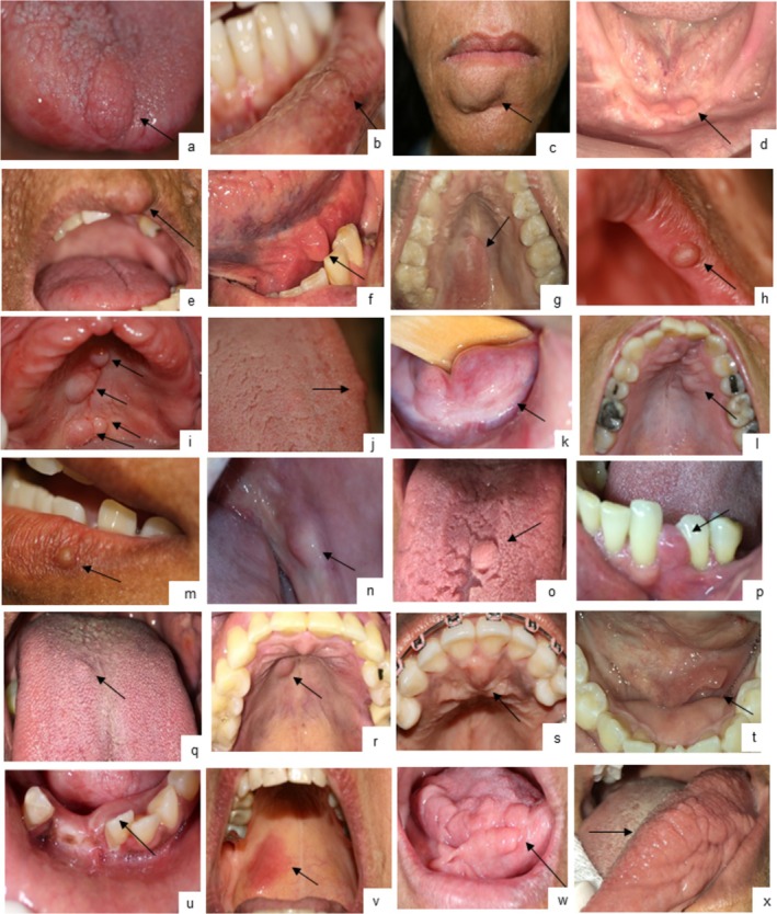 FIGURE 1
| Demographic data | NF1 ( | Controls ( |
|
|---|---|---|---|
| Sex | |||
| Male | 24 (29.6%) | 96 (39%) | 0.128 |
| Female | 57 (70.4%) | 150 (61%) | |
| Age (years) | |||
| Mean (±SD) | 39.7 (±16.5) | 53.5 (±17.7) | < |
| Minimum | 3 | 0.7 | |
| Maximum | 75 | 85 | |
| Oral alterations | NF1 ( | Controls ( |
| Odds ratio (IC 95%) |
|---|---|---|---|---|
| Soft tissue (total) | 75 (92.6%) | 195 (79.3%) |
| 0.306 (0.126–0.743) |
| Exostoses (total) | 10 (12.3%) | 11 (4.5%) |
| 0.332 (0.136–0.815) |
| Enlarged fungiform papillae | 38 (46.9%) | 20 (8.1%) |
| 0.100 (0.053–0.188) |
| Coated tongue | 37 (45.7%) | 72 (29.3%) |
| 0.492 (0.294–0.825) |
| Neurofibromas | 31 (38.3%) | None (—) |
| — |
| Physiological melanin pigmentation | 25 (30.9%) | 26 (10.6%) |
| 0.265 (0.142–0.493) |
| Fissured tongue | 20 (24.7%) | 52 (21.1%) | 0.503 | 0.818 (0.453–1.476) |
| Candidiasis | 17/57 (29.8%) | 80 (32.5%) | 0.694 | 1.34 (0.606–2.123) |
| Geographic tongue | 7 (8.6%) | 22 (8.9%) | — | — |
| Crenated tongue | 5 (6.2%) | 14 (5.7%) | — | — |
| Fordyce granules | 4 (4.9%) | 16 (6.5%) | — | — |
| Unspecific ulcer | 4 (4.9%) | 17 (6.9%) | — | — |
| Ankyloglossia | 3 (3.7%) | None (—) | — | — |
| Recurrent herpes simplex lesion | 2 (2.5%) | 6 (2.4%) | — | — |
| Leukoedema | 2 (2.5%) | 5 (2%) | — | — |
| Lichenoid reaction | 2 (2.5%) | 2 (0.8%) | — | — |
| Enlarged fimbriated fold of tongue | 2 (2.5%) | None (—) | — | — |
| Inflammatory fibrous hyperplasia | 1 (1.2%) | 8 (3.3%) | — | — |
| Unspecific petechiae | 1 (1.2%) | None (—) | — | — |
| Oral condyloma acuminatum | 1 (1.2%) | 3 (1.2%) | — | — |
| Morsicatio buccarum | 1 (1.2%) | 5 (2%) | — | — |
| Focal fibrous hyperplasia | 1 (1.2%) | 9 (3.7%) | — | — |
| Actinic cheilitis | 1 (1.2%) | 26 (10.6%) | — | — |
| Fibrolipoma | 1 (1.2%) | 3 (1.2%) | — | — |
Peer Reviews
No public reviews on file for this paper yet. If you reviewed it on a platform where reviews are public (OpenReview, ICLR, NeurIPS, ICML), you can paste yours below so the community can read it here.
Videos
No videos yet. Explain this paper in a talk, walkthrough, or lecture? Add one.
Taxonomy
TopicsNeurofibromatosis and Schwannoma Cases · Soft tissue tumor case studies · Soft tissue tumors and treatment
Introduction
1
Neurofibromatosis type 1 (NF1) is a common genetic disorder with frequent oral involvement [1]. Enlarged fungiform papillae and oral neurofibromas are the most common findings reported in the literature [2], but large‐scale evaluations remain scarce. Cutaneous neurofibromas affect ~99% of patients, with some developing hundreds or thousands [3], whereas multiple oral neurofibromas are uncommon. We hypothesized that NF1 individuals have additional underrecognized oral alterations and that oral neurofibroma burden correlates with cutaneous tumor counts. We evaluated the prevalence and pattern of oral alterations in NF1 and their relationship with cutaneous neurofibromas.
Material and Methods
2
This retrospective case–control study was conducted at the Oral Diagnosis Outpatient Clinic, Antônio Pedro University Hospital, Brazil (Ethics approval #4.780.584). The NF1 group comprised 81 participants diagnosed according to the revised criteria [1]. Controls (n = 246) were randomly selected from the same database, excluding NF1 or other genetic/systemic conditions affecting oral health.
Photographic documentation and cytopathological examination of the entire oral mucosa, regardless of visible lesions, was recorded in medical files. As NF1 patients had a mean of 2.9 clinical appointments, data from the first three control appointments were analyzed.
Variables for both groups included age, sex, self‐reported skin color, enlarged fungiform papillae (from tongue photographs), other oral alterations, cytopathology results, and denture use. For NF1, additional variables included family history, pregnancies (females), and cutaneous neurofibroma counts using paper frames [4].
Oral candidiasis was diagnosed clinically and cytopathologically; subclinical cases were defined by positive cytology without clinical signs [5]. Suspected oral neurofibromas were biopsied when possible; histopathology and S100 immunohistochemistry confirmed diagnosis. Imaging studies were not included.
Statistical analysis used the Shapiro–Wilk test, chi‐square or Fisher's exact tests, t‐test with Cohen's d or Mann–Whitney U, and Spearman's correlation. Chi‐square was applied only when ≥ 10% of NF1 participants were affected. LOESS modeled the relationship between oral and cutaneous neurofibromas.
Results
3
Table 1 presents demographic data, and Table 2 compares oral lesion prevalence in NF1 and controls. Oral mucosal alterations (92.6% vs. 79.3%; p = 0.006) and exostoses (12.3% vs. 4.5%; p = 0.012) were more prevalent in NF1.
Enlarged fungiform papillae was the most frequent alteration, affecting 46.9% of NF1 vs. 8.1% of controls (p < 0.0001), and was more common in younger participants (mean = 33.7 ± 17.4 years; p = 0.002). Coated tongue, the second most common alteration, affected 45.7% of NF1 vs. 29.3% of controls (p = 0.007).
Oral neurofibromas occurred exclusively in NF1 and ranked third in frequency (38.3%). Most were localized (97.2%; mean = 2.48 ± 2.5; range 1–13); 17.3% had multiple lesions, 20.9% single tumors. Lesions were usually asymptomatic, normochromic papules/nodules (90%), with occasional diffuse swellings (5.7%) and one pruritic erythematous macule (Figure 1). The palate was the most frequent site (35.7%), followed by tongue (24.3%), lip mucosa/semimucosa (12.9%), buccal mucosa (8.6%), alveolar mucosa (8.6%), gingiva (5.7%), and floor of mouth (4.3%). Two participants had oral plexiform neurofibromas (Figure 1), associated with ipsilateral facial plexiform neurofibromas and hemimacroglossia; one progressed to malignant peripheral nerve sheath tumor.
Clinical presentation of suspected and histopathologically confirmed oral neurofibromas in NF1 participants. (a) Nodule on apex of tongue; (b) Papule on inferior labial mucosa; (c) Nodule on lower lip, right side; (d) Papule on anterior inferior alveolar ridge; (e) Nodule on upper lip, left side; (f) Nodule on floor of mouth, left side; (g) Nodule on hard palate mucosa; (h) Papule on upper lip semimucosa, left side; (i) Four nodules on hard and soft palate mucosa; (j) Papule on tongue dorsum; (k) Diffuse nodule on floor of mouth; (l) Nodule on hard palate mucosa, left side; (m) Papule on lower lip semimucosa, left side; (n)* Nodule on buccal mucosa, left side; (o)* Nodule on tongue dorsum; (p)* Nodule on interdental gingiva, left side; (q) Papule on tongue dorsum; (r) Nodule on hard palate mucosa, right side; (s) Nodule on incisive papilla; (t) Diffuse swelling on floor of mouth, left side; (u)* Diffuse swelling on free and attached gingiva; (v)* Erythematous macule on hard and soft palate mucosa, right side; (w) Hemimacroglossia, right side; (x) Hemimacroglossia, left side. Indicates that the lesions were confirmed through histopathological examination.
Nine NF1 participants underwent removal of clinically suspected oral neurofibromas; histopathology confirmed 88%, the remainder being focal fibrous hyperplasia or fibrolipoma.
Oral neurofibromas were more common between ages 41 and 50, youngest case 22 years, and correlated positively with age (Spearman's r = 0.433; p < 0.0001). They were more frequent in those without a family history of NF1 (50% vs. 29%; p = 0.032, Pearson's chi‐square), though counts did not differ (p = 0.068, Mann–Whitney U test).
Cutaneous neurofibroma counts averaged 743.0 ± 821.4; those with oral neurofibromas had higher counts (1133.9 ± 900.6 vs. 489.4 ± 662; p < 0.0001, Mann–Whitney U test). Oral and cutaneous counts correlated (Spearman's r = 0.505; p < 0.0001). LOESS modeling showed ~300 cutaneous neurofibromas corresponded to ~2 oral neurofibromas and ≥ 1750 to > 2. Pregnancy history showed no association with oral neurofibroma presence (p = 0.564, Pearson's chi‐square) or count (Spearman's p = 0.276). Physiological melanin pigmentation was more frequent in NF1 (Table 2) and associated with Black participants (p < 0.0001, Pearson's chi‐square).
Discussion
4
This study revealed a high frequency of oral alterations in NF1, expanding current knowledge. Soft tissue alterations were present in 92.6% of NF1 individuals, exceeding previous reports (66%–74%) [2, 6]. Enlarged fungiform papillae were the most common finding and may serve as an early NF1 indicator, though their pathogenesis remains unclear. Our recent study found reduced sweet and sour taste perception and poor eating habits in NF1, with no association to papillae size [7].
Coated tongue, the second most common finding, showed no association with papillae enlargement and may relate to hyposalivation, diet, or hygiene. However, in a previous study we reported a 55% prevalence of hyposalivation, without direct association with coated tongue [8].
Oral neurofibromas occurred in 38.3% of NF1 individuals, similar to Jouhilahti et al. [2]. Multiple lesions were present in 17.3% of cases, reinforcing the need for regular monitoring. They were more frequent in participants without a family history of NF1, unlike previous findings for cutaneous neurofibromas, suggesting that family history may differently influence their development in skin and oral mucosa [9].
Oral neurofibroma counts correlated with cutaneous lesions, but skin tumors were more numerous, suggesting lower oral mucosa permissiveness. The palate was the most frequent site, unlike prior reports emphasizing the tongue [2, 6, 10], and may impair function, warranting regular evaluation. Unusual presentations, such as erythematous macules, may cause underdiagnosis.
Physiological pigmentation was more frequent in NF1, possibly reflecting altered neurofibromin function in melanocytes. While cutaneous pigmentations are well recognized [11], oral changes remain understudied.
Exostoses prevalence (12.3%) was slightly higher than the 9% reported by Shapiro et al. [6]. Neurofibromin expression in bone cells and high frequency of abnormalities in other bones (up to 70%) [12], suggest a possible association between oral exostoses and NF1, warranting further investigation.
Limitations of this study include unmatched groups, lack of blinding during evaluations, and retrospective design. Biopsies were not performed for all oral neurofibromas, and the onset age of oral neurofibromas was unclear.
In conclusion, oral alterations are frequent in NF1, with papillae enlargement occurring in younger patients and age‐related neurofibromas, which are associated with higher cutaneous burden and NF1 sporadic cases. Other common findings included coated tongue, physiological pigmentation, and oral exostoses. While some alterations may not require treatment, regular oral evaluations are essential for early detection of significant lesions, and hygiene promotion may help manage conditions like coated tongue.
Author Contributions
Pâmella de Pinho Montovani: investigation, formal analysis, writing – original draft. Gabriela Pizão Werneck Moreira da Costa: investigation. Rafaela Elvira Rozza‐de‐Menezes: project administration, writing – review and editing and supervision, formal analysis. Karin Soares Cunha: project administration, writing – review and editing and supervision. All authors read and approved the final manuscript.
Ethics Statement
This work was approved by the research ethics committee of HUAP (#4.780.584) and the procedures performed were in accordance with the Helsinki Declaration of 1975, as revised in 1983.
Conflicts of Interest
The authors declare no conflicts of interest.
The reference list from the paper itself. Each links out to its DOI / PubMed record.
- 1E. Legius , L. Messiaen , P. Wolkenstein , et al., “Revised Diagnostic Criteria for Neurofibromatosis Type 1 and Legius Syndrome: An International Consensus Recommendation,” Genetics in Medicine 23, no. 8 (2021): 1506–1513.34012067 10.1038/s 41436-021-01170-5PMC 8354850 · doi ↗ · pubmed ↗
- 2E. M. Jouhilahti , V. Visnapuu , T. Soukka , et al., “Oral Soft Tissue Alterations in Patients With Neurofibromatosis,” Clinical Oral Investigations 16, no. 2 (2012): 551–558.21301902 10.1007/s 00784-011-0519-x · doi ↗ · pubmed ↗
- 3P. B. Batista , E. M. G. Bertollo , D. d. S. Costa , et al., “Neurofibromatosis: Part 2 – Clinical Management,” Arquivos de Neuro‐Psiquiatria 73, no. 6 (2015): 531–543.26083891 10.1590/0004-282X 20150042 · doi ↗ · pubmed ↗
- 4K. S. Cunha , R. E. Rozza‐de‐Menezes , R. M. Andrade , et al., “Validity and Interexaminer Reliability of a New Method to Quantify Skin Neurofibromas of Neurofibromatosis 1 Using Paper Frames,” Orphanet Journal of Rare Diseases 9, no. 1 (2014): 202.25475340 10.1186/s 13023-014-0202-9PMC 4267434 · doi ↗ · pubmed ↗
- 5B. L. S. Picciani , B. Michalski‐Santos , S. Carneiro , et al., “Oral Candidiasis in Patients With Psoriasis: Correlation of Oral Examination and Cytopathological Evaluation With Psoriasis Disease Severity and Treatment,” Journal of the American Academy of Dermatology 68, no. 6 (2013): 986–991.23384796 10.1016/j.jaad.2012.11.033 · doi ↗ · pubmed ↗
- 6J. A. D'Ambrosio , R. P. Langlais , and R. S. Young , “Jaw and Skull Changes in Neurofibromatosis,” Oral Surgery, Oral Medicine, and Oral Pathology 66, no. 3 (1988): 391–396.3140162 10.1016/0030-4220(88)90252-6 · doi ↗ · pubmed ↗
- 7F. Kleinsorgen , E. B. Luna , P. de Pinho Montovani , et al., “Fungiform Papillae and Gustatory Function in Neurofibromatosis Type 1: A Case‐Control Study,” Oral Diseases 31, no. 2 (2025): 656–671.39402886 10.1111/odi.15148 · doi ↗ · pubmed ↗
- 8K. S. Cunha , R. E. Rozza‐de‐Menezes , E. B. Luna , et al., “High Prevalence of Hyposalivation in Individuals With Neurofibromatosis 1: A Case‐Control Study,” Orphanet Journal of Rare Diseases 10 (2015): 24.25759173 10.1186/s 13023-015-0239-4PMC 4351927 · doi ↗ · pubmed ↗