Orbital Metastasis as the Initial Manifestation of Lung Adenocarcinoma: 18F-FDG PET-CT Findings
Oueriagli Nabih Salah, Aboussabr Meryem, Ait Sahel Omar, Doudouh Abderrahim

TL;DR
A lung cancer patient showed eye symptoms first, and a PET-CT scan helped diagnose the cancer and its spread to the orbit.
Contribution
Highlights the role of 18F-FDG PET-CT in diagnosing lung cancer with orbital metastasis as the initial sign.
Findings
Orbital metastasis was confirmed using 18F-FDG PET-CT and MRI.
PET-CT revealed hypermetabolism in a lung mass and retro-orbital uptake.
Orbital metastasis was the first manifestation of undiagnosed lung cancer.
Abstract
Orbital metastases from malignant tumors are uncommon. In 25% of cases, they are the first sign of an undiagnosed cancer, and they account for approximately 7 to 12% of lung cancer cases. A lack of awareness about this condition can lead to misdiagnosis, distinguishing malignant from benign lesions. We present the case of a 65-year-old patient with orbital metastasis from lung cancer. 18F-fluorodeoxyglucose (18F-FDG) positron emission tomography/computed tomography (PET/CT) was crucial in diagnosing this, revealing hypermetabolism in the left lung mass as well as intense uptake in the right retro-orbital region, which was confirmed as orbital metastasis through cerebro-orbital magnetic resonance imaging. For 2 months, our patient had right eye pain and decreased visual acuity and no attention was paid to these symptoms. Through this clinical case, the authors highlight the utility of…
Genes, proteins, chemicals, diseases, species, mutations and cell lines named across the full text — each resolved to its canonical identifier and authoritative record.
Click any figure to enlarge with its caption.
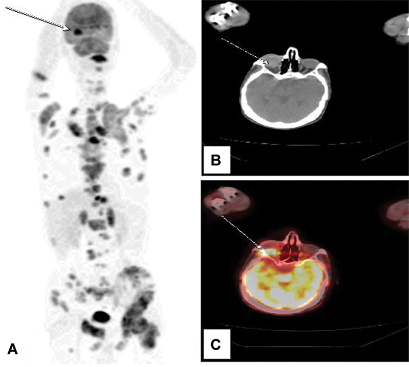 Fig. 1
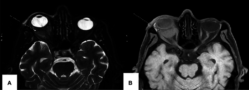 Fig. 2
Peer Reviews
No public reviews on file for this paper yet. If you reviewed it on a platform where reviews are public (OpenReview, ICLR, NeurIPS, ICML), you can paste yours below so the community can read it here.
Videos
No videos yet. Explain this paper in a talk, walkthrough, or lecture? Add one.
Taxonomy
TopicsCancer Diagnosis and Treatment · Ocular Oncology and Treatments · Brain Metastases and Treatment
Introduction
Orbital metastases are relatively uncommon, with breast and lung cancers being the most common primary tumors, accounting for 0.7 to 12% of cases. 1 2 The choroid is the ocular tissue most frequently affected by metastatic disease, followed by the iris and ciliary body. 3 4 In 25% of cases, orbital metastases represent the first manifestation of an unknown primary carcinoma (UPC), where 18F-fluorodeoxyglucose (18F-FDG) positron emission tomography/computed tomography (PET/CT) plays a critical role. We present a case of a patient with orbital metastasis incidentally detected by an 18F-FDG PET/CT with lung cancer.
Case Report
A 65-year-old male, a chronic smoker, presented with right eye pain and decreased visual acuity. Initially, this symptom was overlooked. Two months later, the patient developed a chronic cough with hemoptysis, diffuse bone pain, and weight loss. A whole-body CT scan revealed a left pulmonary mass measuring 32 mm × 25 mm, along with suspicious mediastinal lymph nodes.
A biopsy of the lung mass confirmed invasive, moderately differentiated adenocarcinoma, with immunohistochemical markers (TTF1 + ) and an epidermal growth factor receptor (EGFR) mutation (L858R), indicating a bronchopulmonary origin. An 18F-FDG PET/CT scan showed intense uptake in the left lung mass (maximum standardized uptake value [SUVmax] = 6.5), multiple FDG-avid lymph nodes, and lesions in the liver, adrenal glands, and bones ( Fig. 1 A). Notably, there was also intense and suspicious retro-orbital uptake in the right eye (SUVmax = 10.5) ( Fig. 1 B and C).
( A ) Maximum intensity projection (MIP) of our patient showing normal and pathological uptakes. ( B ) Computed tomography (CT) in axial section of the orbital region showing retro-orbital mass in the right eye (Wight arrow). ( C ) Fusion image in axial section of the orbital region showing a suspect retro-orbital uptake in the right eye related to a retro-orbital metastasis (Wight arrow).
Ophthalmological examination revealed retinal detachment, suggestive of a tumoral origin. Magnetic resonance imaging showed a 30 × 30 × 22 mm retro-orbital mass (isointense on T1, hyperintense on T2, with diffusion restriction), involving the optic nerve, intraconal fat, ocular rectus muscles, and the sphenoid wing ( Fig. 2 A and B), as well as a lesion in the left cerebellum.
Magnetic resonance imaging (MRI) of the orbital region showing a 30 × 30 × 22 mm right retro-orbital mass isointense on T1 ( A ) and hyperintense on T2 with diffusion restriction ( B ) (Wight arrows), involving the optic nerve, intraconal fat, ocular rectus muscles, and the sphenoid wing.
The patient was treated with targeted therapy (erlotinib 150 mg daily) and radiotherapy for the orbital and cerebral metastases, achieving favorable outcomes.
Discussion
Orbital metastases are rare, comprising 10% of orbital tumors and 3 to 7% of orbital lesions. 5 They typically affect the posterior choroid, with only 5 to 11% involving the ciliary body or iris. 5 Approximately in one-third of cases the primary malignancy is unknown. 6 Common primary sources include breast (39–48%), prostate, melanoma (12%), lung (8%), and kidney (7–11%). 7 Metastatic adenocarcinoma is considered as the predominant histological type. 8
Orbital symptoms such as proptosis, pain, and chemosis may indicate an undiagnosed primary cancer in 15% of cases. 9 Ocular metastases are often associated with widespread disease, with average survival ranging from 7.5 to 13 months after diagnosis. 10 For symptomatic patients, ophthalmological screening is recommended. 11
18F-FDG PET/CT is highly sensitive in detecting UPCs and staging disseminated disease. In one study, PET/CT identified primary tumors in 39.5% of cases, with lung cancer being the most common (50%). Sensitivity, specificity, and accuracy were reported as 87, 88, and 87.5%, respectively. 12 13
The treatment for orbital metastases is primarily palliative. Radiotherapy achieves a 79% response rate and preserves vision in 80% of cases. 14 Targeted therapies, such as erlotinib for EGFR-mutated adenocarcinoma, have shown to improve outcomes. Fractionated orbital radiotherapy (30–40 Gy) helps reduce complications like damage to the lacrimal apparatus. 15 The prognosis remains poor, with a 54% mortality rate within 1 year of ocular metastasis diagnosis. 10
Conclusion
Orbital metastasis can be the first sign of carcinoma in up to 25% of cases. Adenocarcinoma represents 92% of lung cancer-related orbital metastases. Early suspicion and the use of multimodal imaging, such as 18F-FDG PET/CT, are essential for prompt diagnosis and intervention to preserve vision and quality of life. This case highlights the importance of PET/CT in detecting primary malignancies in patients presenting with orbital metastasis.
The reference list from the paper itself. Each links out to its DOI / PubMed record.
- 1Manohar K Mittal B R Bhattacharya A Gupta A Les métastases orbitaires comme signe annonciateur du carcinome pulmonaire: détection de la malignité primaire et de la charge de morbidité par TEP/TDM au F-18 FDG Nucl Med Mol Imaging 20124601737524900036 10.1007/s 13139-011-0123-7PMC 4042977 · doi ↗ · pubmed ↗
- 2Singhal N Mundi I K Handa U Punia R P Mohan HFNA dans le diagnostic des lésions orbitaires provoquant une exophtalmie chez l'adulte Diagn Cytopathol 2012401086186421485026 10.1002/dc.21649 · doi ↗ · pubmed ↗
- 3Shields C L Shields J A Gross N E Schwartz G P Lally S E Enquête sur 520 yeux, avec métastases uvéales Ophtalmologie 19971041265127610.1016/s 0161-6420(97)30148-19261313 · doi ↗ · pubmed ↗
- 4Shah S U Mashayekhi A Shields C LMétastase uvéale due au cancer du poumon: caractéristiques cliniques, traitement et résultats chez 194 patients Ophtalmologie 2013•••26
- 5Lieb W E Shields J A Shields C L Spaeth G L Mucinous adenocarcinoma metastatic to the iris, ciliary body, and choroid Br J Ophthalmol 199074063733762165801 10.1136/bjo.74.6.373PMC 1042133 · doi ↗ · pubmed ↗
- 6Char D H Miller T Kroll SMétastases orbitaires: diagnostic et évolution Br J Ophthalmol 1997813863909227204 10.1136/bjo.81.5.386PMC 1722195 · doi ↗ · pubmed ↗
- 7Solari H P Ventura M P Cheema D P Odashiro A N Burnier M NMétastase orbitaire d'un carcinome du sein se présentant comme une kératite neurotrophique Can J Ophthalmol 200641939616462881 10.1016/S 0008-4182(06)80075-X · doi ↗ · pubmed ↗
- 8Amemiya T Hayashida H Dake Y Tumeurs orbitaires métastatiques au Japon: revue de la littérature Ophthalmic Epidemiol 2002901354711815894 10.1076/opep.9.1.35.1718 · doi ↗ · pubmed ↗