A New Labeling Method of 99m Tc-PSMA-HBED-CC
Benchamat Phromphao, Shuichi Shiratori

TL;DR
Researchers developed a new, affordable method to create a prostate cancer imaging drug using 99mTc, which could be more accessible than the current FDA-approved version.
Contribution
The study introduces a novel in-house labeling method for 99mTc-PSMA-HBED-CC as an accessible alternative to 68Ga-PSMA-HBED-CC.
Findings
The optimal labeling conditions achieved a high radiochemical yield of 71.49% and purity of 98.29%.
The radiolabeled compound remained stable with over 95% purity for 4 hours at room temperature.
The method met European Pharmacopoeia quality control standards.
Abstract
68 Ga-PSMA-HBED-CC ( 68 Ga-PSMA-11) was approved by the U.S. Food and Drug Administration as the first prostate-specific membrane antigen (PSMA)-targeted positron emission tomography (PET) imaging drug for patients with prostate cancer. However, the utility of 68 Ga-PSMA-HBED-CC may be limited due to PET/CT or PET/MR accessibility and 68 GaCl 3 availability produced from 68 Ge/ 68 Ga generator or cyclotron. Thus, in-house preparation of 99m Tc-PSMA-HBED-CC was developed as an alternative to 68 Ga-PSMA-HBED-CC to be ubiquitous and affordable in the worldwide population. A solution of 99m Tc-pertechnetate was added to PSMA-HBED-CC and 4% SnCl 2 ·2H 2 O in a 10-mL sterile vial. The mixture was heated at 100°C for 15 minutes and then allowed to cool to room temperature. Labeling conditions were optimized to maximize the radiochemical yield of 99m Tc-PSMA-HBED-CC. The chelation completeness…
Genes, proteins, chemicals, diseases, species, mutations and cell lines named across the full text — each resolved to its canonical identifier and authoritative record.
Click any figure to enlarge with its caption.
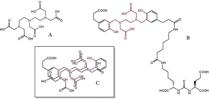 Fig. 1
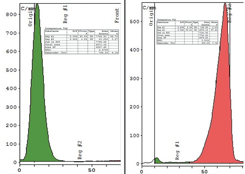 Fig. 2
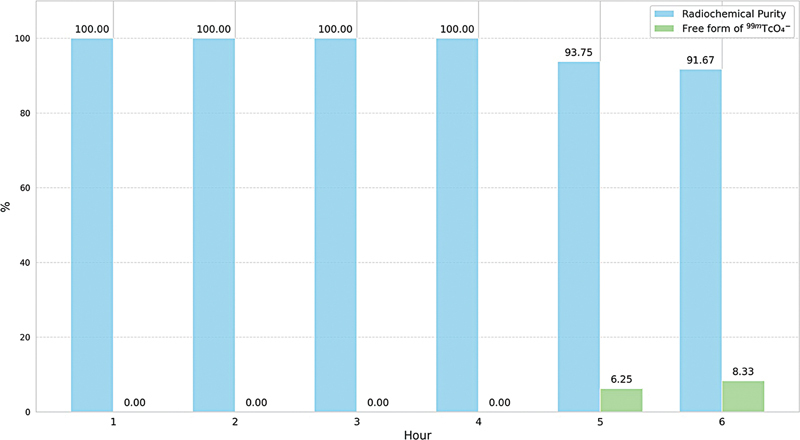 Fig. 3
| Iteration | SnCl 2 ·2H 2 O (µg) | 99m TcO 4 − (mCi) | Labeling condition | Free form of 99m TcO 4 − (%) | 99m Tc-colloid (%) | RCY (%) |
|---|---|---|---|---|---|---|
|
| 0.5 | 10.02 | Room temperature | 99.58 | 0.17 | 0.25 |
|
| 0.5 | 10.32 | 100°C, | 88.89 | 0.00 | 11.11 |
|
| 1.0 | 9.97 | 100°C | 81.82 | 0.00 | 18.18 |
|
| 2.0 | 10.16 | 100°C, | 64.29 | 10.00 | 25.71 |
|
| 2.5 | 10.61 | 100°C, | 20.00 | 20.00 | 60.00 |
|
| 3.0 | 11.02 | 100°C, | 9.09 | 25.00 | 65.91 |
|
| 3.5 | 11.64 | 100°C, | 0.00 | 41.67 | 58.33 |
|
| 4.0 | 11.15 | 100°C, | 0.00 | 50.00 | 50.00 |
Peer Reviews
No public reviews on file for this paper yet. If you reviewed it on a platform where reviews are public (OpenReview, ICLR, NeurIPS, ICML), you can paste yours below so the community can read it here.
Videos
No videos yet. Explain this paper in a talk, walkthrough, or lecture? Add one.
Taxonomy
TopicsRadiopharmaceutical Chemistry and Applications · Prostate Cancer Treatment and Research · Medical Imaging Techniques and Applications
Introduction
Prostate cancer is the most common malignancy found in men and the second leading cause of cancer death worldwide. 1 Over the past two decades, the initial diagnosis and follow-up have been serum prostate-specific antigen levels, digital rectal examination, and some conventional imaging techniques including ultrasound, computed tomography, magnetic resonance imaging, and bone scintigraphy, but none provides highly specific and sensitive detection. Although transrectal ultrasound-guided prostate biopsy is currently accepted as the gold standard to provide the histopathological diagnosis of prostate cancer, 2 it is an invasive procedure that resulted in a risk of side effects and the accuracy of diagnosis. The accurate definition of tumor burden and its staging is particularly important for effective treatment selection. Therefore, molecular imaging, a noninvasive method, has been employed to visualize the tumor in both soft tissue and bone with higher specific and sensitive detection, monitored response to therapy to improve management of prostate cancer, clinical outcome, and patient's quality of life.
The Food and Drug Administration (FDA) approved ^68^ Ga-PSMA-HBED-CC ( ^68^ Ga-PSMA-11, previous name: ^68^ Ga-DKFZ-PSMA-11, generic name: ^68^ Ga-Gazetotide) as the first prostate-specific membrane antigen (PSMA)-targeted positron emission tomography (PET) imaging drug for men with prostate cancer. 3 The development of ^68^ Ga-PSMA-HBED-CC, which targets PSMA, has offered new perspectives for prostate cancer diagnosis and evaluation of therapeutic response. 4 PSMA is a cell surface transmembrane protein with 750 amino acids type II glycoprotein that primarily expresses in normal prostate epithelium and is overexpressed in prostate cancer cells including bone metastasis. 5 X-ray crystal structure analysis of PSMA, also known as N -acetylated L -aspartyl- L -glutamate peptidase (NAALADase I), has identified the critical interaction of potent inhibitors within the hydrophobic active site of the enzyme. 6 Consequently, several classes of NAALADase I inhibitors had been exploited for structure-based design platforms, leading to the novel synthesis of PSMA-HBED-CC. 7 8 9 To date, ^68^ Ga-PSMA-HBED-CC has explicitly demonstrated superior detection of PSMA-positive prostate cancer lesions in recurrent and metastatic sites over conventional imaging methods 10 11 and two other FDA-approved PET tracers, ^18^ F-fluciclovine and ^11^ C-choline that are used in patients with suspected cancer recurrence. 12 Recently, the FDA also approved ^18^ F-piflufolastat as the second PSMA-targeted PET imaging drug with prostate cancer.
Besides ^68^ Ga-PSMA-HBED-CC, the preferential use of ^99m^ Tc-labeled urea-based PSMA inhibitor has received interest as an alternative option to widespread the advantages of PSMA imaging due to a number of prostate cancer patients who are scheduled on PSMA imaging. The hybrid modality of single-photon emission CT (SPECT)/CT offers a wide range of workhorses in nuclear medicine with lower financial access, especially the remote medical center in which PET/CT facility is not available. Although the spatial resolution of ^99m^ Tc is not as good as that of ^68^ Ga, ^99m^ Tc provides a sufficiently long half-life of 6 hours in both preparation and accumulation in the target site. Moreover, the decay range of ^99m^ Tc is short enough to minimize radiation exposure to patients and medical staff.
While some ^99m^ Tc-labeled PSMA tracers have been previously reported using various PSMA ligands with several forms of complexation, for example, [ ^99m^ Tc(CO) 3 (L) 3 ] ^+^ , 13 14 15 MAG3-based ^99m^ Tc-PSMA-I&S, 16 ^99m^ Tc-MIP, 17 18 19 20 ^99m^ Tc-HYNIC-PSMA, 21 22 peptide-chelator-based ^99m^ Tc-DUPA, 23 ^99m^ Tc-PSMA-T4, 24 ^99m^ Tc-PSMA-tricarbonyl-HBED-CC, 25 and ^99m^ Tc-PSMA-HBED-CC, 26 it challenges to develop a convenient labeling method in a single step without coligand for ^99m^ Tc-complexation. According to our experience in theranostics, we adapted the routine standard labeling procedure of ^68^ Ga-PSMA-HBED-CC with the rationale that HBED-CC would serve as a suitable chelator in a mimic manner to diethylenetriamine pentaacetic acid (DTPA) as shown in Fig. 1 . Our attention focused on optimizing the labeling parameters to improve the radiosynthesis of ^99m^ Tc-PSMA-HBED-CC.
Chemical structures of ( A ) diethylenetriamine pentaacetic acid (DTPA), ( B ) PSMA-HBED-CC, and ( C ) imposed chelating motif.
Material and Methods
PSMA-HBED-CC was purchased from ABX advanced biochemical compounds (GmbH, Germany). Sodium ^99m^ Tc-pertechnetate was purchased from Global Medical Solution (Thailand). Stannous chloride dihydrate and hydrochloric acid were purchased from Sigma-Aldrich (Germany). The stock solution of 4% stannous chloride was freshly prepared and kept in a refrigerator. All chemicals and solvents were used without further purification unless otherwise noted. The C18 cartridge (Sep-Pak Light, lot no. 045732200A) was purchased from Waters (United States). The TLC scanner Raytest model MiniGita was used.
Preparation of 4% Stannous Chloride Stock Solution
SnCl 2 ·2H 2 O 0.144 mg was added to 37% HCl 0.75 mL, followed by heating at 100°C for 5 minutes. After cooling down to room temperature, 6 N HCl 2.25 mL was added to 4% SnCl 2 ·2H 2 O, which should be freshly prepared before labeling.
Labeling of PSMA-HBED-CC with Tc-99m
To a mixture of 4% SnCl 2 ·2H 2 O solution calculated as an amount of SnCl 2 and an aliquot of PSMA-HBED-CC (10 µg in H 2 O 100 µL) in 10 mL sterile vial, ^99m^ Tc-pertechnetate 370 MBq was added. The labeling was performed at 100°C for 15 minutes in a heating block, followed by a 10-minute cool down to reach room temperature. The crude product was passed through a C18 cartridge. ^99m^ Tc-PSMA-HBED-CC was slowly purged from a C18 cartridge using EtOH:H 2 O (1:1) 2 mL to the final product vial.
Quality Control
Radiochemical purity (RCP) analyses were performed using instant thin-layer chromatography (iTLC) on silica paper strips as a stationary phase with two different mobile phases. The free form of ^99m^ TcO 4 ^−^ was determined using 0.9% normal saline, whereas ^99m^ Tc-colloid formation was determined using acetone as the mobile phase. The radioactivity distribution on iTLC strips was also determined on a TLC scanner. Subsequently, radiochemical yield (RCY) was calculated. The pH of the final product was determined using a pH indicator.
Stability Test
The chemical stability of ^99m^ Tc-PSMA-HBED-CC was carried out by incubating the final product radioactivity in samples range 10.22 ± 0.40 mCi at room temperature for 6 hours and monitored by iTLC every hour. The radioconcentration used for the stability test was 2.04 mCi/mL. No stabilizer was added.
Results
Labeling of
99m Tc-PSMA-HBED-CC
The labeling parameters were investigated to achieve the highest possible RCY. The quantity of PSMA-HBED-CC used in each experiment remained constant at 10 µg (0.011 µmol). In accordance with the DTPA cold kit formulation, the appropriate radioactivity of ^99m^ Tc-pertechnetate was determined in direct correlation with approximate 370 MBq (10 mCi), while maintaining the solution's pH at 5.0, as indicated in Table 1 .
Table 1: Labeling parameters in 99m Tc-PSMA-HBED-CC
In the preliminary experiment, 10.02 mCi of ^99m^ TcO 4 ^−^ was combined with 0.5 µg of SnCl 2 at room temperature for 15 minutes that resulted in a RCY of 0.25%, with 99.58% of ^99m^ TcO 4 ^−^ remaining unbound. Subsequently, the reaction temperature was increased to 100°C for 15 minutes, in alignment with the standard procedure for labeling ^68^ Ga-PSMA-HBED-CC. This adjustment resulted in a RCY of 11.11%, affirming the choice to set the reaction conditions at 100°C for 15 minutes. In iterations 3 to 8, the amount of SnCl 2 was progressively adjusted to enhance the RCY. By increasing the SnCl 2 quantity by 0.5 µg in each iteration, the highest RCY of 65.91% was achieved in the sixth iteration. However, when the amount of SnCl 2 exceeded 3.5 µg, complete chelation occurred between PSMA-HBED-CC and ^99m^ TcO 4 ^−^ , which also led to a significant rise in the formation of hydrolyzed species, ultimately diminishing the RCY. Total radioactivities of the final product recovered after labeling are 10.12 ± 0.58 mCi.
Chemical Stability of
99m Tc-PSMA-HBED-CC
The chemical stability of ^99m^ Tc-PSMA-HBED-CC was evaluated using the optimized labeling method in the 6th iteration that produced the highest RCY. The compound was incubated at room temperature for 6 hours, and RCP was checked hourly through iTLC ( Fig. 2 ). The findings are shown in Fig. 3 . In general, the RCP of Tc-99m radiopharmaceuticals should meet or exceed 95%. This study demonstrated that the RCP of ^99m^ Tc-PSMA-HBED-CC remained above 95% for up to 4 hours after labeling, and stayed above 90% at the 5th and 6th hours.
Instant thin-layer chromatography (iTLC) chromatogram of 99m Tc-PSMA-HBED-CC; (left) in acetone as mobile phase, (right) in 0.9% normal saline as mobile phase.
Chemical stability of 99m Tc-PSMA-HBED-CC at room temperature.
Discussion
Since the identification of PSMA as an antigen and the discovery of the specific antibody 7E11-C5 (capromab) for both normal and malignant prostate epithelium, as reported by Horoszewicz et al, 27 PSMA has emerged as a crucial target for prostate cancer cells. 28 29 30 In 2012, Eder et al developed the urea-based PET tracer ^68^ Ga-PSMA-HBED-CC (formerly ^68^ Ga-DKFZ-PSMA-11) to address the limitations of the lead antibody J591. 31 The U.S. FDA approved ^68^ Ga-PSMA-HBED-CC as the first PET imaging agent for PSMA-positive lesions in men with prostate cancer in December 2020 3 and ^18^ F-piflufolastat as the second PET imaging agent for PSMA-positive lesions in men with prostate cancer in May 2021. 3 Inspired by this breakthrough, our study aimed to develop ^99m^ Tc-PSMA-HBED-CC as a SPECT imaging analog to increase accessibility for prostate cancer diagnosis.
PSMA-HBED-CC was chosen for this study due to its widespread clinical use and commercial availability. The dose of PSMA-HBED-CC was standardized at 10 μg. To optimize the manual labeling of ^99m^ Tc-PSMA-HBED-CC, various labeling conditions and amounts of calculated SnCl 2 were assessed. The results, presented in Table 1 , indicate that ^99m^ Tc-PSMA-HBED-CC is a thermodynamically favorable product. Using 2.5 to 4.0 μg of SnCl 2 resulted in a RCY exceeding 50%. When the SnCl 2 amount exceeded 3.5 μg, no uncomplexed Tc-99m was detected, indicating effective reduction of TcO 4 ^−^ . However, higher amounts of SnCl 2 also increased colloid formation. Optimal quantitative radiolabelling of 10 μg of PSMA-HBED-CC was achieved with 3.0 μg of SnCl 2 . To prevent colloid formation, both SnCl 2 and PSMA-HBED-CC must be present in the reaction mixture before adding TcO 4 ^−^ to form the desired complex.
The chemical stability of ^99m^ Tc-PSMA-HBED-CC was evaluated by incubating it at room temperature. As shown in Fig. 2 , it retained RCP above 95% for up to 4 hours. After this period, free Tc-99m increased. Therefore, it is recommended to use ^99m^ Tc-PSMA-HBED-CC within 4 hours of preparation or store it in a refrigerator to maintain stability.
Vats et al 26 previously reported the preparation of ^99m^ Tc-PSMA-HBED-CC using 50 mg of PSMA-HBED-CC, 40 mg of SnCl 2 , and TcO 4 ^−^ 740 MBq at pH 5, yielding a RCY of 60 ± 5%, a RCP greater than 98%, and specific activity of 15 ± 5 GBq/µmol. However, they did not conduct stability tests. Economically, our study used 10 µg of PSMA-HBED-CC, 3 µg of SnCl 2 , and 370 MBq of ^99m^ Tc-pertechnetate at 100°C for 15 minutes, achieving a higher RCY (71.49 ± 2.42%), RCP (98.29 ± 2.65%), and specific activity (37.84 ± 1.47 GBq/µmol). This method is more cost-effective and easier to manipulate.
Conclusion
To optimize the labeling of PSMA-HBED-CC with ^99m^ Tc-pertechnetate for prostate cancer imaging, the labeling procedure should be carried out at 100°C for 15 minutes using 3 µg of SnCl 2 , minimizing the presence of free ^99m^ Tc-pertechnetate and colloid formation. Purification with a C18 cartridge is required to achieve RCP that complies with the European Pharmacopoeia standards. The stability of ^99m^ Tc-PSMA-HBED-CC remains robust for up to 4 hours at room temperature, with RCP exceeding 95%. It is recommended to use the labeled product within 4 hours of preparation.
The reference list from the paper itself. Each links out to its DOI / PubMed record.
- 1Bray F Ferlay J Soerjomataram I Siegel R L Torre L A Jemal A Global cancer statistics 2018: GLOBOCAN estimates of incidence and mortality worldwide for 36 cancers in 185 countries CA Cancer J Clin 2018680639442430207593 10.3322/caac.21492 · doi ↗ · pubmed ↗
- 2Moe A Hayne D Transrectal ultrasound biopsy of the prostate: does it still have a role in prostate cancer diagnosis?Transl Androl Urol 20209063018302433457275 10.21037/tau.2019.09.37PMC 7807378 · doi ↗ · pubmed ↗
- 3Hennrich U Eder M [ 68 Ga] Ga-PSMA-11: the first FDA-approved 68 Ga-radiopharmaceutical for PET imaging of prostate cancer Pharmaceuticals 2021140871372534451810 10.3390/ph 14080713 PMC 8401928 · doi ↗ · pubmed ↗
- 4Afshar-Oromieh A Haberkorn U Eder M Eisenhut M Zechmann C M [ 68 Ga]Gallium-labelled PSMA ligand as superior PET tracer for the diagnosis of prostate cancer: comparison with 18 F-FECH Eur J Nucl Med Mol Imaging 201239061085108622310854 10.1007/s 00259-012-2069-0 · doi ↗ · pubmed ↗
- 5Silver D A Pellicer I Fair W R Heston W D Cordon-Cardo C Prostate-specific membrane antigen expression in normal and malignant human tissues Clin Cancer Res 199730181859815541 · pubmed ↗
- 6Davis M I Bennett M J Thomas L M Bjorkman P J Crystal structure of prostate-specific membrane antigen, a tumor marker and peptidase Proc Natl Acad Sci U S A 2005102175981598615837926 10.1073/pnas.0502101102 PMC 556220 · doi ↗ · pubmed ↗
- 7Mesters J R Henning K Hilgenfeld R Human glutamate carboxypeptidase II inhibition: structures of GCPII in complex with two potent inhibitors, quisqualate and 2-PMPA Acta Crystallogr D Biol Crystallogr 200763(Pt 4):50851317372356 10.1107/S 090744490700902 X · doi ↗ · pubmed ↗
- 8Lundmark F Olanders G Rinne S S Abouzayed A Orlova A Rosenström U Design, synthesis, and evaluation of linker-optimised PSMA-targeting radioligands Pharmaceutics 202214051098111735631684 10.3390/pharmaceutics 14051098 PMC 9147442 · doi ↗ · pubmed ↗