Acute Spinal Cord Syndrome As the Initial Manifestation of Multiple Sclerosis: A Case Report
Catarina Pinto Silva, Cristiana Fernandes, Márcia Ribeiro, Dany Cruz

TL;DR
A young man with no prior health issues developed acute spinal cord symptoms, which turned out to be the first sign of multiple sclerosis.
Contribution
This case highlights the rare but important presentation of MS as an isolated spinal cord syndrome.
Findings
MRI showed two spinal cord lesions and a brain lesion consistent with demyelination.
CSF analysis revealed lymphocytic pleocytosis and oligoclonal bands, supporting an MS diagnosis.
Treatment with high-dose steroids led to significant neurological improvement.
Abstract
Multiple sclerosis (MS) typically presents with optic neuritis, brainstem syndromes, or sensory disturbances. Isolated spinal cord involvement as the initial manifestation occurs in fewer than 30% of cases, making early recognition challenging but essential for timely diagnosis and treatment initiation. A previously healthy 22-year-old Caucasian male presented with an acute onset of mid-thoracic pain followed by progressive sensory deficits below the T10 dermatome and lower limb weakness developing over five days. Neurological examination revealed spastic paraparesis (Medical Research Council grade 3/5), sensory level at T10, and hyperreflexia with bilateral Babinski signs. Magnetic resonance imaging (MRI) of the spinal cord demonstrated two focal T2-hyperintense intramedullary lesions at T1-T2 and T11 levels with gadolinium enhancement. Brain MRI revealed a single periventricular…
Genes, proteins, chemicals, diseases, species, mutations and cell lines named across the full text — each resolved to its canonical identifier and authoritative record.
Click any figure to enlarge with its caption.
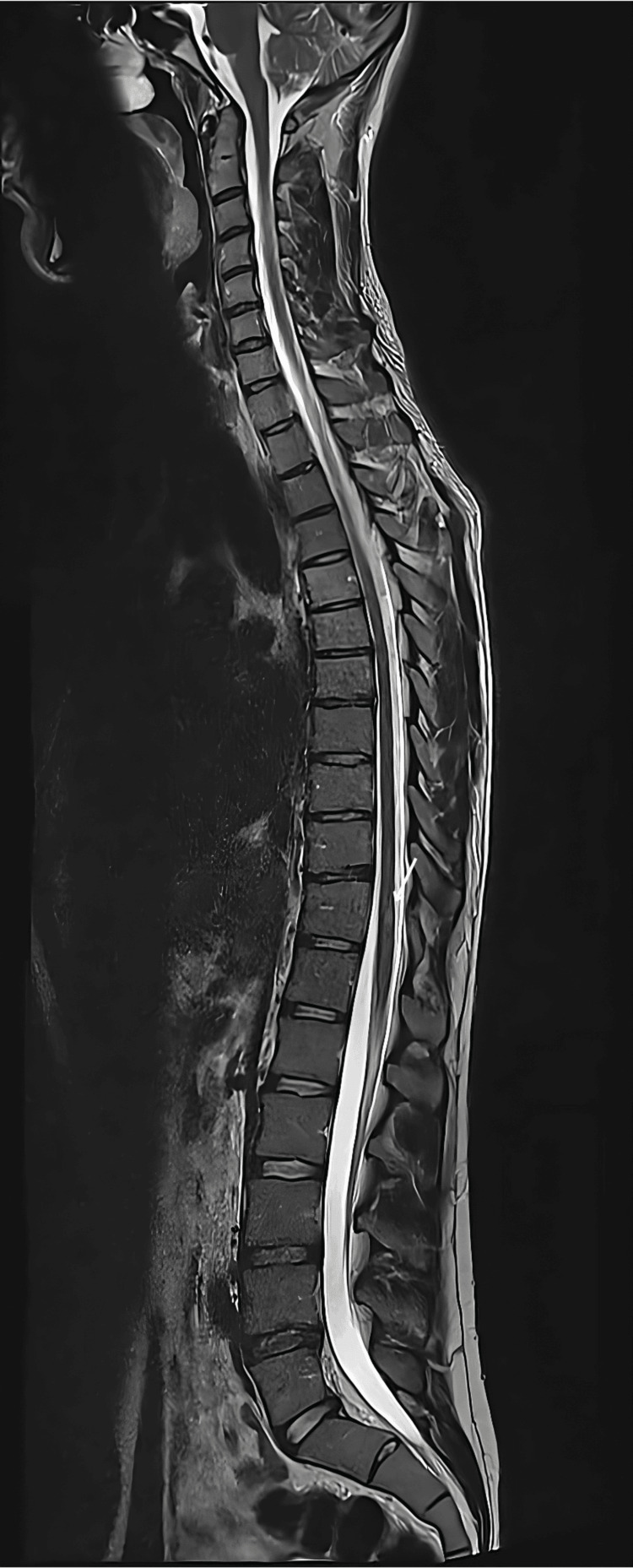 Figure 1
| Parameter | Result | Reference range |
| White blood cells | 11 cells/µL (lymphocytic) | <5 |
| Protein | 52 mg/dL | 15–A45 |
| Glucose | 65 mg/dL (serum glucose: 98 mg/dL) | 45–80 |
| IgG index | 0.85 | <0.7 |
| Oligoclonal bands | Positive (CSF-restricted) | Negative |
| Vitamin B12 | 485 pg/mL | 200–900 |
| ESR | 12 mm/h | <20 |
| CRP | 0.8 mg/L | <5 |
| HIV serology | Negative | – |
| Syphilis serology | Negative | – |
| Aquaporin-4 antibody | Negative | – |
| MOG antibody | Negative | – |
Peer Reviews
No public reviews on file for this paper yet. If you reviewed it on a platform where reviews are public (OpenReview, ICLR, NeurIPS, ICML), you can paste yours below so the community can read it here.
Videos
No videos yet. Explain this paper in a talk, walkthrough, or lecture? Add one.
Taxonomy
TopicsMultiple Sclerosis Research Studies · Peripheral Neuropathies and Disorders · Autoimmune Neurological Disorders and Treatments
Introduction
Multiple sclerosis (MS) is a chronic, immune-mediated demyelinating disorder of the central nervous system (CNS) and a leading cause of neurological disability in young adults. According to the most recent Atlas of MS, approximately 2.8 million people are affected worldwide [1].
Typical initial manifestations include optic neuritis, brainstem syndromes, and sensory disturbances [2,3]. Spinal cord involvement may occur in up to one-third of first presentations but is less frequently isolated, making early diagnosis more challenging [4].
Spinal cord lesions in MS typically appear as short, ovoid, and peripherally located, most often involving less than two vertebral segments [5]. Under the 2017 revision of the McDonald criteria, demonstrating dissemination in space across the brain and spinal cord is essential for diagnosis [6].
Early recognition of spinal cord presentations is critical, as lesion burden strongly correlates with long-term disability and risk of conversion to secondary progressive MS [7,8]. According to the 2017 revision of the McDonald criteria, MS may be diagnosed after a first clinical event when dissemination in space (lesions in at least two typical CNS regions, such as the brain and spinal cord) and dissemination in time (simultaneous presence of enhancing and non-enhancing lesions or cerebrospinal fluid (CSF)-specific oligoclonal bands) are demonstrated [6]. However, when the spinal cord is the sole site of clinical involvement, diagnosis may be delayed due to the rarity and diagnostic overlap with other inflammatory myelopathies. We report a young man who presented with acute spinal cord syndrome as the first clinical manifestation of MS, highlighting the diagnostic challenges, differential considerations, and therapeutic implications.
Case presentation
A 22-year-old Caucasian male with no prior medical or family history presented with five days of progressive neurological symptoms. He initially developed sharp, band-like thoracic pain at the T6-T8 dermatomes. Within 48 hours, ascending paresthesias appeared in both feet and spread to the umbilical level. By day three, he experienced bilateral lower limb weakness that rapidly progressed, requiring assistance for ambulation.
On admission, his vital signs were stable, and the general physical examination was unremarkable. Neurological examination showed normal strength in the upper limbs (Medical Research Council (MRC) grade 5/5), while the lower limbs demonstrated weakness (MRC grade 3/5) with increased tone and spasticity. A well-defined sensory level was noted at T10, with absent light touch and pinprick sensation below this level. Vibration sense was reduced in the lower extremities, and proprioception was impaired in the toes. Deep tendon reflexes were brisk in the lower limbs (3+ to 4+) with bilateral Babinski signs, while upper limb reflexes were normal. Abdominal reflexes were absent below T10. Cranial nerves were intact, and there were no visual or cerebellar deficits. The patient's gait was spastic and paraparetic, requiring bilateral support. The CSF and serum laboratory investigations are shown in Table 1.
Spinal MRI revealed two short-segment intramedullary hyperintense lesions on T2-weighted imaging: an ovoid lesion at T1-T2 in the left posterolateral cord and another at T11 in the right posterolateral cord (Figure 1). Both lesions enhanced with gadolinium, without cord swelling or longitudinal extension. Brain MRI demonstrated a small periventricular lesion near the left frontal horn, consistent with demyelination.
Spinal MRI, sagittal T2-weighted sequence. White arrow highlights focal hyperintense lesion at T11
A diagnosis of clinically isolated syndrome highly suggestive of MS was made. The patient received intravenous methylprednisolone (1 g daily for three days) followed by a 10-day taper of oral prednisone. Inpatient neurological rehabilitation included physiotherapy, occupational therapy, bladder retraining, and vitamin D supplementation.
At one month, lower limb strength improved to MRC grade 4/5 with partial sensory recovery and independent ambulation using a walking aid (Expanded Disability Status Scale (EDSS) score 3.0) [9]. At three months, strength further improved to 4+/5 with mild residual sensory deficits and an EDSS score of 2.0. MRI surveillance was scheduled, and initiation of disease-modifying therapy was discussed in a multidisciplinary setting.
Discussion
This case illustrates an uncommon initial presentation of MS in which acute spinal cord syndrome constituted the first and dominant clinical manifestation. While spinal cord involvement is frequent throughout the disease course, it is less commonly the sole initial presentation, occurring in fewer than one-third of patients [4]. Unlike typical longitudinally extensive lesions seen in neuromyelitis optica spectrum disorders (NMOSD) or myelin oligodendrocyte glycoprotein antibody-associated disease (MOGAD), our patient presented with short-segment, peripherally located intramedullary lesions at two separate spinal levels, a pattern that is more characteristic of MS [5].
The detection of a single periventricular brain lesion on MRI is particularly relevant. This finding suggests prior subclinical disease activity and fulfills the criterion for dissemination in space according to the 2017 McDonald criteria when combined with spinal cord lesions [6]. Moreover, the presence of CSF-restricted oligoclonal bands fulfills the criterion for dissemination in time, allowing an earlier diagnosis after a first clinical event. It is therefore plausible that a previous minor central nervous system inflammatory event may have occurred without being clinically recognized by the patient. If the brain lesion had not been present, the diagnosis would likely have remained classified as a clinically isolated syndrome with a lower immediate probability of conversion to definite MS, influencing both prognostic counseling and therapeutic strategy.
Differential diagnoses included NMOSD and MOGAD, which were excluded based on characteristic radiologic patterns, negative aquaporin-4 and MOG antibodies, and the absence of longitudinally extensive transverse myelitis [10,11]. These features reinforce the specificity of the diagnostic workup in distinguishing MS from other inflammatory myelopathies.
Prognostically, spinal cord involvement and early cord atrophy are associated with worse disability trajectories compared with brain lesion burden alone [12-14]. These insights underscore the value of MRI surveillance of both brain and spinal cord, as well as early rehabilitation to maximize functional recovery.
High-dose corticosteroids remain the standard of care for acute inflammatory demyelinating episodes [15,16]. Contemporary evidence supports early use of highly effective disease-modifying therapies in appropriate patients to reduce relapse rates and delay disability accrual [16,17]. Our patient's favorable short-term recovery after corticosteroids and rehabilitation aligns with published experience, while highlighting the need for long-term monitoring and timely initiation of disease-modifying therapy.
Conclusions
MS should be considered in young adults presenting with acute spinal cord syndromes, particularly when associated with short-segment spinal lesions and inflammatory CSF findings. In this case, the coexistence of spinal cord lesions, a periventricular brain lesion, and CSF-restricted oligoclonal bands fulfilled the 2017 McDonald criteria for early diagnosis. Prompt corticosteroid therapy was associated with significant neurological recovery, supporting early treatment in similar presentations. MRI surveillance and timely initiation of disease-modifying therapy remain essential to reduce the risk of future relapses and disability progression.
The reference list from the paper itself. Each links out to its DOI / PubMed record.
- 1Rising prevalence of multiple sclerosis worldwide: Insights from the Atlas of MS, third edition Mult Scler Walton C King R Rechtman L 181618212620203317447510.1177/1352458520970841 PMC 7720355 · doi ↗ · pubmed ↗
- 2Diagnosis of multiple sclerosis: progress and challenges Lancet Brownlee WJ Hardy TA Fazekas F Miller DH 1336134638920172788919010.1016/S 0140-6736(16)30959-X · doi ↗ · pubmed ↗
- 3Multiple sclerosis Lancet Compston A Coles A 1502151737220081897097710.1016/S 0140-6736(08)61620-7 · doi ↗ · pubmed ↗
- 4Spinal cord abnormalities in recently diagnosed MS patients: added value of spinal MRI examination Neurology Bot JC Barkhof F Polman CH 2262336220041474505810.1212/wnl.62.2.226 · doi ↗ · pubmed ↗
- 5Magnetic resonance imaging in multiple sclerosis: the present and the future Neurol Sci Filippi M Preziosa P Meani A Rocca MA 25312537412020
- 6Diagnosis of multiple sclerosis: 2017 revisions of the Mc Donald criteria Lancet Neurol Thompson AJ Banwell BL Barkhof F 1621731720182927597710.1016/S 1474-4422(17)30470-2 · doi ↗ · pubmed ↗
- 7Magnetic resonance imaging correlates of physical disability in relapse onset multiple sclerosis of long disease duration Mult Scler Kearney H Rocca MA Valsasina P 72802020142381228310.1177/1352458513492245 PMC 4107776 · doi ↗ · pubmed ↗
- 8Spinal cord MRI in multiple sclerosis--diagnostic, prognostic and clinical value Nat Rev Neurol Kearney H Miller DH Ciccarelli O 3273381120152600900210.1038/nrneurol.2015.80 · doi ↗ · pubmed ↗