Morphological and Optical Characterization of NRAS‐Mutant Melanoma Cells and Primary Melanocytes via Quantitative Phase Imaging With Digital Holographic Microscopy
Ayah A. Farhat, Yazan A. Almahdi, Fatima Z. Alshuhani, Besa Xhabija

TL;DR
This study uses holographic microscopy to identify differences between normal skin cells and melanoma cells with NRAS mutations, offering a new tool for early cancer detection.
Contribution
The study introduces a label-free method using digital holographic microscopy and data analysis to distinguish NRAS-mutant melanoma cells from normal melanocytes.
Findings
Distinct morphological and optical differences were found between NRAS-mutant melanoma cells and melanocytes.
Optical properties emerged as key features separating the two cell types.
QPI with DHM shows potential as a non-invasive diagnostic tool for melanoma.
Abstract
Early detection of melanoma, a highly variable and aggressive form of skin cancer, is crucial for improving patient outcomes. It is essential to distinguish malignant cells from normal melanocytes, and therefore, label‐free imaging methods that can do so are needed. Given the genotoxic effect of UV radiation, these mutations are numerous and affect many genes, including NRAS; therefore, therapeutic strategies can be directed toward these recurrent mutations. The aggressive nature of NRAS‐mutant melanoma contributes to poor patient prognosis, highlighting the need for early diagnosis. This study utilizes quantitative phase imaging (QPI) with digital holographic microscopy (DHM) to differentiate the morphology of NRAS‐mutant SK‐MEL‐2 cells from melanocytes using holographic microscopy; dimensionality reduction techniques, including Principal Component Analysis (PCA),t‐distributed…
Genes, proteins, chemicals, diseases, species, mutations and cell lines named across the full text — each resolved to its canonical identifier and authoritative record.
Click any figure to enlarge with its caption.
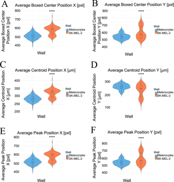 Figure 1
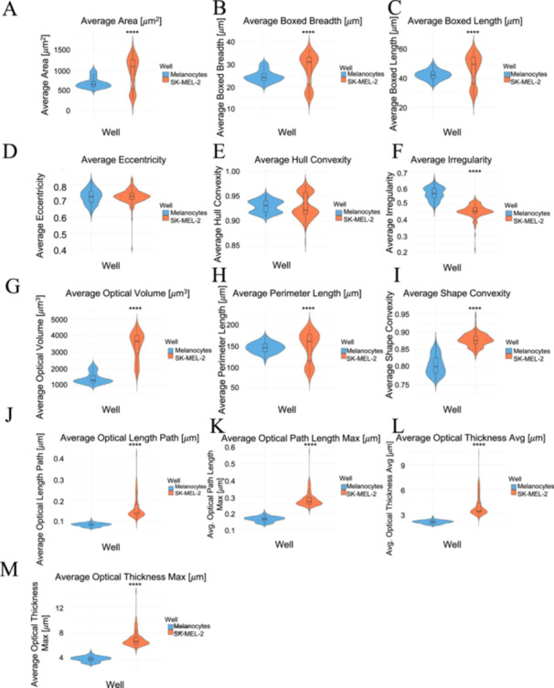 Figure 2
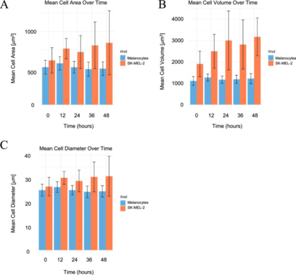 Figure 3
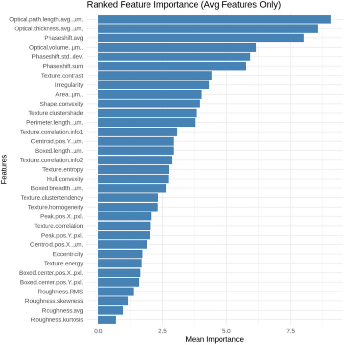 Figure 4
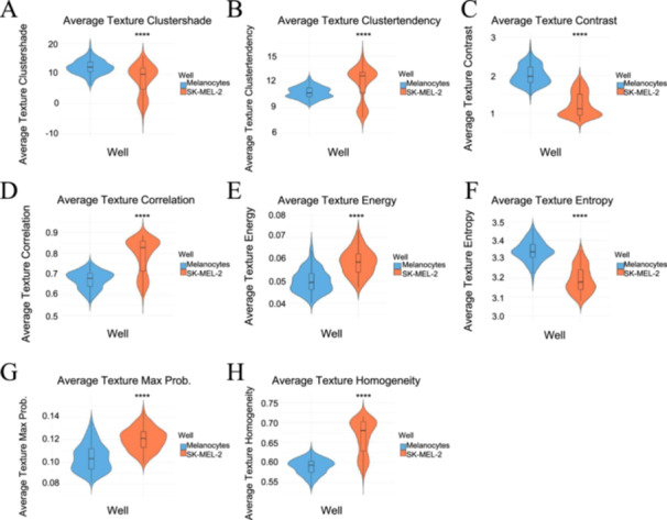 Figure 5
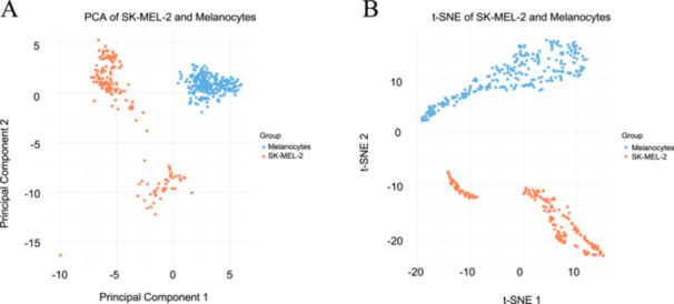 Figure 6
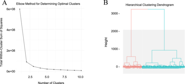 Figure 7
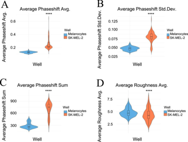 Figure 8
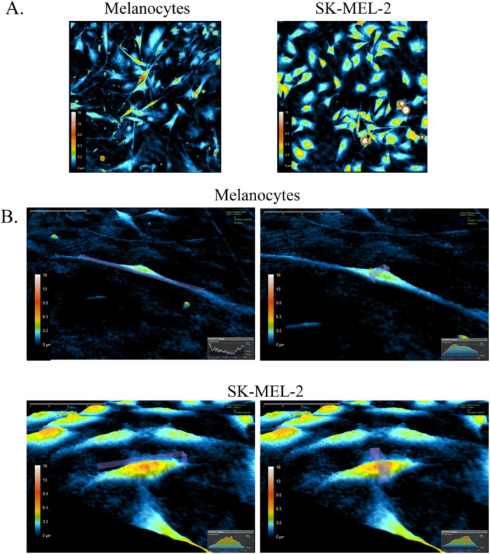 Figure 9
Peer Reviews
No public reviews on file for this paper yet. If you reviewed it on a platform where reviews are public (OpenReview, ICLR, NeurIPS, ICML), you can paste yours below so the community can read it here.
Videos
No videos yet. Explain this paper in a talk, walkthrough, or lecture? Add one.
Taxonomy
TopicsDigital Holography and Microscopy · Optical measurement and interference techniques · Advanced X-ray Imaging Techniques