Monitoring Acute Posterior Multifocal Placoid Pigment Epitheliopathy Disease Progression Using Non-invasive Multimodal Imaging: A Case Series
Andrew Palmier, Alastair Bezzina

TL;DR
This case series shows how non-invasive imaging can track the progression and treatment response of a rare eye disease called APMPPE.
Contribution
The study demonstrates the use of non-invasive multimodal imaging to monitor APMPPE without dye-based angiography.
Findings
Non-invasive imaging showed reduced hyper-reflectivity in retinal layers as the disease resolved.
Changes in autofluorescence and choriocapillaris flow voids correlated with clinical improvement.
Imaging biomarkers can detect disease activity and treatment effects in APMPPE.
Abstract
In this case series, we present two cases of acute posterior multifocal placoid pigment epitheliopathy (APMPPE): a patient presenting for the first time with headaches and prodromal symptomatology associated with mild photophobia who was later diagnosed with APMPPE, and a patient who presented with a relapse of the same disease entity who was recently started on mycophenolate mofetil in view of previous macular involvement. In both cases, disease progression and therapeutic effect were assessed using non-invasive imaging, including optical coherence tomography (OCT) and fundus photography. Imaging biomarkers in keeping with disease resolution included a reduction in hyper-reflectivity in the outer plexiform layer (OPL) and outer nuclear layer (ONL) overlying the active placoid lesions on OCT, a progression from heterogeneous autofluorescent foci to smaller hypo-autofluorescent ones…
Genes, proteins, chemicals, diseases, species, mutations and cell lines named across the full text — each resolved to its canonical identifier and authoritative record.
Click any figure to enlarge with its caption.
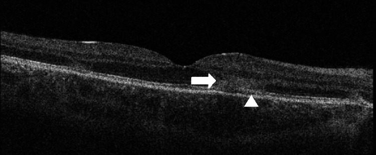 Figure 1
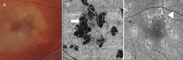 Figure 2
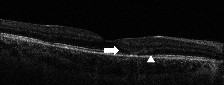 Figure 3
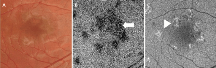 Figure 4
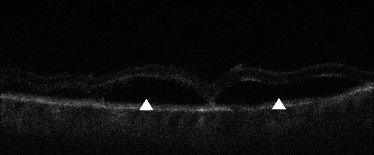 Figure 5
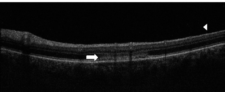 Figure 6
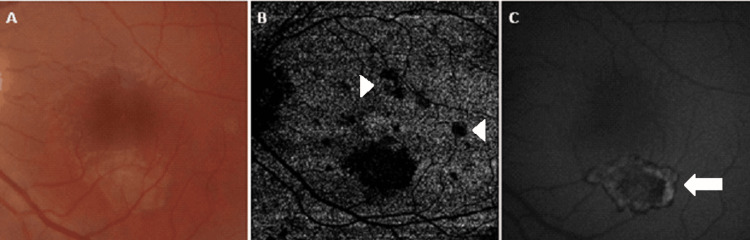 Figure 7
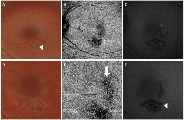 Figure 8
Peer Reviews
No public reviews on file for this paper yet. If you reviewed it on a platform where reviews are public (OpenReview, ICLR, NeurIPS, ICML), you can paste yours below so the community can read it here.
Videos
No videos yet. Explain this paper in a talk, walkthrough, or lecture? Add one.
Taxonomy
TopicsOcular Diseases and Behçet’s Syndrome · Retinal Diseases and Treatments · Sarcoidosis and Beryllium Toxicity Research