Surpassing the diffraction limit for improved lateral resolution in adaptive optics optical coherence tomography of the living human eye
Andrew J. Bower, Furu Zhang, Tao Liu, Joanne Li, Nancy Aguilera, Sarah Abouassali, Jonathan Krynitsky, Randy Pursley, Tom Pohida, Bartlomiej Kowalski, Rongwen Lu, Alfredo Dubra, Johnny Tam

TL;DR
Researchers improved the resolution of retinal imaging in the human eye beyond the diffraction limit, enabling clearer visualization of photoreceptor cells.
Contribution
A modular strategy for sub-diffraction lateral resolution in adaptive optics optical coherence tomography is introduced.
Findings
Annular illumination and sub-Airy disk detection improved lateral resolution by 36% in vivo.
The method enables better visualization of foveal cone and rod photoreceptor mosaics.
The approach is compatible with existing and new AOOCT instruments.
Abstract
Advances in adaptive optics optical coherence tomography (AOOCT) have facilitated the three-dimensional assessment of structural and functional properties of individual retinal cells in the living human eye. However, even with diffraction-limited AOOCT systems, some cells in the living human retina can be difficult to resolve, especially when using near-infrared wavelengths of light (~1000 nm). We demonstrate that modifying the traditional AOOCT instrument design to enable annular illumination and sub-Airy disk detection results in improved imaging resolution beyond fundamental limits imposed by diffraction. We successfully applied this approach to in vivo human retinal imaging, achieving on average 36% improvement in lateral resolution beyond conventional imaging conditions, enabling improved visualization of the foveal cone and rod photoreceptor mosaics using AOOCT. These results…
Genes, proteins, chemicals, diseases, species, mutations and cell lines named across the full text — each resolved to its canonical identifier and authoritative record.
Click any figure to enlarge with its caption.
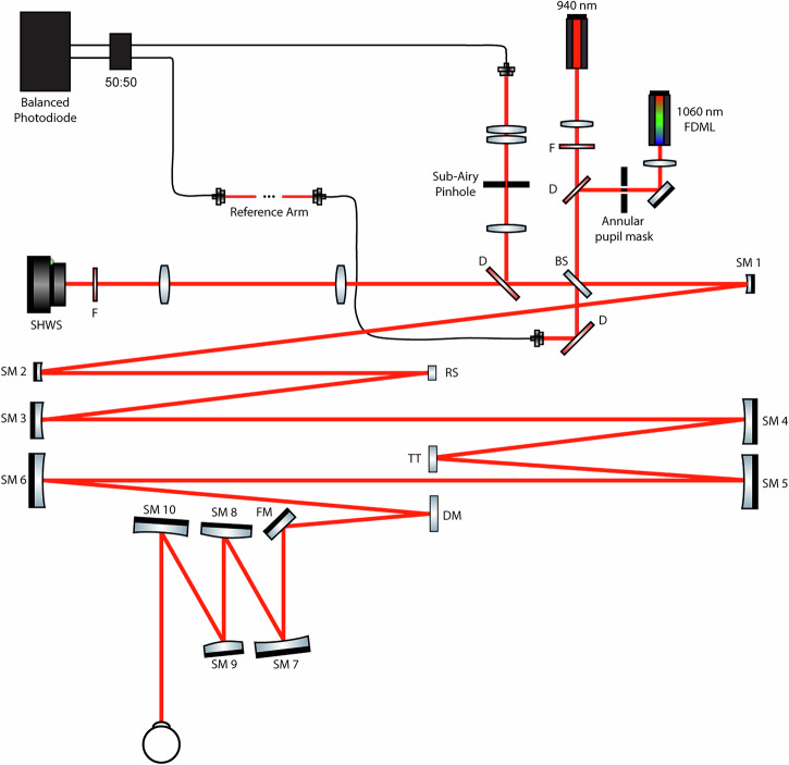 Figure 1
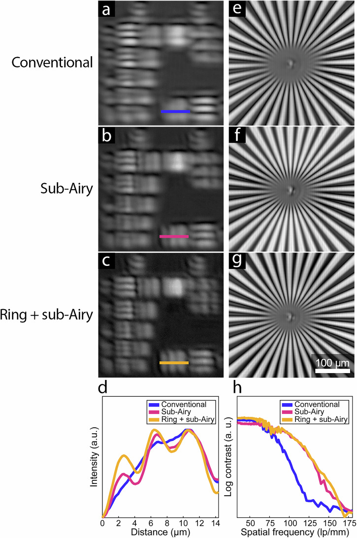 Figure 2
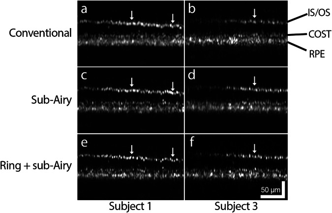 Figure 3
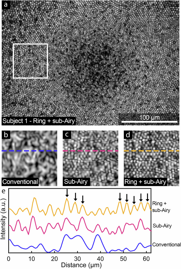 Figure 4
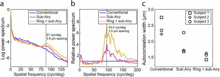 Figure 5
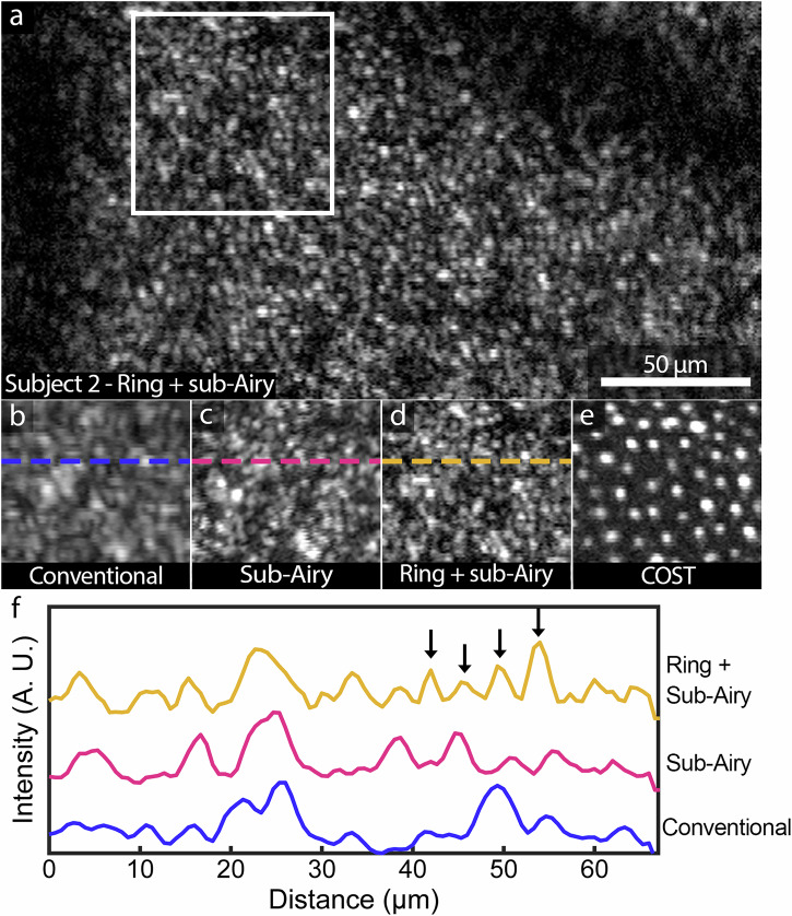 Figure 6
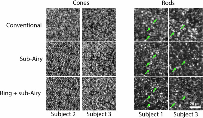 Figure 7
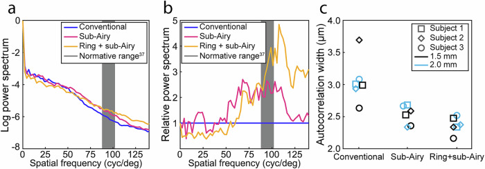 Figure 8
Peer Reviews
No public reviews on file for this paper yet. If you reviewed it on a platform where reviews are public (OpenReview, ICLR, NeurIPS, ICML), you can paste yours below so the community can read it here.
Videos
No videos yet. Explain this paper in a talk, walkthrough, or lecture? Add one.
Taxonomy
TopicsOptical Coherence Tomography Applications · Ophthalmology and Visual Impairment Studies · Retinal and Macular Surgery