The structure, redox chemistry and motor neuron toxicity of heterodimeric zinc-deficient SOD1-implications for the toxic gain of function observed in ALS
Victor A. Streltsov, Katherine E. Ganio, Stewart D. Nuttall, J. Andres Hernandez, Cassandra N. Dennys, Peter J. Crouch, Alvaro G. Estevez, Maria Clara Franco, Joseph S. Beckman, Blaine R. Roberts

TL;DR
This paper explores how a zinc-deficient form of SOD1, when paired with normal SOD1, becomes toxic and contributes to motor neuron disease in ALS.
Contribution
The study genetically tethers and structurally characterizes a heterodimeric SOD1 complex to explain its toxic role in ALS.
Findings
Zinc-deficient SOD1 forms a stable heterodimer with wild-type SOD1 and adopts a normal-like structure.
The heterodimer produces peroxynitrite and is highly toxic to motor neurons.
Wild-type SOD1 may contribute to disease progression through heterodimer formation with mutant SOD1.
Abstract
A subset of familial cases of amyotrophic lateral sclerosis (fALS) are caused by mutations to copper, zinc superoxide dismutase (Cu, Zn SOD1). Over 200 mutations to SOD1 that have been associated with fALS and the majority of these mutations are dominantly inherited. Thus, individuals are heterozygous and express both wild-type SOD1 and the mutant form of the protein. Paradoxically, the motor neuron disease accelerates in rodent models that mimic the co-expression of wild-type SOD1 with mutant fALS SOD1. Previously, we have shown that the loss of zinc from SOD1 triggers motor neuron death in culture due to a gained, redox activity catalyzed by the active-site copper. Furthermore, motor neuron toxicity of zinc-deficient SOD1 is enhanced by wild-type Cu, Zn SOD1. Because SOD1 exists as a non-covalent dimer, the enhanced toxicity might result from stabilization of the heterodimeric…
Genes, proteins, chemicals, diseases, species, mutations and cell lines named across the full text — each resolved to its canonical identifier and authoritative record.
Click any figure to enlarge with its caption.
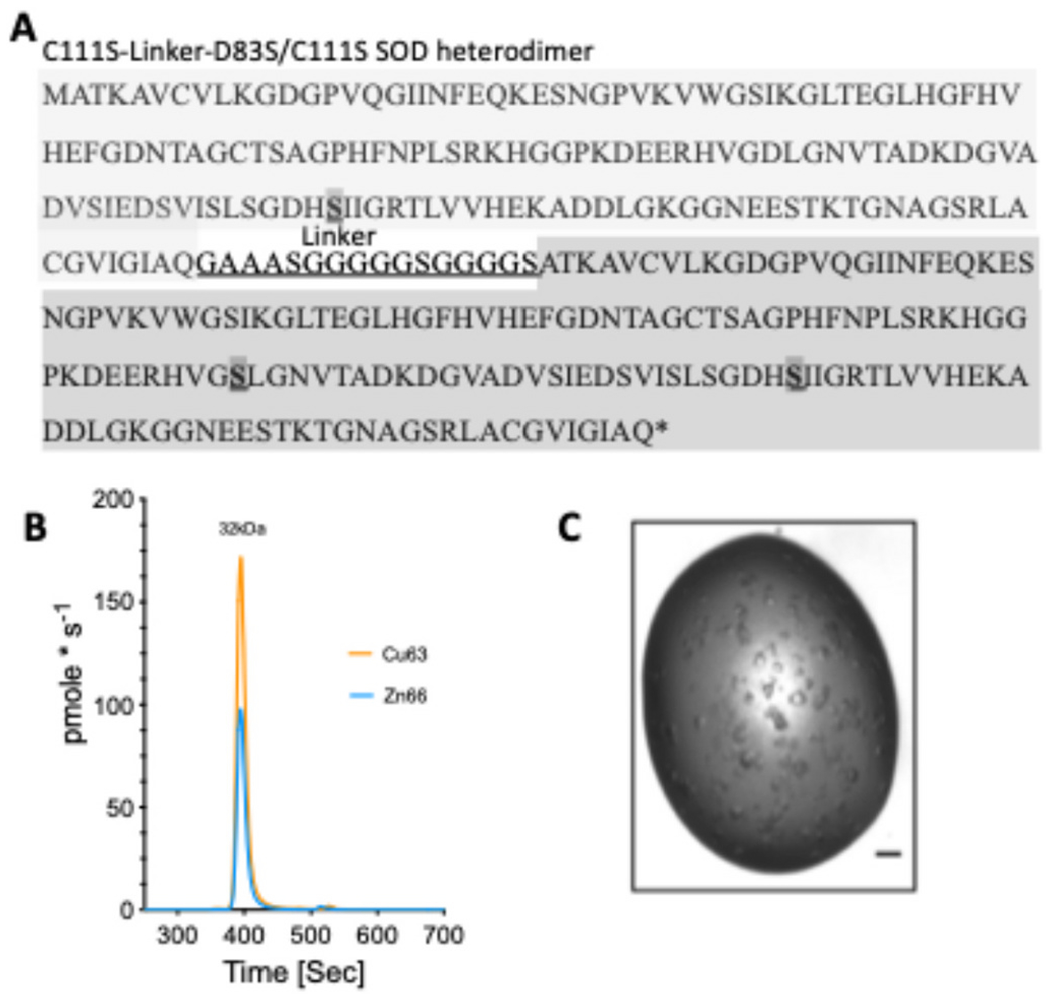 Figure 1
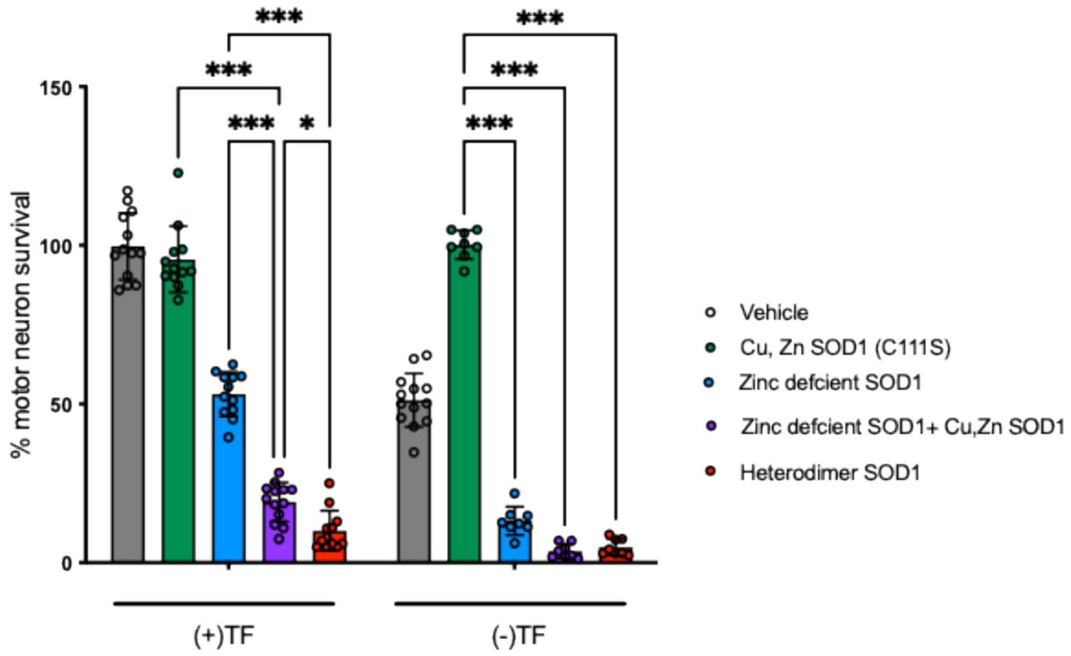 Figure 2
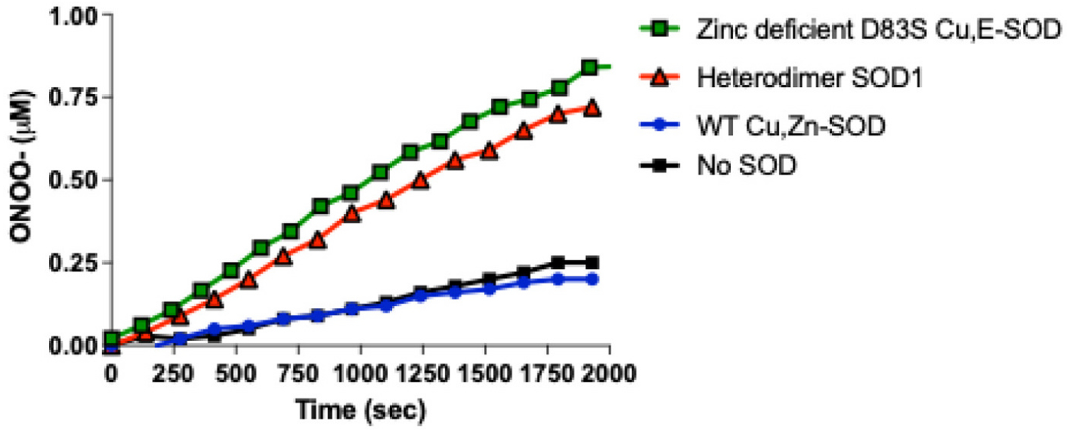 Figure 3
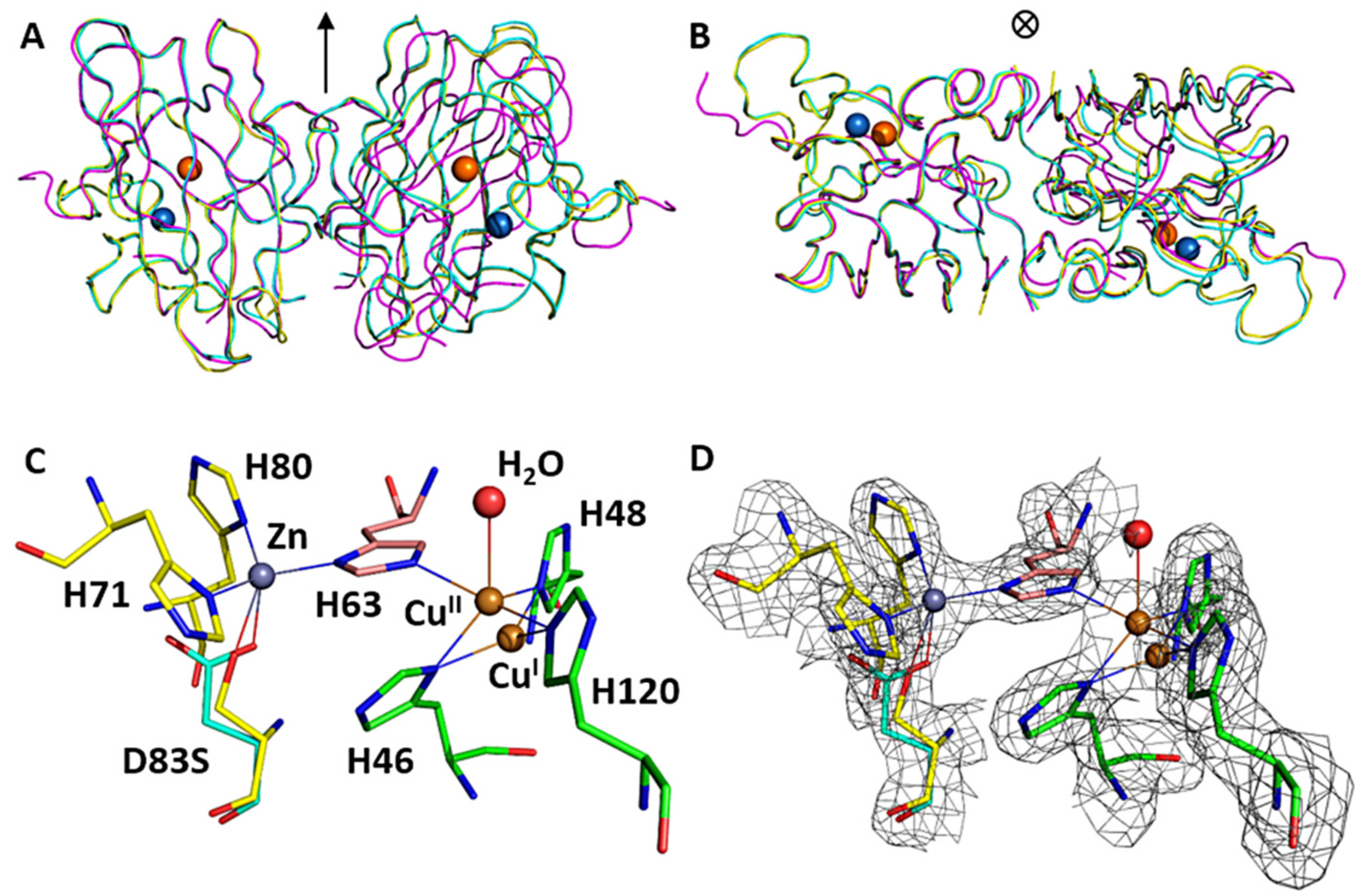 Figure 4
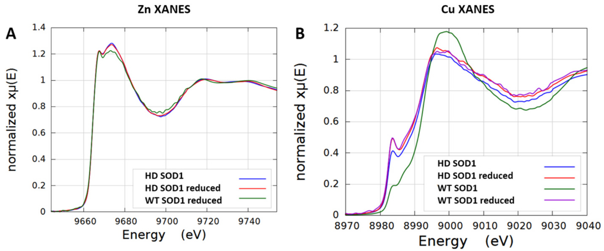 Figure 5
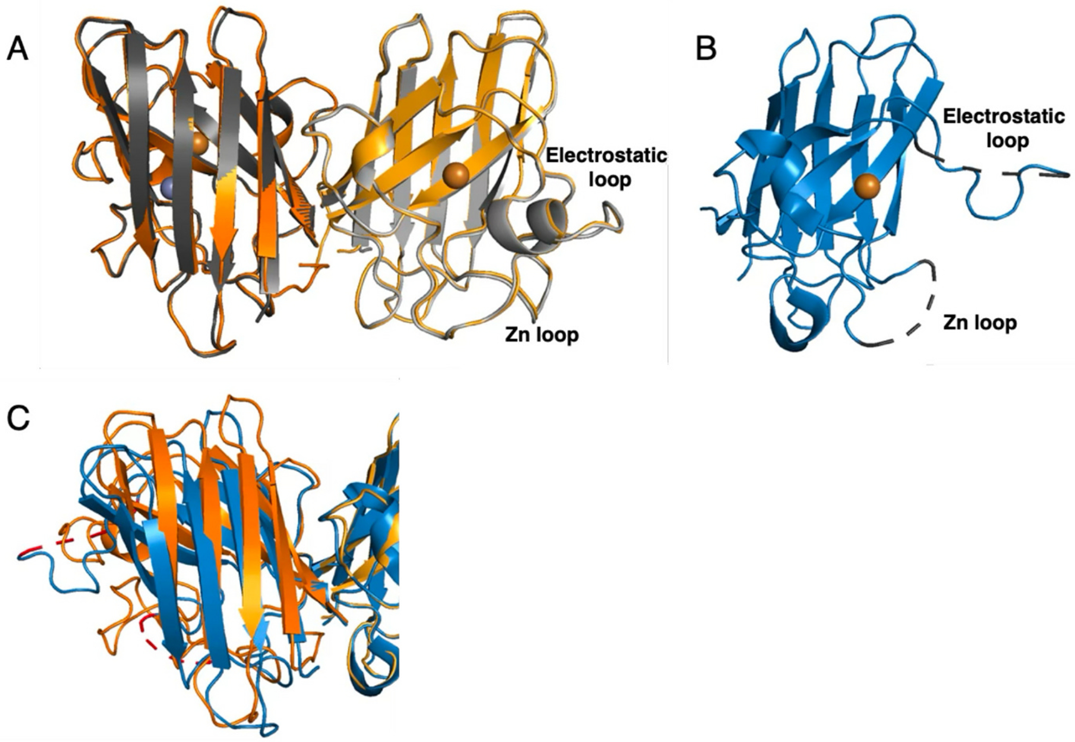 Figure 6
Peer Reviews
No public reviews on file for this paper yet. If you reviewed it on a platform where reviews are public (OpenReview, ICLR, NeurIPS, ICML), you can paste yours below so the community can read it here.
Videos
No videos yet. Explain this paper in a talk, walkthrough, or lecture? Add one.
Taxonomy
TopicsAmyotrophic Lateral Sclerosis Research · Genetic Neurodegenerative Diseases · Cervical and Thoracic Myelopathy
Introduction
Amyotrophic lateral sclerosis (ALS) is an adult-onset disease involving the progressive death of lower motor neurons in the spinal cord and upper motor neurons in the brain stem and cortex, with consequent muscular paralysis (Rowland and Shneider, 2001). In 1993, thirteen missense mutations to the Cu, Zn superoxide dismutase (SOD1) gene were linked to familial ALS (fALS) (Rosen et al., 1993). Structural analysis of the these variant showed an impact on the β-barrel fold and dimer interface (Deng et al., 1993). Since this discovery more than 200 dominant missense mutations and a dozen C-terminal frameshift and truncation mutations to the SOD1 gene have been identified in ALS patients. Approximately 2–7 % of ALS cases result from SOD1 mutations with the percentages varying in different regions around the world (Benatar et al., 2025). For many of the fALS SOD1 mutations, the structural changes produce subtle localized shifts in structure and many mutant SOD1s can retain full enzymatic activity. Virtually all fALS SOD1 patients have one remaining wild-type allele that produces enzymatically active Cu, Zn SOD1 protein.
Despite a lack of obvious structural or enzymatic effects, overexpression of fALS mutant SOD1 in mice and rats results in the progressive motor neuron death. These animal models remain the most used models of human ALS. Overall, there is a consensus that SOD1 mutations confer a toxic gain-of-function, rather than the loss of superoxide-scavenging activity. This is further supported by the acceleration of the onset of ALS-like paralysis when wild-type (WT) SOD1 is co-expressed with fALS SOD1 in transgenic animal models (Jaarsma et al., 2000; Witan et al., 2008). Accelerated death by WT SOD1 is also observed in motor neuron cultures isolated from fALS SOD1 mice (Garner et al., 2010).
We and others have hypothesized that the intermediates in the SOD unfolding process provide the foundation for toxicity that may explain the gain-of-function (Rakhit et al., 2004; Tiwari and Hayward, 2005). Indeed, careful biophysical studies have shown that the mutations to SOD1 result in a subtle decrease in protein stability affecting the dimer interface, and the degree of destabilization correlates with disease severity (Wang et al., 2008). Many fALS SOD variants are known to decrease in zinc binding affinity (Crow et al., 1997; Lyons et al., 1996; Smirnova et al., 2022; Souza et al., 2019), that is associated with a loss of dimer stability (Kumar et al., 2017). Loss of zinc has one of the most dramatic effects on the quaternary structure, distorting the dimer interface by 9° and disordering the electrostatic and zinc-binding loops (Roberts et al., 2007).
The absence of zinc results in disorder of the electrostatic loop, opening the narrow ~4 Å-wide active site channel that protects the reactive copper. The restrictive active site channel normally allows only superoxide to reach the mostly buried copper. The widening of the channel in zinc-deficient SOD1 allows copper to become reduced to Cu (I) by low molecular weight reductants ~3000 times faster than Cu, Zn SOD1 and thereby produce superoxide through the reduction of oxygen (Estevez et al., 1999). In the presence of a low concentration of nitric oxide, zinc-deficient SOD1 catalyzes the formation of peroxynitrite that activates a cell-death pathway leading to the death of primary motor neurons and makes astrocytes reactive (Barbeito et al., 2004; Estevez et al., 1999). The death pathway in motor neurons involves the nitration of heat shock protein 90 (HSP90) and subsequent activation of the purinergic receptor P2X7 receptor (Franco et al., 2013; Gandelman et al., 2013; Gandelman et al., 2010).
We have previously shown that the toxicity of zinc-deficient SOD1 in vitro is enhanced by the addition of WT Cu, Zn SOD1 (Garner et al., 2010). The increase in toxicity has been attributed to increased stabilization of zinc-deficient SOD1 through the dimer interface with Cu, Zn SOD1. Indeed, the disulfide bond of constitutively zinc-deficient SOD1 is less susceptible to reduction in vitro in the presence of WT Cu, Zn SOD1 and more resistant to aggregation (Garner et al., 2010; Roberts et al., 2007). We hypothesized that the Cu, Zn holo form of the enzyme structurally stabilizes the zinc-deficient SOD1 subunit and thereby protects this subunit from losing copper. To test this hypothesis, we made a tethered heterodimeric protein to prevent swapping of SOD1 subunits. The carboxyl-terminus of WT like Cu, Zn SOD1 (C111S) was tethered with a 16-residue linker comprised of glycine, serine, and alanine residues to the amino-terminus of zinc-deficient SOD1 (D83S/C111S) to produce a single chain protein modeling the heterodimer (Fig. 1). The linker was chosen from the well-known approach used for tethering heavy and light antibody chain fragments to create recombinant single-chain antibodies (Holliger et al., 1993). The x-ray structure, metal binding properties, redox activity, and toxicity to primary motor neurons were assessed using the tethered SOD1 heterodimer to determine how the dimer interface might contribute to toxicity.
Materials and methods
Cloning and protein expression
2.1.
The gene sequence for the wild-type-like SOD1 (C111S) tethered to zinc-deficient SOD1 (D83S/C111S) heterodimer (Heterodimer-SOD1) is shown in Fig. 1. The D83S mutation alters one of the zinc ligands to produce a constitutively zinc-deficient SOD1 (Bahadorani et al., 2013; Estevez et al., 1999). Both subunits also contained the thermostable substitution at cysteine 111 to serine (C111S), which is known to be reactive in vitro and able to bind metals and complicate metal binding studies (Parge et al., 1992). The C111S mutation also improves expression of soluble SOD1 in E. coli (Garner et al., 2010; Leinweber et al., 2004). The sequence including 5’ NdeI and 3’ BamH1 sites was synthesized at Geneart AG (Regensburg, Germany, www.geneart.com), sub-cloned into the expression vector pET3d (Novagen) and the sequence validated by complete sequencing. This wild-type-like SOD1 C111S + zinc-deficinet SOD1 D83S/C111S tethered heterodimer was expressed in E. coli strain BL21(DE3) pLysS. Briefly, a single colony was cultured overnight in 300 mL Luria broth (LB) containing 0.1 mg/mL ampicillin at 37 °C and 180 rpm. The inoculate was added to three flasks, each containing 1 L of LB media and 0.1 mg/mL ampicillin and grown at 37 °C at 150 rpm to an OD_600_ = 0.75. Protein expression was induced by addition of 1 mM (final) isopropyl-β-D-thiogalactoside. To obtain Cu, Zn-bound protein, CuSO_4_ (0.2 mM final) and ZnSO_4_ (0.1 mM) were added 1 h post-induction, the temperature decreased to 24 °C, and expression continued for 16 h. Bacteria were pelleted by centrifugation (10,000 xg for 20 min) and stored at −20 °C prior to processing.
Protein purification of tethered WT SOD1 + D83S (Heterodimer-SOD1)
2.2.
Bacteria were lysed by thawing with 1× tris-buffered saline (TBS, 50 mM Tris pH 8.0, 150 mM NaCl) at a ratio of 0.5 mL of 1× TBS per g of pellet). DNase I bovine pancreas (3 mg) was added followed by incubation at 37 °C for 2 h. Lysates were clarified by centrifugation (10,000 g for 20 min) at room temperature (RT), the supernatant adjusted to 35 % ammonium sulfate followed by a further clarification by centrifugation as above. The solution was then brought to a final concentration of 70 % ammonium sulfate followed by centrifugation as above. The supernatant was applied to a Phenyl Sepharose column (GE Healthcare, 5 × 50 mm GL) equilibrated in 2 M NH_4_SO_4_ + 150 mM NaCl and a linear gradient over 10 column volumes from 0 to 100 % buffer B (50 mM KP_i_, pH 8). SOD1 positive fractions, determined by SDS-PAGE, were pooled and exhaustively dialyzed against 10 mM KP_i_, pH 8 at 4 °C. Dialyzed material was loaded on a 5/50 GL Mono Q anion exchange column (GE Healthcare, 5 × 50 mm) with an elution gradient from 10 mM KP_i_, pH 8 to 10 mM KP_i_ + 100 mM NaCl, pH 8 over 20 column volumes. Fractions from the anion exchange column containing SOD1 were pooled and concentrated using 5 K MWCO Millipore Amicon Ultra-50 Centrifugal Filter Units. The concentrated material was loaded on to a 10/300 GL Superdex 75 size exclusion column developed with 1× phosphate buffered saline (PBS). SOD1 fractions corresponding to the heterodimer molecular mass of 32 kDa were pooled and concentrated using 3 K MWCO Millipore Amicon Ultra-50 Centrifugal Filter Units. We also observed a small fraction of a tetrameric SOD species (64 kDa) that we ascribe to formation of a dimer of two tethered heterodimers in the crude lysates that was purified away (data not presented). Protein concentration was calculated using an extinction coefficient of 5810 M^−1^ cm^−1^ at 280 nm, which is based on the absorbance calculated for the denatured SOD protein sequence (Gill and von Hippel, 1989).
Peroxynitrite generation assay
2.3.
The boronate-based fluorogenic probe, coumarin-7-boronic acid (CBA), was used to detect peroxynitrite formed during the production of superoxide via SOD1 re-oxidation in the presence of nitric oxide. CBA was a kind gift from Drs. Zielonka and Kalyanaraman at the Medical College of Wisconsin. CBA was dissolved in dimethylformamide and stored at −80 °C until use. Re-oxidation experiments were performed at 37 °C in 20 mM HEPES buffer, pH 7.4 containing 100 μM CBA, 5 μM ascorbate, 10 μM SOD, and 5 μM PAPA-NONOate as steady-state source of nitric oxide. Catalase (100 U/mL) was added to prevent CBA oxidation due to H_2_O_2_. Fluorescence of coumarin was measured at excitation/emission wavelengths of 330/455 nm using a Gemini fluorescence microplate reader (Molecular Devices, Sunnyvale CA). Standard curves for quantifying peroxynitrite generation were determined by reacting a known amount of peroxynitrite with CBA. Peroxynitrite was synthesized from nitrite and hydrogen peroxide using previously described methods (Robinson and Beckman, 2005). The concentration of peroxynitrite was determined by measuring absorbance at 302 nm (ε = 1700 M^−1^ cm^−1^). Peroxynitrite working stocks 100× were prepared in cold, degassed water and a small aliquot was immediately added to degassed 20 mM HEPES buffer, pH 7.4 containing 100 μM CBA and 100 U/mL catalase. Samples were measured in triplicate. Relative fluorescence was converted to peroxynitrite concentration using non-linear regression fitted as a single exponential. Standard curves for CBA were applied to experiments to quantify peroxynitrite generation.
Size-exclusion chromatography with inductively coupled plasma mass spectroscopy (SEC-ICP-MS) metal assay
2.4.
The metal status of the SOD1 heterodimer was determined using a SEC column (BioSEC3 4.6 × 300 mm, Agilent) connected directly to an ICP-MS (Agilent 7700) as previously described(Lothian and Roberts, 2016). Briefly, the column flow rate was 0.4 mLmin^−1^ with 200 mM ammonium nitrate pH 7.7–7.8. The column temperature was maintained at 30 °C using a thermostatically controlled column compartment. The ICP-MS was run under standard multi-elemental conditions and bovine SOD1 (Sigma) was used as a molecular mass standard and a standard for copper and zinc. A standard curve for copper and zinc was generated using bovine SOD1 (200 ppb Cu and zinc) injection volumes ranged from 3 to 30 μL. The integrated area for SOD1 was determined for copper and zinc traces and the response for the ICP-MS in picograms**sec^−1^ was determined by dividing picograms of copper or zinc injected on the column over the integrated peak area in counts per second (CPS). The picograms per count constant was then used to convert CPS to picograms of metal per second. Sample was measured in duplicate.
ESI-MS determination of mass and validation of metal status
2.5.
The metal content and mass of the D83S + WT heterodimer SOD1 was determined by native electrospray mass spectrometry using a size exclusion column (AdvanceBio SEC 200 Å, 1.9 μm 2.1 × 50 mm) coupled to a QTOF with Jet stream source (Agilent 6545XT). The column was developed with 100 mM ammonium acetate pH 7.0. Instrument setting was as described in Supplemental Table 1. Data were collected in profile mode and deconvoluted with Bioconfirm software (Agilent Technologies) with the mass range 600–5000, mass step of 0.05 and baseline factor 7.00. The observed deconvoluted mass for the heterodimer with metals bound was 32,759.14 g/mol (5.5 ppm mass error, most probable mass 32,758.96). The most probable mass for the heterodimer C111S + D83s/C111S SOD1 was calculated with iMass application (v1.4, Mobile Science Apps.).
Heterodimer SOD1 crystallization
2.6.
Crystallization screening was performed at the CSIRO Collaborative Crystallization Centre (www.csiro.au/c3). C111S + D83S/C111S SOD1 (Heterodimer-SOD1) concentrated to ~10 mg/mL in PBS buffer was set up as 0.4 μL sitting drops at 20 °C against a customized 96-well screen, based upon successful previous crystallization conditions for SOD1 (Roberts et al., 2007), heavily biased toward 2–3 M ammonium sulfate or sodium malonate at pH range 4.5–7. Crystals grew under a variety of conditions; the best diffracting crystals were 100–200 μm in size grown in sodium malonate for 7–21 days.
X-ray crystallography
2.7.
X-ray diffraction data sets for single crystals of Heterodimer-SOD1 in well solution plus ca 15 % (v/v) glycerol as cryo-protectant were collected at 100 K using the MX2 beamline at the Australian Synchrotron, a micro-focused in-vacuum undulator beamline. The data sets were processed with HKL2000 (Otwinowski and Minor, 1997). Further data collection and processing statistics are given in Table 1. The locations of SOD1 monomers were identified in the asymmetric units by PHASER (McCoy et al., 2007) molecular replacement using the structure of SOD (PDB ID: 2R27) (Roberts et al., 2007) without including water molecules. Five independent heterodimers were identified in the asymmetric unit of the Heterodimer-SOD1 crystal. The structures with protein molecules alone were initially refined and visible parts of linker sequences plus water molecules were subsequently added. Occupancies of metal sites were carefully refined with constrained atomic B-factors. Iterative refinement and model building were conducted using REFMAC (Murshudov et al., 1997) and XFIT/MIFit (McRee, 1999). Progress of the refinement was monitored using the Rfree statistics based on a test set encompassing 5 % of the observed diffraction amplitudes (Brunger, 1992). Deposition codes and further structure refinement details are given in Table 1. The figure was produced using PyMol (Schrödinger Software).
X-ray absorption spectroscopy (XAS)
2.8.
XAS spectra of frozen solution samples of <0.5 mM concentration of wtSOD1, Heterodimer-SOD1 without and with 20-fold concentration of sodium ascorbate as a reductant were collected at the Australian synchrotron XAS beamline (1.9 T Wiggler). The beamline was equipped with liquid nitrogen (LN_2_) cooled Si double crystal monochromator (ΔE/E 1.5 × 10^−4^) with a Rh-coated focusing mirror to produce a focused X-ray beam with a harmonic content better than 1 part in 105. The incident and transmitted x-ray intensity was monitored using ionization chambers with a continuous stream of He gas. Fluorescence measurements were obtained using a 100-element LN_2_-cooled Ge detector (Canberra). Energy calibration was achieved by the simultaneous accumulation of a Cu foil spectrum (transmittance) where the inflection point of the first absorption feature was set to an energy of 8980.4 eV. Ice formation was inhibited by addition of glycerol (~15 %) to samples immediately prior to their injection into the 40 μL cavity of polycarbonate cells (2 mm × 2 mm × 10 mm) with Kapton (Goodfellow Cambridge, Cambridge, UK) front and back windows. Samples were frozen and stored in liquid N^2^ until transfer to the beamline closed-cycle pulse tube He cryostat (“Optisat”, Oxford Instruments).
A series of Zn (9659 eV) and Cu (8979 eV) K-edge XANES (X-ray Absorption Near Edge Spectroscopy) measurements scans up to k = 10 Å^−1^ were obtained from samples in a fluorescence mode at 5-10 K. Radiation damage of samples was tested by quick XANES measurements from the same sample position with 30 min exposure intervals. The spectra recorded from each sample position were averaged to obtain the final spectra. The XANES spectra were pre-processed by the software package SAKURA at the Australian synchrotron XAS beamline. Data points affected by monochromator glitches were also removed. Edge step normalization for each spectrum was performed by subtracting the pre-edge and post-edge backgrounds in program ATHENA (Ravel and Newville, 2005) based on the IFEFFIT library on numerical and XAS algorithms (Newville, 2001).
Motor neuron culture and survival assays.
Motor neurons were purified from E15 rat embryos by a combination of density centrifugation and immunoaffinity using an antibody to the p75 low affinity neurotrophin receptor as previously described (Raoul et al., 2002). Purified motor neurons were plated in 96 well plates at a density of 500 cells per well in Neurobasal media supplemented with B27, β-mercaptoethanol, glutamine, glutamate, horse serum, brain-derived neurotrophin factors (BDNF), glial-derived neurotrophic factor (GDNF) and cardiotrophin-1. After 24 h in culture, SOD1 was delivered utilizing the membrane permeant carrier agent Chariot (Active Motif, Carlsbad, CA). SOD1 was diluted in H_2_O at a final concentration of 5 μg/mL at room temperature. SOD1 was incubated at room temperature before mixing with Chariot and incubated for additional 30 min for complexes formation. The medium was aspirated and replaced by Opti-MEM transfection medium (Invitrogen) containing the mixture Chariot and SOD1. The cells were incubated for 1 h, at which time 100 μL of Neurobasal medium containing twice the concentration of supplements and penicillin-streptomycin plus or minus trophic factors was added to each well and further incubated for 24 h. Motor neuron survival was determined by high throughput image capture and analysis in 96-well plates using a Runner^™^ (Trophos, Marseilles, France) as reported previously (Franco et al., 2013; Garner et al., 2010). Survival was standardized between experiments by considering the survival in the presence of trophic factors as 100 %.
Results
Protein expression, characterization, and crystallization
3.1.
The heterodimer SOD1 (Heterodimer-SOD1) composed of wild-type like SOD1 (C111S) tethered to zinc-deficient SOD1 (D83S/C111S) expressed well in E. coli at yields greater than 5 mg/L (Fig. 1A). Following purification, size exclusion, inductively coupled mass spectrometry (SEC-ICP-MS) was used to determine metal content (Fig. 1B). The observed ratio of 1.76 (0.03, SD) Cu per Zn in the SOD1 heterodimer is slightly less than the expected ratio of two coppers and one zinc per mole of heterodimer SOD1.
Native mass spectrometry confirmed that the major Heterodimer-SOD1 protein had two coppers and one zinc. The experimentally determined mass of 32,759.14 Da closely matched the calculated most-abundant-isotope theoretical mass of 32,758.96 Da (C_1395_H_2220_N_422_O_467_S_6_Cu_2_Zn_1_), corresponding to a mass error of 5.5 ppm, indicating excellent agreement between the observed and theoretical values. A less intense peak with a mass of 32,695.2 Da differed from the main peak by a mass of 62.8 Da, which is consistent with the loss of one additional metal. Other small peaks are consistent with one or two sodium adducts as well as a minor peak corresponding to the loss of two metals. Together, the ICP-MS and native results show that the Heterodimer-SOD1 protein was predominantly one peptide chain containing two coppers and one zinc, with a smaller fraction most likely missing a copper based on the Cu/Zn ratio determined by ICP-MS.
Motor neuron survival
3.2.
Previously, we have shown that SOD1 protein can be delivered intracellularly with Chariot^™^ (active Motif, Carlsbad, CA)(Sahawneh et al., 2010). Consistent with prior results, the delivery of zinc-deficient D83S SOD1 in the presence of trophic factors induce motor neuron by a nitric oxide-dependent oxidative mechanism that resulted in 53 ± 7 % cell survival of motor neurons in 24 h post-delivery (p-value<0.001). In contrast, Cu, Zn bound-SOD1 did not diminish or increase survival (Fig. 2). However, addition of an equal concentration of Cu, Zn bound SOD1 with zinc-deficient D83S SOD1 decreased motor neuron survival to 19 ± 6.1 %, and Heterodimer-SOD1 further decreased cell survival to 10 ± 6.3 %.
Motor neurons intentionally deprived of trophic factors undergo cell death with 51 ± 8.4 % survival after 24 h. Consistent with prior results (Garner et al., 2010), delivery of Cu, Zn bound SOD1 completely protected motor neurons from trophic factor deprivation-induced cell death (100. ± 4.6 % survival). Conversely, zinc-deficient SOD1 (D83S) delivered alone decreased survival to 13 ± 2.4 % and the co-delivery with Cu, Zn bound SOD1 further decreased survival to 3.5 ± 2.3 %. Heterodimer-SOD1 also resulted in a significant decrease in survival 4.9 ± 2.6 % versus control. Thus, holo Cu, Zn SOD1 by itself protected motor neurons from trophic factor deprivation, but zinc-deficient SOD1 alone or as a tethered heterodimer decrease survival consistent with previous reports (Estevez et al., 1999; Garner et al., 2010).
Peroxynitrite generation by zinc-deficient SOD
3.3.
Because we have previously shown that zinc-deficient SOD1 activates motor neuron death by an oxidative mechanism requiring both nitric oxide and superoxide, we next compared the peroxynitrite generation activity of the tethered heterodimer-SOD1 (D83S-WT SOD1) to that of zinc-deficient SOD1. The coumarin boronate probe has proved to be more specific and sensitive assay for peroxynitrite generation than other fluorescent oxidation probes (Zielonka et al., 2012). Under the current assay conditions (Fig. 3), the probe slowly hydrolyzed at a rate equivalent to 8.3 ± 0.2 nM·min^−1^ and this rate was only slightly decreased with the addition of 10 μM WT-Cu, Zn SOD1 to 6.8 ± 0.1 nM·min^−1^. Zinc-deficient WT SOD1 produced peroxynitrite at an apparent rate of 25.6 ± 0.3 nM·min^−1^ per μmol SOD1, while the Heterodimer-SOD1 heterodimer produced peroxynitrite at a similar rate of 23.5 ± 0.3 nM·min^−1^.
X-ray crystal structure of D83S + WT heterodimer
3.4.
Initial crystallization conditions for SOD1 homodimers had a marked preference for solutions rich in ammonium sulfate. Accordingly, a customized 96-well screen was designed around ammonium sulfate and sodium malonate. Multiple wells containing cuboidal crystals typically of 100–200 nm in length were found between 7 and 21 days (Fig. 1).
The D83S + WT heterodimer SOD1 readily crystallized in a form (space group C 2 2 1 with a = 162.7 Å, b = 201.7 Å, c = 143.8 Å). The asymmetric unit contained five SOD1 heterodimers with a high solvent content (66.3 %), and is highly similar to the asymmetric subunits of WT SOD1 that have been previously reported (DiDonato et al., 2003; Parge et al., 1992). The structure was solved by molecular replacement which identified ten subunits, or five heterodimers. The 16-residue linker between monomers were partially visible in heterodimers and were built into available density. The structure was refined to 2.0 Å resolution with final R/R^free^ factors of 0.156/0.190 (Table 1) with well-ordered parts and coordinate accuracy of ~0.1 Å. The ten monomeric subunits were refined individually without the use of non-crystallographic symmetry restraints to assess subtle structural changes between the heterodimer and wild-type dimer. Overall, these results suggest insignificant structural changes compared to the wild type variants and the linker region had minimal impact on the overall SOD1 structure.
The basic fold of zinc-deficient D83S subunit remained unchanged. The main chain conformations for the 153 residues were well defined as previously characterized (Fig. 4). The average RMSD between all five heterodimers in the asymmetric unit was 0.41 Å. The average RMSD between the D83S-C111S heterodimer (6DTK) and the homodimer of the wild type SOD1 (PDB ID: 1PU0) (DiDonato et al., 2003) was 0.30 Å. Comparison of the Heterodimer-SOD1 with the homodimeric zinc-deficient D83S SOD1 structure (PDB ID: 2R27) (Roberts et al., 2007) produced a RMSD of 0.66 Å. Similarly, the RMSD of alignment between wild type SOD1 (1PU0) and zinc-deficient SOD1 (2R27) was 0.67 Å. Hence, the structure of D83S + WT SOD1 heterodimer was more similar to wild-type Cu, Zn homodimeric structure than the Zn-deficient homodimeric structure. This is most pronounced in the structure of the electrostatic and zinc-binding loops were ordered in the heterodimer structure (Fig. 4).
Furthermore, the C-alpha backbones of heterodimeric subunits exhibit near two-fold symmetry with the average RSMD of 0.25 Å, further supporting the closer similarity to the wild-type SOD. Previously, we showed that the loss of zinc results in local unfolding of the electrostatic and zinc binding loops. This was associated with a shift in the quaternary structure that is expressed as a 9.9° rotation of the dimer due to an opening of the dimer interface (Roberts et al., 2007). Due to the inherent link of the dimer interface with the zinc-binding loop, we proposed that a heterodimer consisting of one monomer of Cu, Zn bound-SOD1 with one monomer of zinc-deficient SOD1 was expected to correct the shift in the quaternary structure. This was based on the observations that mixing zinc-deficient SOD1 with Cu, Zn bound-SOD1 resulted in protection of the disulfide bond to reduction and resistance to protein aggregation (Garner et al., 2010). The crystal structure presented here demonstrates that the heterodimer corrects the quaternary structure of the dimer with a rotation angle between the two subunits of only − 0.3° different compared to wild type enzyme (1PU0) (Fig. 4A, B). This angle was nearly identical to the wild type SOD1 (1PU0) (DiDonato et al., 2003; Tainer et al., 1983).
Active site structure.
The electron density map confirmed the presence of mutated side chain D83S and C111S. However, the electron density of the D83S side chain in the zinc coordination spheres showed a mixed population of the mutant sites (Fig. 4 C, D). This might possibly be due to crystal averaging of subunits, because the heterodimers exhibit near two-fold symmetry. Therefore, the mutant site was specifically refined using mixed fractional population of two residues D83 and S83 while keeping the atomic B-factor of two amino acids restrained to similar values. The population of zinc at this site was also refined with a fixed, physically reasonable atomic B-factor. The resulting deficiency of zinc site in the range of 0.3–0.8 (0.64 on average) was proportional to the population of Serine-83 in the mutated site. The quasi-tetrahedral coordination of zinc with D83 ligand remained similar to that found in WT SOD1 with the distance between zinc and D83 of 1.97–1.99 Å and with histidine’s H63, H71 and H80 at 2.0–2.1 Å. The zinc coordination site varied in subunits with mutant D83S ligand being at 2.3–3.5 Å and with occasional additional waters located 2.3 Å from zinc.
The electron density of the copper site showed that fractions of copper can be modeled at alternate positions shifted by 1.1 Å in average over the structure (Fig. 4 C, D). The coordination of copper in oxidized (Cu^II^) site involved residues: His46, His48, His120 and His63 that is the bridging ligand for both copper and zinc. An additional one or two waters at the average distance of 2.5 Å resulting in five- or even six-coordinated site. The bridging His63 – Cu^II^ distance tends to be somewhat longer of ~2.2 Å and the increased chemical-reactivity measured in the peroxynitrite generation assay stance tends to be somewhat longer of ~2.2 Å and the imidazole plane is slightly tilted relative to the Cu–N bond. The coordination of copper in reduced (Cu^I^) site was approximately trigonal with binding residues: His46, His48, His120. Importantly, the population of Cu^I^ site always refined to be significantly greater than that of the Cu^II^ site with the ratio of Cu^I^ / Cu^II^ of 2.61 averaged over all heterodimers in the unit cell. The ratio of total copper to zinc was of 1.57, which agrees with the SEC-ICP-MS estimate of ~1.78. Even though all of the C57-C146 disulfide bonds were in place, indicating absence of marked radiation damage, the proportion of Cu^I^ vs Cu^II^ could have been influenced by radiation-induced reduction of copper by a high flux of X-ray synchrotron radiation during data collection (Burmeister, 2000; Deng et al., 1993; Hart et al., 1999; Stroppolo et al., 1998; Weik et al., 2000). This was confirmed by X-ray absorption spectroscopy as described below. The bound copper redox potentials in SOD1 are within the range of x-ray absorption energy changes that occur due to alterations in protein conformations (Hart et al., 1999; Hart et al., 1998).
In contrast to the highly disordered zinc-binding section of loop IV (residues 68–78) and adjacent residues 132–139 of the electrostatic loop VII reported in the zinc-deficient homodimer structure (Roberts et al., 2007), the residues in these loops assumed their wild type conformation, but with higher atomic displacement parameters (B-factors) compared to the rest of the structure in D83S subunit. Therefore, the active-site channel was less disrupted compared to Zn-deficient homodimer (Roberts et al., 2007). The conformations of conserved Arg143 in heterodimer remained similar to those seen in wild-type Cu, Zn SOD1 and the disulfide subloop was not significantly distorted and shifted.
X-ray absorption near-edge spectroscopy (XANES)
3.5.
To investigate the redox status and coordination of the Cu and Zn metals and whether photoreduction played a role in our crystallography experiments at the synchrotron source, the X-ray absorption edges of frozen solution samples were collected at 10 K. Previous reports demonstrated that metal coordination geometry is unaffected by the solution or by the crystalline state of SOD1 (Ascone et al., 1997). XANES data were obtained for Cu and Zn K edge for four samples: WT SOD1, WT SOD1 reduced with ascorbate, D83S-C111S heterodimer with and without reduction by ascorbate (Fig. 5). The experiments were performed at the Australian Synchrotron XAS beamline, where the photon flux was two orders of magnitude lower than in the MX2 beamline used for crystallography. No significant radiation damage or photoreduction was detected during irradiation, as judged by comparing individual XANES scans at the start and finish to the measurements.
The XANES spectra for Zn K-edges for D83S-C111S heterodimer in both its untreated and chemically reduced states (Fig. 5A) overlay well, supporting the structural observation that only small changes took place in the geometry of the bridging His63 ligand relative to Zn in the Zn- containing subunit of the heterodimer upon reduction of Cu. The reduced WT SOD1 XANES spectrum was similar to that reported before (Hasnain et al., 1987), but the Zn XANES was markedly different from the D83S-C111S heterodimer indicating a shift in the electronic structure in the heterodimer consistent with long range changes in the WT monomer coordination environment around Zn due to the absence of Zn in the Zn-deficient subunit.
All Cu K-edge spectra for oxidized and reduced forms of D83S-C111S heterodimer and WT SOD1 (Fig. 5B) exhibit a peak at ~8984 eV which is attributed to the 1 s - 4p transition of Cu^I^ (8980–8985 eV) (Kau et al., 1987; Streltsov and Varghese, 2008). The relatively high intensity of this pre-edge peak for reduced forms was consistent with a 3-coordinate Cu^I^ species (Blackburn et al., 1989; Kau et al., 1987). This is consistent with the bridging imidazole His 63 becoming protonated and the Cu moving to form a trigonal geometry with the remaining three His46, His48, His120 ligands. The reduced forms of both heterodimer and wild type SOD1 overlay quite well, suggesting that both may be reduced completely and the mutation in the zinc site only minimally affected the Cu^I^ trigonal binding site. The lower heights of pre-edge peaks at ~8984 eV for untreated form of heterodimer and wild type SOD1 suggest that mixed Cu oxidation states exist with high content of Cu^I^ in the heterodimer.
The XANES data were collected and analyzed with the ATHENA package (Ravel and Newville, 2005) and Linear Combination Fitting (LCF) was used to estimate the reduction of copper in the heterodimer. Wild-type oxidized and reduced SOD1 were used as standards to approximate the complete oxidation and reduction of Cu. The XANES normalized μ(E) data converged to a Cu^I^:Cu^II^ ratio of 0.74 / 0.26 (Fig. S1, Supplemental information). This corresponds to a Cu^I^/Cu^II^ ratio in the heterodimer of 2.8, which is close to the estimate of 2.6 based on structural refinement reported above and confirms the propensity of zinc-deficient SOD1 to be more easily reduced. The greater amount of the Cu^I^ reduced state in Zn-deficient D83S-C111S SOD1 heterodimer is consistent with the crystallographic fitting of increased Cu^I^/Cu^II^ in the x-ray structure and the increased chemical-reactivity measured in the peroxynitrite generation assay reported above.
Discussion
The tethering of SOD1 subunits allowed visualization of how the dimer interface of the Cu, Zn SOD1 subunit affects the zinc-deficient subunit. Remarkably, the structure of the tethered heterodimeric SOD1 was essentially identical to the previously reported holo Cu, Zn SOD1 structure (Fig. 6A), with both the zinc-binding and the electrostatic loops in the zinc-deficient subunit assuming a native configuration. This structural order in the tethered zinc-deficient subunit is in sharp contrast to the disorder observed in the fully zinc-deficient structure. In the zinc-deficient homodimer, the zinc and electrostatic loops in both subunits are highly disordered with 11 and 8 residues respectively being unstructured in the structure (Fig. 6B). Because each zinc loop forms 30 % of the dimer interface, the absence of zinc in both subunits understandably results in the dimer interface twisting by nine degrees (Fig. 6C). In addition, the disulfide bridge between C57-C146 is far more susceptible to reduction in the homodimeric zinc-deficient SOD structure (Roberts et al., 2007). As a consequence, homodimeric zinc-deficient SOD1 is more conformationally mobile than the heterodimer and therefore likely to further unfold, lose copper, and eventually undergo irreversible aggregation.
Loss of zinc is recognized as an early step in the unfolding of Cu, Zn SOD1 (Crow et al., 1997; Mulligan et al., 2008), leading to further misfolded species hypothesized to be toxic and prone to aggregation. However, the heterodimer, missing just one zinc, was structurally native and not misfolded, as shown in Fig. 6A. Yet, it was substantially more toxic to cultured primary motor neurons than an equivalent amount of homodimeric zinc-deficient SOD1 (Fig. 2). The enhanced toxicity of the heterodimer reported here aligns with our previous results showing delivery of Cu, Zn SOD1 subunits mixed with zinc-deficient SOD1 was more damaging to motor neurons than the equivalent amount of SOD1 that was entirely zinc-deficient (Garner et al., 2010). These results further align with multiple studies showing that transgenic mice co-expressing wild-type SOD1 and familial ALS SOD1 suffer from an accelerated disease progression (Fukada et al., 2001; Jaarsma et al., 2000). Similarly, increased toxicity without aggregation has been reported for tethered SOD1 heterodimers in C. elegans (Witan et al., 2008). These convergent findings from multiple experimental system strong suggest that the wild-type allele in human familial-SOD1 ALS patients can physically contribute to the dominant inheritance of SOD1 mutations through heterodimer formation. If heterodimeric, zinc-deficient SOD1 is indeed a toxic species driving disease pathogenesis, it should be detectable in the affected tissues of ALS patients.
Measuring zinc-deficient SOD1 in vivo has been a daunting task as the remaining copper is labile (Ellerby et al., 1996; Pantoliano et al., 1982) and may be lost during the ante-mortem interval and sample preparation. However, growing in vivo evidence now supports the metal loss from SOD1 with its malfunction in ALS. Zinc-deficient, copper-containing SOD1 has now been measured specifically in the ventral horn in autopsied spinal cord both sporadic and familial ALS patients (Trist et al., 2022). Notably, zinc-deficient SOD1 was not found in non-ALS control patient samples nor in non-disease affected CNS regions in the same patients. The zinc-deficient SOD1 was present mostly in a ratio of 1.67 copper atoms per zinc atom. This ratio is consistent with zinc-deficient SOD1 forming heterodimers with Cu, Zn SOD1.
Using native nano-DESI imaging mass spectrometry, Hale et al. (2025) have shown a striking localization of dimeric G93A-SOD1 missing one metal in the ventral horn of the spinal cord at an early symptomatic stage of motor neuron degeneration. The imaging of SOD1 with its bound metals provides strong evidence for a functional change in mutant SOD1 that is specifically associated with disease affected regions that is not present in transgenic WT SOD1-overexpressing mice. The resolution of the mass spectrometer could not distinguish whether G93A-SOD1 lost zinc or copper. However, the presence of dimeric three-metal G93A-SOD1 localized specifically in ventral spinal cord is suggestive that heterodimeric zinc-deficient SOD1 could be a significant toxic species and is present at disease onset in G93A SOD1 transgenic mice. The metal loss reported by Trist et al. (2022) and by Hale et al. (2025) provide important clues as to how the ubiquitous expression of mutant SOD1 throughout the whole body from birth can lead to the restricted degeneration of motor neurons in ALS.
Independent support for zinc-deficient SOD1 being localized in motor neurons comes from the immunological studies by Tokuda and colleagues (Fujisawa et al., 2012; Fujisawa et al., 2015; Tokuda et al., 2024). As noted in Tokuda et al. (2024), early antibody-based detection of zinc-deficient SOD1 was challenging to interpret because the antibodies recognized both zinc-deficient and apo-SOD1 but not aggregated SOD1. However, the mass spectrometry data from Hale et al. (2025) demonstrate that apo-SOD1 is undetectable in G93A SOD1 transgenic mice—a finding consistent with our own measurements in both SOD1 transgenic mice and rats (Rhoads et al., 2011; Rhoads et al., 2013). Taken together, with the Tokuda studies showing that zinc-deficient SOD1 is predominantly localized to motor neurons. These data further support zinc-deficient forms as the pathogenic species.
The presence of zinc-deficient SOD1 specifically in disease-affected motor neurons helps address a fundamental question: why does disease onset occur decades after birth if mutant SOD1 is expressed ubiquitously from early development? This paradox might be resolved by recognizing that the loss of zinc from SOD1 within motor neurons could be the triggering event that initiates selective toxicity—a hypothesis we have maintained since the mid-1990s (Crow et al., 1997). The vast majority of ALS-associated SOD1 mutants retain high affinity for zinc and form fully metallated, enzymatically active SOD1 in most tissues. Consequently, mutant SOD1 functions normally in most cells throughout life. The critical question is not why mutant SOD1 is toxic, but rather what causes zinc loss from SOD1 specifically in motor neurons at later stages of life.
Motor neurons are uniquely vulnerable to zinc dysregulation for both biochemical and anatomical reasons. As we demonstrated in Crow et al. (1997), neurofilament L possesses exceptionally high affinity for zinc. The pathological accumulation of abnormally assembled neurofilaments—a hallmark of ALS—creates a potent intracellular zinc sink that can sequester zinc away from SOD1 and other zinc-dependent proteins. This vulnerability is compounded by the extreme length of motor neuron axons, which can exceed one meter in humans. These extraordinary dimensions create challenges for maintaining zinc homeostasis and efficient trafficking of zinc to distal axonal compartments where SOD1 is present. The combination of neurofilament-mediated zinc sequestration and the anatomical constraints of long-distance zinc transport renders motor neurons particularly susceptible to progressive zinc depletion over time.
The delayed and focal nature of ALS onset thus reflects the progressive dysregulation of zinc homeostasis specifically within motor neurons, rather than the sudden appearance or constitutive presence of a toxic SOD1 species. This model provides a coherent explanation for why disease manifests decades after birth and why it selectively targets motor neurons despite ubiquitous expression of mutant SOD1 throughout the body. The findings of zinc-deficient SOD1 localized to the ventral horn in human ALS patients (Trist et al., 2022) and in the spinal cord of symptomatic G93A mice (Hale et al., 2025) provide direct in vivo evidence supporting this mechanism of selective motor neuron vulnerability through spatially restricted zinc loss from SOD1.
The loss of zinc from SOD1 has two important consequences: it enables aberrant redox chemistry that generates peroxynitrite, and it de-stabilizes the protein structure, rendering SOD1 more prone to misfolding and aggregation. A central question in ALS pathogenesis is whether the misfolding and aggregation of zinc-deficient SOD1 drives toxicity or represents a cellular defense mechanism that sequesters the catalytically active, toxic species. The discovery that WT SOD1 accelerates disease progression when co-expressed with mutant SOD1 in transgenic mice (Deng et al., 1993; Wang et al., 2008) provides a critical test of these competing hypotheses.
The misfolding and aggregation hypotheses predict heterodimer formation would slow disease progression by decreasing aggregation. Conversely, the pro-oxidant SOD1 hypothesis (Garner et al., 2010) suggests heterodimers can help drive motor neuron degeneration by prolonging the lifetime of zinc-deficient SOD1. Our findings support the latter pro-oxidant SOD1 hypothesis. This challenges the idea that aggregation is the primary driver of toxicity in ALS (Benatar et al., 2025; Wang et al., 2008). Instead, the present results support the view that zinc-deficient SOD1 stabilized as a heterodimer increases toxicity and can contribute to the dominant inheritance of SOD1 mutations.
This work establishes that wild-type Cu, Zn SOD1 is an active participant in the dominant gain-of-function responsible for motor neuron loss in familial ALS by stabilizing toxic zinc-deficient SOD1 in heterodimers. The tethered D83S + WT SOD1 heterodimer recapitulated our previous findings demonstrating that Cu, Zn SOD1 greatly enhances the toxicity of zinc-deficient SOD1which we showed was mediated by a superoxide and nitric oxide-dependent oxidative mechanism. (Drechsel et al., 2012; Garner et al., 2010). Here we extended on this using a boronate probe, which are a more selective detector for peroxynitrite ((Zielonka et al., 2012), we validated the generation of peroxynitrite during copper reoxidation in the zinc-deficient subunit.
The mechanistic role of peroxynitrite generation in zinc-deficient SOD1 toxicity has been comprehensively established through multiple independent studies over three decades. Inhibition of nitric oxide synthesis protects motor neurons and reduces tyrosine nitration, while low, sustained nitric oxide generation via DETA-NONOate restores both toxicity and nitration (Estevez et al., 1999; Estevez et al., 1998). These findings have been reproduced across different experimental systems, including combinations of zinc-deficient SOD1 with Cu, Zn SOD1 (Garner et al., 2010), and the downstream death pathway involving HSP90 nitration and P2X7 receptor activation has been well characterized (Franco et al., 2013; Gandelman et al., 2010; Pehar et al., 2004; Raoul et al., 2002). The present study demonstrates that this established oxidative cascade applies equally to the stabilized heterodimeric form of zinc-deficient SOD1.
Thus, this work implicates WT Cu, Zn SOD1 can be an active participant in the dominant gain-of-function responsible for motor neuron loss in ALS. The tethered D83S + WT SOD1 heterodimer recapitulated our previous findings that demonstrated Cu, Zn SOD1 greatly enhances the toxicity of zinc-deficient SOD1 (Drechsel et al., 2012; Garner et al., 2010). We also used a boronate probe, which is a more selective detector for peroxynitrite (Zielonka et al., 2012), to further validate the generation of peroxynitrite during the reoxidation of SOD1. In addition, XANES measurements demonstrated that zinc-deficient SOD1 shifts the copper redox equilibrium toward the pro-oxidant Cu (I) state. This oxidative death cascade catalyzed by zinc-deficient SOD1 has been elucidated in multiple prior studies over the past three decades (Beckman et al., 1993; Estevez et al., 2000; Estevez et al., 1998; Franco et al., 2013; Pehar et al., 2004; Raoul et al., 2002).
Supplementary Material
1
The reference list from the paper itself. Each links out to its DOI / PubMed record.
- 1Ascone I, , 1997. Evidence of his 61 imidazolate bridge rupture in reduced crystalline Cu,Zn superoxide dismutase. Biochem. Biophys. Res. Commun 241, 119–121.9405243 10.1006/bbrc.1997.7777 · doi ↗ · pubmed ↗
- 2Bahadorani S, , 2013. Expression of zinc-deficient human superoxide dismutase in Drosophila neurons produces a locomotor defect linked to mitochondrial dysfunction. Neurobiol. Aging 34, 2322–2330.23601674 10.1016/j.neurobiolaging.2013.03.024PMC 4145400 · doi ↗ · pubmed ↗
- 3Barbeito LH, , 2004. A role for astrocytes in motor neuron loss in amyotrophic lateral sclerosis. Brain Res. Brain Res. Rev 47, 263–274.15572176 10.1016/j.brainresrev.2004.05.003 · doi ↗ · pubmed ↗
- 4Beckman JS, , 1993. ALS, SOD and peroxynitrite. Nature 364, 584.8350919 10.1038/364584 a 0 · doi ↗ · pubmed ↗
- 5Benatar M, , 2025. Amyotrophic lateral sclerosis caused by SOD 1 variants: from genetic discovery to disease prevention. Lancet Neurol. 24, 77–86.39706636 10.1016/S 1474-4422(24)00479-4 · doi ↗ · pubmed ↗
- 6Blackburn NJ, , 1989. X-ray absorption-edge spectroscopy of copper(I) complexes - coordination geometry of copper(I) in the reduced forms of copper proteins and their derivatives with carbon-monoxide. Inorg. Chem 28, 1349–1357.
- 7Brunger AT, 1992. Free R value: a novel statistical quantity for assessing the accuracy of crystal structures. Nature 355, 472–475.18481394 10.1038/355472 a 0 · doi ↗ · pubmed ↗
- 8Burmeister WP, 2000. Structural changes in a cryo-cooled protein crystal owing to radiation damage. Acta Crystallogr. D Biol. Crystallogr 56, 328–341.10713520 10.1107/s 0907444999016261 · doi ↗ · pubmed ↗