FLI1 Expression in Invasive Breast Carcinoma: Clinicopathological Correlations and Prognostic Implications
Nusrat Jahan Doly, Dong Yeul Lee, Kazi Nafisa Tahsin, Jhuma Akhter, Shahana Sultana, Julekha Khatun, Sue-zann Chua, A. Tasleema Banu, Qingfeng Chen, Jabed Iqbal

TL;DR
This study explores how FLI1 expression in breast cancer relates to tumor aggression and response to LSD1 inhibitors, suggesting FLI1 could be a marker for treatment response.
Contribution
The study reveals FLI1's dual role in tumor progression and its regulation by LSD1, offering new insights into breast cancer aggressiveness and epigenetic therapy response.
Findings
High FLI1 expression correlates with advanced histological grade in invasive breast carcinoma.
FLI1-high tumors show elevated stromal and immune scores, indicating tumor microenvironment remodeling.
LSD1 inhibition downregulates FLI1 target genes involved in angiogenesis and invasion.
Abstract
Background: The E26 transformation-specific (ETS) transcription factor Friend Leukemia Integration 1 (FLI1) has been linked to breast cancer aggressiveness, stromal remodeling, and immune modulation, yet the regulatory mechanisms governing its activity remain poorly defined. Of note, various studies have shown that EWS-FLI1-mediated transcription programs are facilitated via direct recruitment and binding of the NuRD-LSD1 complex, regulating its associated gene targets. Furthermore, LSD1 inhibition exhibited reverse transcriptional profiles driven by ETS-FLI and reduced in vivo tumorigenesis in cancers. Methods: We evaluated FLI1 expression across multiple invasive breast carcinoma (IBC) cohorts to determine its prognostic significance and associations with stromal features. In parallel, we investigated FLI1 regulation in humanized breast cancer mouse models treated with an LSD1…
Genes, proteins, chemicals, diseases, species, mutations and cell lines named across the full text — each resolved to its canonical identifier and authoritative record.
Click any figure to enlarge with its caption.
 Figure 1
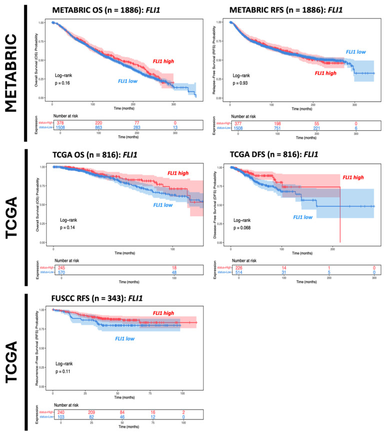 Figure 2
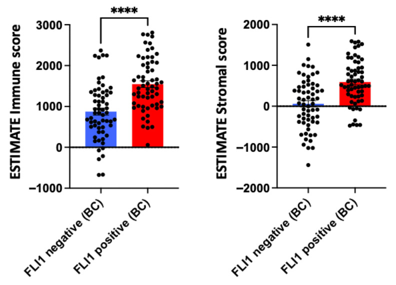 Figure 3
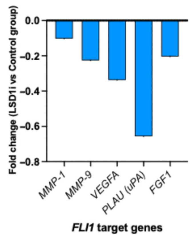 Figure 4
Peer Reviews
No public reviews on file for this paper yet. If you reviewed it on a platform where reviews are public (OpenReview, ICLR, NeurIPS, ICML), you can paste yours below so the community can read it here.
Videos
No videos yet. Explain this paper in a talk, walkthrough, or lecture? Add one.
Taxonomy
TopicsHistone Deacetylase Inhibitors Research · Epigenetics and DNA Methylation · Acute Myeloid Leukemia Research
1. Introduction
Triple-negative breast cancer (TNBC) remains one of the most aggressive breast cancer subtypes due to the lack of hormone receptors and human epidermal growth factor receptor 2 (HER2) amplification, which limits therapeutic options and contributes to poor prognosis [1]. Emerging evidence underscores the tumor microenvironment (TME) as a key determinant of TNBC progression and therapeutic response. Integrative strategies combining epigenetic modulators with immunotherapy are being investigated to overcome immune evasion and therapeutic resistance [2].
FLI1, an ETS transcription factor, is critical for hematopoietic and endothelial differentiation but has also been implicated in breast cancer aggressiveness [3]. Elevated FLI1 expression correlates with higher histological grade, stromal remodeling, and poor clinical outcomes in TNBC [4]. FLI1 activity is influenced by chromatin regulation, as ETS factors, including FLI1, frequently co-occupy regulatory regions with LSD1 and other chromatin remodelers, indicating a functional intersection between epigenetic repression and ETS-driven transcriptional programs [5].
Lysine-specific demethylase 1 (LSD1) is a histone demethylase that regulates H3K4 and H3K9 methylation to maintain repressive chromatin states [6]. In cancer, LSD1 suppresses interferon signaling, antigen presentation, and immune-stimulatory endogenous retroviral elements, whereas its inhibition reactivates type I interferon pathways, enhances T cell infiltration, and sensitizes tumors to immune checkpoint blockade (ICB) [7]. Mechanistically, LSD1 has been shown to co-occupy genomic loci with ETS family transcription factors, including FLI1, suggesting direct regulatory interplay in transcriptional programs relevant to tumor progression [8,9].
Given the central role of FLI1 in cellular regulation, understanding its expression patterns in breast cancer and its contributions to tumor progression and metastasis is critical. This study aims to investigate FLI1 expression in invasive breast carcinoma (IBC) and evaluate its association with histopathological grading. Specifically, it seeks to determine whether FLI1 expression correlates with aggressive tumor characteristics and clinical outcomes, thereby providing insights into its potential as a prognostic marker. The study addresses the following research objectives: (a) the expression pattern of FLI1 in IBC, (b) the association of FLI1 expression with histopathological grading and prognosis in IBC, and (c) the influence of FLI1 on the tumor microenvironment in IBC. To address these objectives, we employed a comprehensive approach integrating immunohistochemical analysis of FLI1 expression in a well-annotated cohort of invasive breast carcinoma cases, correlation with clinicopathological parameters including histological grade and survival outcomes, and profiling of immune and stromal components within the tumor microenvironment.
2. Materials and Methods
2.1. Study Design and Population
A cross-sectional observational study was carried out on female patients diagnosed with invasive breast carcinoma (IBC) through histopathological examination at the Department of Pathology, Dhaka Medical College, between September 2022 and August 2024. A total of 100 IBC cases, obtained from wide local excision and mastectomy specimens, were included in the study. Immunohistochemical analysis was performed on 60 of these cases. The study protocol was approved by the Institutional Review Board of Dhaka Medical College. Informed consent was obtained from all participants, and confidentiality of their information was maintained.
2.2. List of Variables Examined
Demographic variables (age), pathological characteristics (laterality, tumor type, site, size, focality, presence of in situ component, lymphovascular invasion, lymph node involvement, microcalcification, necrosis, hormonal status, tumor grade, pT stage, and pN stage), and FLI1 expression parameters (percentage of positively stained tumor area, staining intensity, and overall expression level) were recorded. Patient demographic and clinical information was recorded using a structured proforma. Informed written consent was obtained, along with relevant clinical history.
2.3. Selection Criteria
This study enrolled female patients of any age, including all mastectomy and wide local excision specimens diagnosed with invasive breast carcinoma, with available immunohistochemical reports for receptor status (ER, PR, HER2). Exclusion criteria included patients who received neoadjuvant chemotherapy or radiotherapy, cases of recurrent breast carcinoma, HER2-equivocal cases, and those with incomplete clinical or pathological information.
2.4. Histopathological Evaluation
Gross examination documented tumor size and axillary nodal status. Paraffin-embedded sections were stained with hematoxylin and eosin (H&E) and evaluated for histologic subtype, grade, staging, and lymphovascular invasion.
2.5. Immunohistochemical Procedure
Paraffin-embedded sections (3–4 µm thick) were immunostained using a standard labeled streptavidin–biotin system (Genemed, South San Francisco, CA, USA) with Anti-FLI-1 polyclonal antibody (Chongqing Biospes Co., Ltd., Chongqing, China) at a 1:50 dilution. Sections were deparaffinized, followed by antigen retrieval in 10 mmol/L citrate monohydrate buffer (pH 6.0) heated in a microwave for 20 min. Diaminobenzidine (DAB) served as the chromogen, and immunodetection was performed using commercial detection kits (Dako, Glostrup, Denmark).
2.6. Immunohistochemical Analysis
Cells were considered immunohistochemically positive for FLI1 when a brownish nuclear staining was observed in tumor cell nuclei [10]. To allow for a semi-quantitative and reproducible assessment, immunohistochemical staining was scored on a scale of 0–9, based on the percentage of positive tumor cells and staining intensity (Table 1 and Table 2). The FLI1 score was calculated by multiplying the staining intensity by the percentage of positive cells, yielding possible scores of 0, 1, 2, 3, 4, 6, and 9. A cutoff score of 4 was used to define FLI1 expression: scores ≥ 4 indicated high expression, while scores ≤ 3 indicated low expression. Normal tonsillar tissue was used as an external positive control for the staining procedure.
2.7. Follow-Up and Statistical Analysis
Follow-up data were obtained from electronic medical records. Survival outcomes were estimated with Kaplan–Meier analysis and compared between subgroups with log-rank statistics. Multivariate Cox Regression was carried out to evaluate the effect of FLI1 tumor cell expression level, with survival adjusted for the effects of age, grade, tumor size, lymph node stage, and lymph node positivity. Descriptive statistics, Chi-square tests, and Fisher’s exact tests were performed, with a p-value < 0.05 considered statistically significant.
2.8. Database
Data were obtained from multiple publicly available breast cancer genomic resources. Information from the Molecular Taxonomy of Breast Cancer International Consortium (METABRIC) and the Cancer Genome Atlas (TCGA) was accessed via cBioPortal (https://www.cbioportal.org/, accessed on 24 September 2022) [11,12]. Data from the Fudan University Shanghai Cancer Center (FUSCC) were obtained from BioSino (https://www.biosino.org/node/, Project ID: OEP00015, accessed on 24 September 2022) [12] and the Fudan Data Portal for Cancer Genomics (https://doi.org/10.6084/m9.figshare.19783498.v5, accessed on 24 September 2022) [13,14]. For invasive breast carcinoma (IBC) cohorts, the case numbers were METABRIC (n = 1886), FUSCC (n = 343), and TCGA (n = 816). Mass spectrometry-based proteogenomic data of for IBC samples were also obtained from cBioPortal [11,12,15]. The ESTIMATE (Estimation of Stromal and Immune cells in Malignant Tumor tissues using Expression data) algorithm was applied to calculate stromal scores (reflecting stromal content) and immune scores (reflecting immune cell infiltration), which were compared between FLI1-high and FLI1-low samples.
2.9. Humanized Breast Cancer Mouse Model
Humanized immune system (HIS) mice were transplanted with 1 × 10^6^ MDA-MB-231 cells. Tumor volumes were monitored twice weekly until reaching approximately 80–100 mm^3^, at which point mice were treated intraperitoneally with the LSD1 inhibitor phenelzine (40 mg/kg daily) [16]. All injections were performed under sterile conditions, and mice were monitored daily for treatment-related effects throughout the study. Tumors from the endpoint were harvested and submitted for bulk-RNA sequencing. Raw counts were subjected to differential gene expression analysis using DESeq2, with fold change estimation values (between treatment group and control group) used for downstream analysis.
3. Results
3.1. Demographics
The mean age of patients was 46.9 years, with the highest frequency observed in the 46–55-year age group (Table 3), consistent with previous studies reporting peak breast cancer incidence in late middle age.
3.2. Tumor Type and Stage
Infiltrating duct carcinoma (IDC) was the predominant tumor type, accounting for 93.0% of cases (Table 4). Representative images of IDC (grades 2–3) and invasive lobular carcinoma (grades 1–2) with varying FLI1 immunostaining intensity from low to high are provided in the supplementary material. Most tumors were classified as pT2 (57.0%) (Table 5), with sizes ranging from 2.1 to 5.0 cm (Table 6). Lymphovascular invasion (LVI) was observed in 44.0% of cases (Table 7), possibly due to differences in sampling.
3.3. Distribution of Nodal Involvement and Hormonal Receptor Status in Study Cohort
Nodal metastasis was present in 42.0% of cases, whereas 52.0% showed no nodal involvement. Hormonal receptor analysis revealed estrogen receptor (ER) positivity in 60.0% of cases and progesterone receptor (PR) positivity in 45.0%, with a minority of HER2-enriched tumors (6.0%) (Table 8).
3.4. Histological Grading and Its Association with FLI1 Expression
Within the study cohort, Grade 2 tumors were the most prevalent (50.0%), followed by Grade 3 (37.0%) and Grade 1 (13.0%) (Figure 1), consistent with previous studies [17,18,19].
A significant correlation was observed between elevated FLI1 expression and higher histological grade (p < 0.001), with 60% of Grade 3 tumors exhibiting high FLI1 levels (Table 9). Although not reaching statistical significance, cases with high FLI1 expression tended to demonstrate increased nodal involvement.
3.5. Kaplan–Meier Survival and Stromal/TIL Analyses
Within IBC cohorts, FLI1 mRNA expression levels were shown to not be significantly associated with survival outcomes (Figure 2). This is concordant with existing studies with conflicting results over the prognostic function of FLI1 in breast cancer [20]. Interestingly, FLI1 expression in breast cancer samples was positively associated with both immune and stromal infiltration, underscoring FLI1’s role in tumor stroma and immune infiltration (Figure 3) [3].
3.6. FLI1-LSD1 Axis in Oncogenesis-Associated Transcriptional Programs
In a humanized breast cancer mouse model, LSD1 inhibition resulted in reduced FLI1 target gene signatures related to angiogenesis (VEGFA, FGF-1, MMP-1, MMP-9), migration, and invasion (PLAU) (Figure 4).
4. Discussion
This study characterized FLI1 protein expression in invasive breast carcinoma and assessed its association with histopathological grade, clinicopathological features, and patient survival. The results indicate that FLI1 may play a role in tumor progression, aggressiveness, and prognosis. Additionally, we explored the modulation of FLI1 in breast cancer under LSD1 inhibition. Using breast cancer mouse models, LSD1 inhibition influenced FLI1 target genes related to tumor progression. These findings suggest that LSD1 inhibition may regulate tumor and endothelial programs regulated by FLI1.
Kaplan–Meier analyses revealed variable associations between FLI1 expression and clinical outcomes across datasets. While higher FLI1 immunostaining correlated with higher histological grading, FLI1 mRNA levels did not present a significant association with prognostic trends across invasive breast cancer cohort samples. These observations suggest that the prognostic value of FLI1 may be context-dependent, influenced by factors such as tumor subtype, genetic background, and treatment strategies. Our findings align with prior studies linking FLI1 expression to higher histological grade, consistent with the role of ETS factors in regulating proliferation, differentiation, and apoptosis in breast cancer [6].
No significant correlation was observed between FLI1 expression and pathological T or N stage (p = 0.604 and 0.125, respectively), in contrast to Song et al. (2015), who reported an association between FLI1 score and lymph node metastasis, potentially reflecting differences in sample characteristics [7].
FLI1 expression was not significantly associated with ER, PR, or HER2 status, suggesting that its role in breast cancer progression may be independent of these canonical molecular markers and could serve as a complementary biomarker. This contrasts with Lafta (2017), who reported reduced FLI1 expression across all stages of primary breast carcinoma and correlations with ER, PR, and HER2 [8].
FLI1 positivity appeared to influence immune and stromal infiltration in breast cancer [4]. FLI1-positive tumors displayed significantly higher stromal scores, indicating greater abundance of stromal components such as fibroblasts and enriched extracellular matrix and vasculature. This enrichment may reflect a complex tumor microenvironment, potentially contributing to immune modulation, therapeutic resistance, and tumor progression. The association between FLI1 and stromal content suggests FLI1’s role in stromal remodeling or fibroblast activation, warranting further mechanistic investigation.
Given that ETS-FLI transcriptional activity can be regulated by LSD1 in other cancers, our results similarly demonstrated that LSD1 inhibition could repress FLI target genes related to angiogenesis and migration, thereby promoting cancer progression or higher histological grading [21]. Overall, our results suggest the role of the FLI1-LSD1 axis in the induction of tumor migration, angiogenesis, and tumor progression through direct regulation of ECM-degrading proteases in cancer and stromal cells [4,22,23]. However, given reports of tumor-intrinsic FLI1 promoting proliferation and invasion [6], some of this signal may represent adaptive responses to epigenetic stress. These dual roles underscore the complexity of FLI1 biology in TNBC. These findings suggest two translational opportunities: (i) FLI1 could be used as a pharmacodynamic biomarker of breast cancer metastastatic potential, capturing stromal and immune remodeling, and (ii) combining direct FLI1 inhibition and LSD1 blockade may yield stnergistic therapeutic effects. Single-cell and spatial transcriptomic analyses will be critical to delineate the cellular origin of FLI1 and determine whether its elevation predominantly reflects immune activation or malignant adaptation.
This study has several limitations: (a) The assessment of lymphovascular invasion was based solely on histopathology, potentially introducing bias. (b) Pathological staging included only tumor size (pT) and lymph node status (pN), excluding metastasis (pM). (c) Immunohistochemistry was performed in a limited subset of 60 cases. (d) ER, PR, and HER2 data were obtained from various laboratory reports, and molecular subtyping was not conducted. (e) Long-term patient follow-up data were unavailable, precluding outcome analyses. (f) Newly defined subtypes based on immune and stromal infiltration were not externally validated. And lastly, (g) not all histological subtypes of invasive breast carcinoma were represented in this study.
5. Conclusions
This study highlights the potential role of FLI1 in promoting tumor progression and stromal remodeling in invasive breast carcinoma. Higher FLI1 protein expression was associated with increased histological grades and enriched stromal features, suggesting a role in tumor aggressiveness and microenvironment modulation. Although FLI1 expression was not correlated with patient survival, its modulation under LSD1 inhibition points to a regulatory axis with therapeutic relevance. Collectively, these findings support the consideration of FLI1 as a context-dependent biomarker for breast cancer and a potential target in epigenetic therapy strategies. Limitations such as a limited sample size, lack of molecular subtyping, and absence of longitudinal outcome data underscore the need for further validation in larger, well-characterized cohorts. Future studies leveraging multi-omics will be essential to disentangle the tumor-intrinsic and microenvironmental contributions of FLI1 expression.
The reference list from the paper itself. Each links out to its DOI / PubMed record.
- 1Won K.A. Spruck C. Triple-negative breast cancer therapy: Current and future perspectives (Review)Int. J. Oncol.2020571245126110.3892/ijo.2020.513533174058 PMC 7646583 · doi ↗ · pubmed ↗
- 2Singh D.D. Haque S. Kim Y. Han I. Yadav D.K. Remodeling of tumour microenvironment: Strategies to overcome therapeutic resistance and innovate immunoengineering in triple-negative breast cancer Front. Immunol.202415145521110.3389/fimmu.2024.145521139720730 PMC 11666570 · doi ↗ · pubmed ↗
- 3Kim D. Kim K.I. Baek S.H. Roles of lysine-specific demethylase 1 (LSD 1) in homeostasis and diseases J. Biomed. Sci.2021284110.1186/s 12929-021-00737-334082769 PMC 8175190 · doi ↗ · pubmed ↗
- 4Sheng W. La Fleur M.W. Nguyen T.H. Chen S. Chakravarthy A. Conway J.R. Li Y. Chen H. Yang H. Hsu P.-H. LSD 1 Ablation Stimulates Anti-tumor Immunity and Enables Checkpoint Blockade Cell 2018174549563.e 1910.1016/j.cell.2018.05.05229937226 PMC 6063761 · doi ↗ · pubmed ↗
- 5Sankar S. Theisen E.R. Bearss J. Mulvihill T. Hoffman L.M. Sorna V. Beckerle M.C. Sharma S. Lessnick S.L. Reversible LSD 1 inhibition interferes with global EWS/ETS transcriptional activity and impedes Ewing sarcoma tumor growth Clin. Cancer Res.2014204584459710.1158/1078-0432.CCR-14-007224963049 PMC 4155010 · doi ↗ · pubmed ↗
- 6Scheiber M.N. Watson P.M. Rumboldt T. Stanley C. Wilson R.C. Findlay V.J. Anderson P.E. Watson D.K. FLI 1 expression is correlated with breast cancer cellular growth, migration, and invasion and altered gene expression Neoplasia 20141680181310.1016/j.neo.2014.08.00725379017 PMC 4212256 · doi ↗ · pubmed ↗
- 7Song W. Li W. Li L. Zhang S. Yan X. Wen X. Zhang X. Tian H. Li A. Hu J.-F. Friend leukemia virus integration 1 activates the Rho GT Pase pathway and is associated with metastasis in breast cancer Oncotarget 20156237642377510.18632/oncotarget.435026156017 PMC 4695150 · doi ↗ · pubmed ↗
- 8Lafta I.J. FLI 1 Expression in Breast Cancer Cell Lines and Primary Breast Carcinomas is Correlated with ER, PR and HER 2Int. J. Med. Res. Health Sci.201768795