Plant-Derived Modifiers for Antimicrobial Soft Denture Liners: A Review
Patrycja Kula, Grzegorz Chladek, Izabela Barszczewska-Rybarek

TL;DR
This paper reviews how plant-based compounds can improve antifungal properties of soft denture liners while maintaining mechanical performance.
Contribution
The paper systematically reviews plant-derived modifiers for antimicrobial denture liners and identifies key limitations and future directions.
Findings
Plant-derived compounds like aloe vera and neem inhibit Candida albicans growth and improve antifungal properties of soft denture liners.
Most formulations maintain acceptable mechanical properties and improve surface smoothness.
Key limitations include rapid leaching of active compounds and lack of in vivo and cytotoxicity data.
Abstract
This review examines strategies to enhance the antifungal properties of commercial soft lining materials (SLMs) through modification with plant-derived oils, extracts, and powders. These natural bioactive compounds act via multiple mechanisms, including disruption of fungal cell membranes, inhibition of biofilm formation, and interference with Candida albicans metabolism, the pathogen causing denture-associated candidiasis. Their incorporation into SLM provides localized antifungal activity at the denture–mucosa interface. The review highlights Aloe vera (aloe), Azadirachta indica (neem), Ocimum basilicum (basil), Melaleuca alternifolia (tea tree), Cocos nucifera (coconut), Allium sativum (garlic), Thymus vulgaris (thyme), and chitosan as notable sources of phytotherapeutics that consistently inhibit C. albicans growth. In addition to antimicrobial effects, studies assessed both…
Genes, proteins, chemicals, diseases, species, mutations and cell lines named across the full text — each resolved to its canonical identifier and authoritative record.
Click any figure to enlarge with its caption.
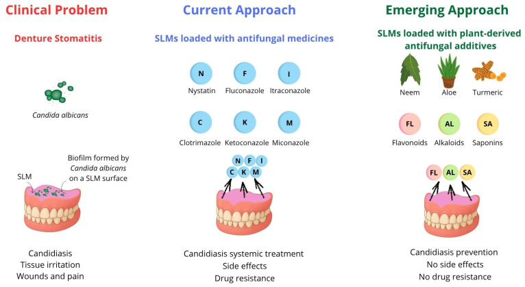 Figure 1
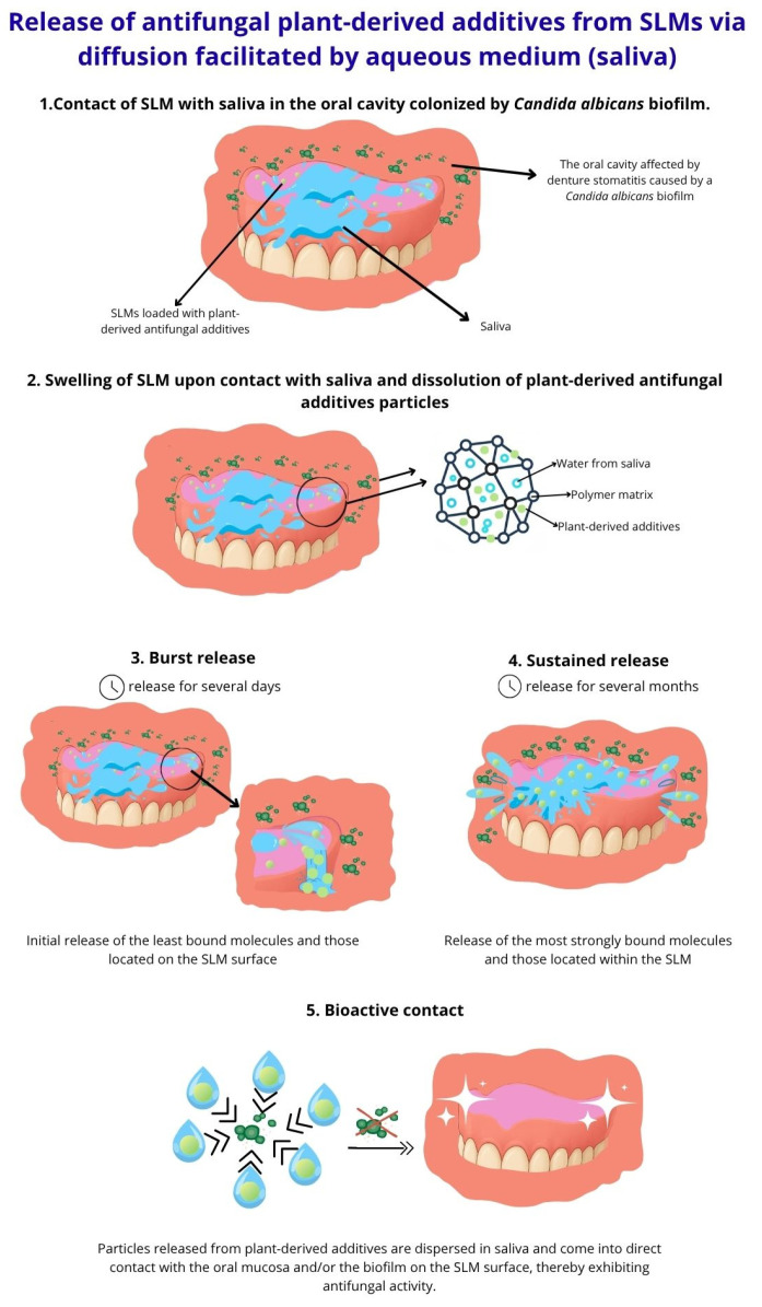 Figure 2
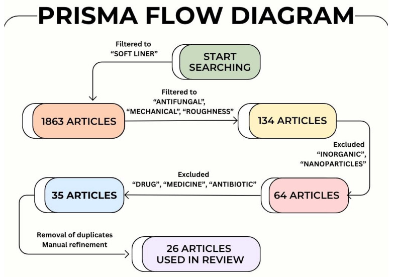 Figure 3
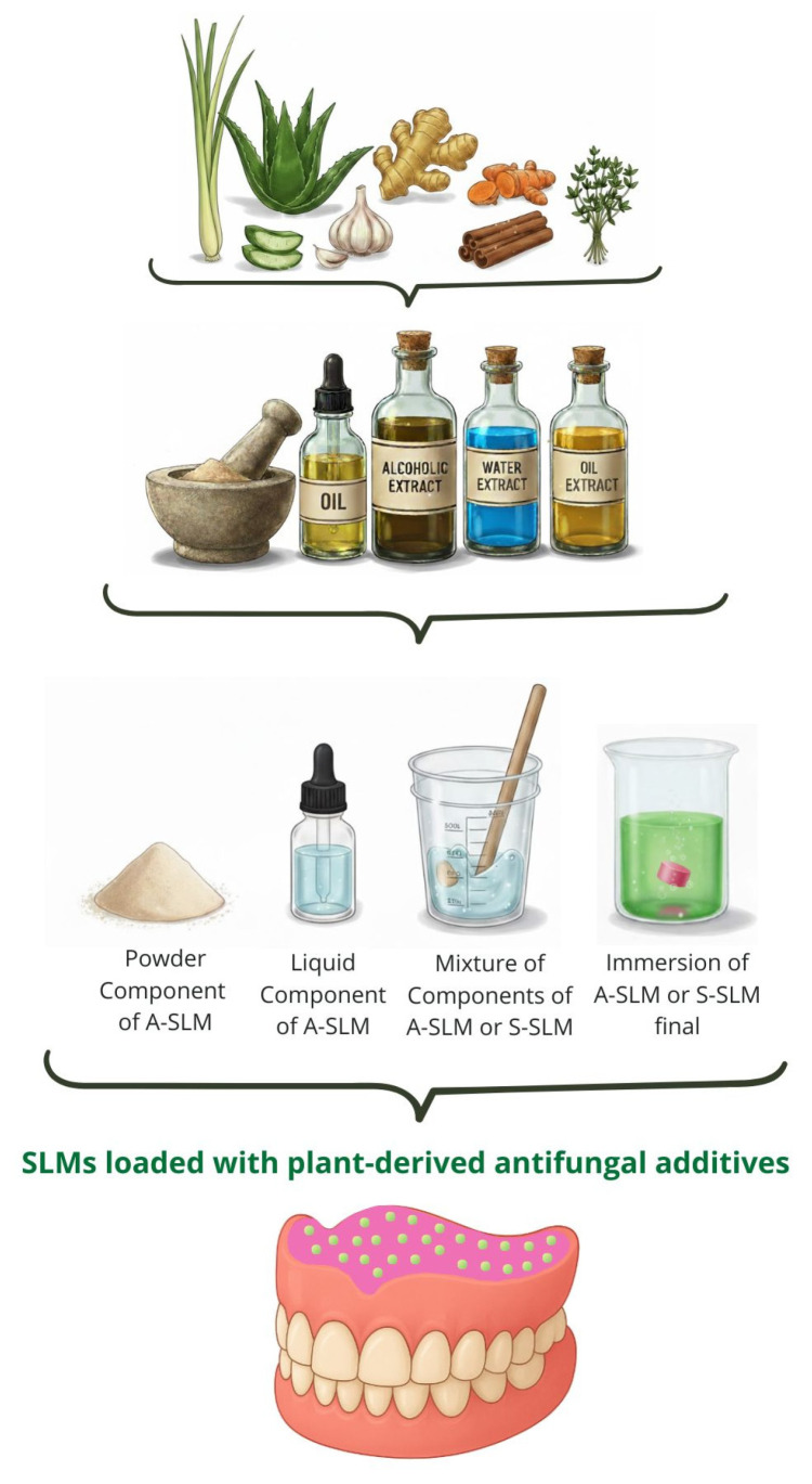 Figure 4
- —Polish Budget Funds for Scientific Research in 2025
- —Silesian University of Technology
- —European Funds for Silesia 2021–2027
Peer Reviews
No public reviews on file for this paper yet. If you reviewed it on a platform where reviews are public (OpenReview, ICLR, NeurIPS, ICML), you can paste yours below so the community can read it here.
Videos
No videos yet. Explain this paper in a talk, walkthrough, or lecture? Add one.
Taxonomy
TopicsEndodontics and Root Canal Treatments · Essential Oils and Antimicrobial Activity · Dental Erosion and Treatment
1. Introduction
Denture-related infections remain a significant challenge in prosthetic dentistry. Soft lining materials (SLMs), commonly used to enhance denture fit and comfort, do not inhibit the growth of pathogenic microorganisms, contributing to a high incidence of denture stomatitis, predominantly caused by Candida albicans [1,2,3]. Candidiasis frequently affects denture wearers with poor oral hygiene, prolonged denture use, unhealthy diet, smoking habits, micro-injuries, reduced salivary flow, hormonal imbalances, or compromised immunity. High-risk groups include older adults, diabetics, chemotherapy patients, and individuals with HIV [4,5,6,7]. Over the past two decades, its prevalence has risen significantly, now affecting even healthy individuals [8]. Conventional approaches to managing this condition rely on incorporating antifungal medicines, such as nystatin, clotrimazole, miconazole, fluconazole, and itraconazole, into SLMs [2]. However, the limitations of conventional antifungal therapeutics, including systemic side effects and emerging drug resistance [9], have prompted interest in plant-derived bioactive compounds. These natural additives offer broad-spectrum antimicrobial activity with lower cytotoxicity and minimal side effects. Phytochemicals such as flavonoids, terpenes, saponins, and alkaloids have demonstrated promising antifungal efficacy and may serve as safer, effective alternatives for modifying SLMs (Figure 1).
Unlike previous reviews, which focused on the incorporation of synthetic antifungal medicines into SLMs, this review offers a comprehensive and comparative analysis of current advancements in the development of SLMs modified with plant-derived additives. This is the first literature review on this topic so far. It highlights the influence of plant-derived additives on antifungal efficacy, antibacterial efficiency, biocompatibility, mechanical properties and roughness of commercial SLMs. By emphasizing the unique benefits of plant-derived additives, this article aims to establish their role as a viable and sustainable solution in the research of modern prosthodontic biomaterials.
2. Classification of SLMs
SLMs are classified based on service time into temporary soft lining materials (T-SLMs) and long-term soft lining materials (LT-SLMs) [2].
T-SLMs include tissue conditioners (TCs) and short-term soft lining materials (ST-SLMs), both representing acrylic-based SLMs (A-SLMs), typically comprising two components: a powder and a liquid. TCs are classified as self-curing systems. The gelation, occurring at oral cavity temperature, involves the plasticization of poly(ethyl methacrylate) (PEMA) or poly(methyl methacrylate) (PMMA) with the plasticizer-solvent system. In contrast, ST-SLMs contain an initiating system and have a more complex composition of a liquid component, being a mixture of a plasticizer, a solvent (commonly ethanol), and methacrylate monomers such as methyl methacrylate (MMA), ethyl methacrylate (EMA), n-butyl methacrylate (BuMA), ethylene glycol dimethacrylate (EGDMA), and triethylene glycol dimethacrylate (TEGDMA). Additionally, an accelerator for initiating free radical polymerization, typically N,N-dimethyl-p-toluidine (DMPT), is included. The initiator for the free radical polymerization, usually benzoyl peroxide (BPO), is mixed into the powder. ST-SLMs are heat-curing systems, which undergo gelation at elevated temperature through the simultaneous plasticization of PEMA (alternatively PMMA) and the polymerization of the methacrylate monomers, forming a crosslinked structure when dimethacrylates are present, thereby extending service time. LT-SLMs usually are crosslinked silicone elastomers (S-SLMs) available as one- or two-component systems. Single-component S-SLMs consist of vinyl-terminated polydimethylsiloxanes and organic peroxide (typically BPO). Their polymerization requires high temperatures. In contrast, two-component S-SLMs consist of a catalyst paste that combines vinyl-terminated polydimethylsiloxanes with a platinum catalyst, and a base paste made of vinyl-terminated and hydride-terminated polydimethylsiloxanes. The reaction between the catalyst and the base occurs at room temperature [2].
3. The Concept of Antimicrobial SLMs with Plant-Derived Additives
The concept of antimicrobial SLMs with plant-derived additives is based on their ability to release bioactive compounds into saliva, similar to controlled drug-release systems, allowing direct action on the oral soft tissues without toxic effects (Figure 2). To prevent denture-related candidiasis, these additives must primarily exhibit antifungal activity against C. albicans.
A wide variety of plant-derived compounds exhibit potent antifungal activity against C. albicans through diverse bioactive compounds and mechanisms. Aloe vera (aloe) [10,11], Azadirachta indica (neem) [12,13], and Triphala (three fruits) [14,15] contain anthraquinones, alkaloids, tannins, and flavonoids that disrupt fungal membranes, inhibit biofilm formation, and suppress hyphal growth. Origanum vulgare (oregano) [16,17], Thymus vulgaris (thyme) [18], and Ocimum species (basil, tulsi) [19,20] owe their activity mainly to terpenes and phenolics such as thymol, carvacrol, and eugenol, which damage cell membranes and inhibit ergosterol synthesis. The main bioactive compound in Cocos nucifera (coconut), lauric acid, exerts antifungal effects by disrupting the fungal cell membrane and causing leakage of cellular contents [21,22]. The major bioactive component in Allium sativum (garlic) is allicin, which damages the cell wall, inhibiting key enzymes and disrupting metabolism, leading to cell lysis [23,24]. Melaleuca alternifolia (tea tree) and Cymbopogon citratus (lemongrass) oils, rich in terpenoids like terpinen-4-ol and citral, disrupt membrane integrity and induce oxidative stress [25,26]. Curcuma longa (turmeric), Zingiber officinale (ginger), and Cinnamomum (cinnamon) contain polyphenols (curcumin, gingerol, cinnamaldehyde) that interfere with fungal signaling, enzyme activity, and cell wall synthesis [27,28,29]. Nigella sativa (black seed) and Linum usitatissimum (flaxseed) alter ergosterol and lipid metabolism [30,31], while Centratherum anthelminticum (black cumin) and Litsea cubeba show similar effects via terpenoids [32,33]. Piper betle demonstrates antifungal action through membrane disruption and inhibition of ergosterol biosynthesis [34,35]. Recently, Rana et al. demonstrated that anticancer flavonoids such as prunin and isorhamnetin also exhibit notable antifungal activity. These compounds disrupt fungal cell membranes and induce oxidative stress, positioning them as mechanistic analogues to other antifungal flavonoids [36,37]. Finally, chitosan, a natural polymer, exhibits antifungal activity through electrostatic interactions with the fungal cell surface [38,39].
4. Methodology
This narrative systematic literature review combines a systematic methodology with a narrative synthesis approach.
The literature regarding the main topic, i.e., SLMs modified with plant-derived additives, was systematically reviewed in accordance with the PRISMA guidelines. A comprehensive literature search was conducted across four scientific databases: Scopus, Lens, Web of Science, and PubMed, covering the period from January 2015 to September 2025. Search queries were constructed using combinations of keywords and Boolean operators (AND, OR) associated with the review’s main concepts. The principal search terms included: “soft liner”, “antifungal”, “mechanical”, “roughness”, “inorganic”, “nanoparticles”, “drug”, “antibiotic”, and “medicine.” To ensure completeness, manual searches were also conducted to identify additional relevant publications not indexed in the selected databases.
Studies were included if they met the following criteria: (i) published in peer-reviewed journals between 2015 and 2025, (ii) written in English, (iii) directly addressed the core topic and objectives of this review, (iv) contained experimental data, systematic analyses, or comprehensive discussions relevant to antimicrobial SLMs modified with plant-derived compounds.
Studies were excluded if they: (i) were conference abstracts, editorials, or book chapters, (ii) lacked sufficient methodological or analytical detail, (iii) were duplicates or unavailable in full text, (iv) focused on modifications involving inorganic additives or medicines, pharmaceutics, or antiseptics.
Figure 3 illustrates the selection process and keyword co-occurrence flowchart, summarizing the identification and screening stages and highlighting research clusters on SLMs modified with plant-derived antifungals in current literature.
The initial database search yielded 1863 records. Following keyword refinement using “soft liner” AND “antifungal” AND “mechanical” AND “roughness”, and excluding “inorganic”, “nanoparticles”, “drugs”, “medicine”, and “antibiotics”, 35 potentially relevant articles were identified. Additionally, 8 supplementary records were retrieved through manual searches and cross-referencing. After the removal of 10 duplicate records, the remaining 33 full-text articles were screened for eligibility. Following manual refinement by limiting the studies to findings strictly related to SLMs modified with plant-derived additives, a total of 26 studies were ultimately included in the final review (The Excel file containing these articles can be found in the Supplementary Materials). The data from these selected studies were then systematically organized according to major research themes, including plant-derived extracts, essential oils, herbal formulations, SLMs type, methods of modification, in vitro and in vivo antifungal assays, in vitro antibacterial assays, biocompatibility assessment, mechanical properties, roughness, limitations and future perspectives. Each study was carefully reviewed to identify recurring trends, experimental outcomes, knowledge gaps, and limitations to outline emerging challenges and prospective directions for future research on SLMs modified with plant-derived antimicrobial additives.
In addition to the directly identified studies on SLMs modified with plant-derived antifungals, a further 130 publications were manually searched and critically reviewed. This broader literature base supported and contextualized the findings, and allowed for a critical assessment of trends, gaps, and inconsistencies in the field.
5. Discussion
The reviewed studies indicate that plant-derived additives can enhance the antifungal potential of SLMs. However, this effect varies considerably depending on the plant species, the bioactive form used, the method of incorporation, and the type and composition of the SLM.
The plant-derived additives used for SLM modification included essential oils, solutions, and liquid or dry extracts. These bioactive substances were incorporated into the material by adding them to the powder component, the liquid component, or a mixture of both immediately before polymerization. Alternatively, a post-fabrication approach was employed in which ready-to-use SLMs were immersed in phytochemical solutions, extracts, or oils to impart antifungal properties (Figure 4).
Table 1 provides a comparative overview of acrylic-based SLMs, including TCs and ST-SLMs, modified with plant-derived compounds via addition to the powder, liquid, or powder–liquid mixture of the SLM. The table is organized according to the plant species and family, plant-derived additive content and form (powder, oil, or solution), the SLM type, trade name, manufacturer, information on processing, component enriched, and the corresponding in vitro antimicrobial and mechanical properties tested.
Table 2 provides a comparative overview of silicone-based SLMs (LT-SLMs) modified with plant-derived compounds via addition to the powder–liquid mixture of the SLM. The table is organized according to the plant species and family, plant-derived additive content and form (powder, oil, or solution), the SLM type, trade name, manufacturer, information on processing, component enriched, and the corresponding in vitro antimicrobial and mechanical properties tested.
Table 3 provides a comparative overview of tissue conditioners (TCs) modified with plant-derived compounds via immersion method. The table is organized according to the plant species and family, immersion liquid (volume), the SLM type, trade name, manufacturer, information on processing, and the corresponding in vitro antimicrobial and mechanical properties tested.
While Table 1, Table 2 and Table 3 summarize the antifungal performance of SLMs according to material type, plant species, and method of incorporation, it should be noted that multiple sources of methodological bias may influence the analysis.
The following general issues in methodological variability and potential bias can be distinguished:
- SLM type (TC, ST-SLM, LT-SLM).
- SLM composition, including polymer type (A-SLM, S-SLM), plasticizer, and ethanol content.
- Differences in extraction methods for bioactive compounds.
- Variations in the form of bioactive compounds (essential oil, powder, or dried plant parts).
- Additive incorporation method, such as direct admixing, immersion, or nanoparticle dispersion.
- Additive-enriched component in direct admixing (added to liquid, powder, or pre-formulated mixture).
- Inconsistent reporting of additive concentration, rarely expressed as weight or volume fractions relative to total sample mass. Often, fixed amounts were added to liquid or powder according to manufacturer-recommended ratios, preventing standardized comparison.
- Additive concentration range tested.
- Immersion medium, e.g., pure additive oil or solvent.
This structure highlights the multiple sources of variability that can influence the reproducibility and comparability of results in studies on plant-modified SLMs.
Despite this variability, a stepwise analysis of the available studies enabled the identification of important trends and facilitated a discussion of the results, as presented in the following sections.
5.1. In Vitro Antifungal Efficacy
The following section discusses the in vitro antifungal properties of SLMs modified with plant-derived additives against C. albicans, highlighting trends associated with plant species, incorporation method, and SLM type (Table 4). Nineteen plant-derived species and compounds were identified. The most frequently tested were M. alternifolia (6 times), A. indica (4 times), and chitosan (3 times). Others, including A. vera, A. sativum, O. vulgare, T. vulgaris, O. basilicum, O. sanctum, C. nucifera, Cinnamomum sp., C. longa, Z. officinale, N. sativa, L. cubeba, C. citratus, L. usitatissimum, C. anthelminticum, and P. betle, were each evaluated once.
Songsang et al. [53] investigated L. cubeba oil (5–30 vol./vol.%) in three TCs (A-SLMs, Table 1 and Table 4): GC Soft Liner, Coe-Comfort, and Visco-gel. No activity was observed at 5 vol./vol.%, whereas concentrations of 10 vol./vol.% and higher produced increasing inhibition zones. IZD was also dependent on the TC and decreased followingly: Visco-gel > Coe-Comfort > GC Soft Liner, with IZDs at 10, 20, and 30 vol./vol.% of 12.09, 16.56, 24.68 mm (Visco-gel); 10.22, 14.07, 22.61 mm (Coe-Comfort); and 7.61, 13.19, 22.42 mm (GC Soft Liner), all exceeding the nystatin control (12 mm). This highlighted both the antifungal potential of L. cubeba oil and the influence of SLM composition, particularly the plasticizer type. The highest antifungal activity of Visco-gel can be explained by synergistic interaction between the triethyl citrate plasticizer and ethanol. Nikawa et al. [64] reported that ethanol content in SLM can significantly affect antifungal activity depending on the plasticizer, showing a positive correlation with benzyl salicylate but an inverse correlation with benzyl n-butyl phthalate. Supporting this, Truhlar et al. [65] observed that nystatin incorporated into Visco-gel exhibited greater antifungal activity than in Lynal, indicating that material composition itself influences antifungal performance. This highlights that both plasticizer type and its interaction with ethanol modulates antifungal efficacy. Benzyl benzoate plasticizer possesses intrinsic antifungal properties, contributing to intermediate activity of Coe-Comfort, which is the opposite of inert butyl phthalyl butyl glycolate, explaining the weakest activity of GC Soft Liner [66].
Vankadara et al. [50] consistently found higher antifungal activity of Visco-gel than GC Soft Liner due to compositional differences. They investigated M. alternifolia oil (0.5–2.0 mL; 10–40 vol./vol.%) in these two TCs (A-SLM, TC, Table 1 and Table 4), observing a clear dose-dependent response for both. For Visco-gel, the IZDs increased from 19.0 mm (0.5 mL) to 20.8 mm (2 mL) at 10 vol./vol.% and from 21.6 mm (1.5 mL) to 26.4 mm (2 mL) at 40 vol./vol.%. The maximum inhibition (26.4 mm at 2 mL, 40 vol./vol.%) slightly exceeded the nystatin control (22.6 mm). In contrast, GC Soft Liner exhibited weaker activity at each condition, with IZDs increased from no inhibition zone observed (0.5 mL) to 17.4 mm (2 mL) at 10 vol./vol.% and from 15.2 mm (0.5 mL) to 23.2 mm (2 mL) at 40 vol./vol.%. The latter was comparable to the control. These results were supported by CFU analysis. For Visco-gel, C. albicans colonization decreased from 4.0 ×10^5^ CFU/mL (0.5 mL) to 2.24 ×10^5^ CFU/mL (2 mL) at 10 vol./vol.%, and from 1.72 ×10^5^ to 0.88 ×10^5^ CFU/mL at 40 vol./vol.%. For GC Soft Liner, the corresponding reductions were from 4.88 ×10^5^ to 2.86 ×10^5^ CFU/mL (10 vol./vol.%) and from 3.8 ×10^5^ to 1.08 ×10^5^ CFU/mL (40 vol./vol.%). Overall, Visco-gel consistently exhibited larger IZDs and lower colonization than GC Soft Liner at corresponding doses, confirming that matrix composition strongly affects antifungal performance. However, it is worth noting that highly effective C. albicans reduction was achieved for both TCs, which reached 88% for Visco-gel and 86% for GC Soft Liner (for 2 mL, 40 vol./vol.%).
Pachava et al. [60] confirmed the notable antifungal activity of M. alternifolia oil (15 wt.%) in the silicone-based GC Reline Extra Soft (S-SLM, LT-SLM, Table 2 and Table 4). The modified SLM exhibited markedly lower log CFU/mm^2^ values (2.1, 2.8, and 3.1 at 1, 30, and 60 days, respectively) compared to the unmodified control (7.1, 6.5, and 6.8), corresponding to fungal growth reductions of 70.4%, 56.9%, and 54.4%. Although antifungal activity declined gradually over time, the activity against C. albicans remained evident after 60 days, indicating the strong and sustained antifungal activity of S-SLMs.
On the contrary, Patil et al. [63] reported only limited antifungal activity when incorporating M. alternifolia into GC Soft Liner (A-SLM, TC, Table 1 and Table 4) using an immersion method (50 vol./vol.% DMSO solution). This result indicates that the antifungal efficacy of M. alternifolia depends strongly on its concentration, incorporation method, and the TC. Consequently, the immersion method appears least effective, and using undiluted or higher-concentration oil formulations with more compatible TCs could be recommended to achieve stronger SLM antifungal performance. In the same study, M. alternifolia showed lower antifungal activity than C. citratus (MIC 0.03125 vol./vol.%, 50% inhibition) and T. vulgaris (MIC 0.0625 vol./vol.%, 30% inhibition), with M. alternifolia exhibiting only minimal biofilm reduction at 0.25 vol./vol.%. This is consistent with the high citral (C. citratus) and thymol, and carvacol (T. vulgaris) content. The lowest antifungal activity of M. alternifolia oil can be attributed to the high volatility of terpinen-4-ol, which probably reduces potency over time, particularly during extended incubation.
AlHamdan et al. [62] compared M. alternifolia and A. indica extracts incorporated into GC Soft Liner (A-SLM, TC, Table 3 and Table 4) by immersion. A. indica achieved slightly higher antifungal efficacy (log CFU/mL = 2.15) than M. alternifolia (2.31), both acting comparably to the chlorhexidine control (2.36). These close values suggest that both extracts impart similar fungicidal effects, with A. indica demonstrating slightly stronger activity, which can be attributed to its more hydrophobic bioactive components (azadirachtin, nimbidin, gedunin) [12,13]. In contrast, the comparatively weaker activity of M. alternifolia may be attributed to the high volatility of terpinen-4-ol, which diminishes in potency over time [67]. The authors concluded that undiluted immersion can be effective if extract composition is compatible with the liner and the bioactive component has sufficient stability.
Singhania et al. [61] further showed that the combination of M. alternifolia with A. indica can produce a synergistic antifungal effect. Using the immersion technique with GC Soft Liner (A-SLM, TC, Table 3 and Table 4), M. alternifolia oil alone reduced fungal growth to a log CFU/mL of 2.30, whereas the combination of M. alternifolia with A. indica resulted in a near-complete inhibition, with a log CFU/mL of 0.40.
Kumar et al. [45] extended the comparative analysis beyond previous studies by evaluating the effects of A. indica, M. alternifolia, and C. nucifera oils (5–40 vol./vol.%) on the antifungal properties of Visco-gel (A-SLM, TC, Table 1 and Table 4). At 5 vol./vol.%, all plants exhibited low but measurable antifungal activity. The IZDs measured after 48 h were 6.35 mm for A. indica and 5.0 mm for both M. alternifolia and C. nucifera. At the highest tested concentration, 40 vol./vol.%, all materials achieved their maximum IZDs, with A. indica showing 20.10 mm, C. nucifera 16.55 mm, and M. alternifolia 15.35 mm. After 7 days, the same concentration-dependent trend persisted but with slightly reduced IZDs, indicating decreased long-term activity. At 40 vol./vol.%, A. indica maintained the highest inhibition (19.15 mm), followed by C. nucifera (16.20 mm) and M. alternifolia (14.75 mm). Regarding the optimum concentrations, these were determined based on the most pronounced increase in IZDs. For A. indica, the most significant improvement was observed at 15 vol./vol.%, where the IZD reached 12.20 mm after 48 h and remained high at 11.35 mm after 7 days. C. nucifera exhibited optimum at 20 vol./vol.%, with an IZD of 11.0 mm after 48 h and 9.70 mm after 7 days. M. alternifolia showed its most effective concentration at 25 vol./vol.%, achieving an IZD of 8.8 mm after 48 h and 8.0 mm after 7 days. These results indicate that all plant-derived oils maintained antifungal activity over time, though with reductions after prolonged incubation. The observed decreasing trend reflects the influence of oil composition and antifungal mechanism. The strongest performance of A. indica results from the richness of bioactive compounds such as azadirachtin, nimbidin, and gedunin [12,13]. C. nucifera showing moderate activity, is composed of medium-chain fatty acids [21,22]. Finally, M. alternifolia, appearing to be the weakest fungicide, contains highly volatile terpinen-4-ol [67,68].
In another study, Kumar et al. [44] confirmed complete inhibition of C. albicans with A. indica powder (50–500 µg) incorporated into GC Soft Liner (A-SLM, TC, Table 1 and Table 4), which persisted through 7 days. In the same study, A. sativum powder demonstrated significantly weaker activity, with complete inhibition persisting through 2 days. The lower antifungal efficacy of A. sativum can be attributed to allicin instability [69].
Mohammed et al. [47] compared A. sativum, O. vulgare, N. sativa, and Z. officinale (50 µg/mL) in Acrosoft (A-SLM, TC, Table 1 and Table 4), observing the following order of decreasing activity: A. sativum (IZD = 8.00 mm) > O. vulgare (IZD = 7.75 mm) > Z. officinale (negligible effect) ≈ N. sativa (no inhibition). These findings showed that less hydrophobic compounds, such as allicin (A. sativum, water solubility 24 g/L at 10 °C [70]), exhibit high activity due to facilitated diffusion into the aqueous oral environment. In contrast, poorly water-soluble compounds, such as thymoquinone (N. sativa, water solubility 0.1 g/100 mL (20 °C) [71]) and gingerol (Z. officinale, water solubility < 0.0001 mole fraction at < 130 °C [72]), showed negligible activity, likely due to limited release. Interestingly, O. vulgare exhibited strong antifungal activity despite its predominantly hydrophobic constituents, thymol and carvacrol. This effectiveness can be attributed to their synergistic interaction, which enhances membrane disruption and antifungal potency even at low concentrations [73,74].
Regarding antifungal mechanisms of the aforementioned bioactive compounds, one can conclude that hydrophilic enzyme-targeting compounds (e.g., allicin) typically show stronger activity in SLMs due to improved diffusion through the polymer matrix, while certain combinations of phenolics (e.g., thymol and carvacrol) maintain potency through membrane-disruptive synergy [73].
Consistent with this, Baygar et al. [59] observed a high sensitivity of C. albicans cells to the silicone-based Ufi Gel P (S-SLM, LT-SLM, Table 2) modified with carvacrol (10 mL), the principal monoterpenoid in O. vulgare oil. A notable IZD of 38.33 mm and a 98% biofilm reduction were recorded for the modified SLM.
Analysis of results for S-SLM: Ufi Gel P [59] and GC Reline Extra Soft [60] leads to the conclusion that silicone-based SLMs provide stronger inhibition than acrylic ones. It can be explained by the silicone hydrophobic character, which minimizes interactions with antifungal compounds, promoting their diffusion and release.
Muttagi et al. [46] evaluated C. anthelminticum, O. sanctum, and L. usitatissimum (600–1000 µL) in Visco-gel (A-SLM, TC, Table 1 and Table 4). Among the tested formulations, C. anthelminticum exhibited the strongest and most durable antifungal effect, with complete inhibition of fungal growth at 600–800 µL during the first 48 h, and for 800 µL even up to 72 h. After 72 h, the IZD decreased in a dose-dependent trend (800 µL—complete inhibition; 700 µL—49.33 mm; 600 µL—46.00 mm). By day 7, IZDs dropped to 31.66, 21.67, and 18.33 mm, respectively, indicating that higher oil content prolonged both intensity and duration of activity. O. sanctum-modified SLMs showed moderate antifungal activity. Complete inhibition was noted during the first 48 h for all concentrations except 600 µL (IZD = 40 mm). After 72 h, inhibition persisted for 600 µL (38.33 mm) and 700 µL (43.66 mm), with complete inhibition maintained only at 800 µL. By day 7, a notable reduction was observed (21.00, 28.00, and 29.66 mm for 600, 700, and 800 µL, respectively). In contrast, L. usitatissimum oil demonstrated only transient antifungal activity. Inhibition zones were observed for higher concentrations (700–1000 µL) at 24 h (20.33–21.00 mm), decreased slightly at 48 h (15.00–17.00 mm), and disappeared by 72 h, matching the control. Increasing concentrations beyond 800 µL did not enhance inhibition. These results show the following order of decreasing fungicidal activity: C. anthelminticum > O. sanctum > L. usitatissimum, with overall performance showing both concentration- and time-dependent behavior [46]. The strongest and most sustained activity of C. anthelminticum can be explained by the high diversity of its bioactive compounds, including sterols, terpenes, tannins, fatty acids, lactones, phenolics, flavonoids, cyclic polyols, and benzoic acid. Among these, terpenes are primarily hydrophobic, sterols are amphiphilic, and tannins possess both hydrophilic and hydrophobic regions, allowing multiple mechanisms of action [32]. O. sanctum exhibited moderate activity due to eugenol and other phenolics, which are generally hydrophobic and interfere with ergosterol synthesis [19]. In contrast, L. usitatissimum showed weak and short-lived antifungal effects, likely due to its chemical composition dominated by hydrophobic fatty acids, in the combination of the lack of strong membrane-disruptive or enzyme-targeting antifungal properties [30].
Muttagi et al. [46] supported their findings with a detailed analysis of mass reduction, as a parameter, which is closely related to the controlled release of bioactive compounds from SLMs into the oral environment [75]. The release rate determines the SLM antifungal performance, with rapid initial release producing strong initial activity and slower diffusion providing prolonged effectiveness [76]. Mass-loss analysis revealed faster initial release (24 h) for O. sanctum (6.7%) and slower, sustained release for C. anthelminticum (4.0%). After this stage, C. anthelminticum-modified samples reached a plateau, suggesting stabilization of the polymer–fungicide system and minimal further leaching. In contrast, O. sanctum-modified specimens showed a modest increase by 72 h (3.2%), likely due to water absorption. Comparison with antifungal activity indicates that mass reduction within the first 24 h is not directly correlated with antifungal efficacy. C. anthelminticum exhibited the strongest and most sustained antifungal effect, even though its mass loss was smaller than that of O. sanctum. The observed plateau in C. anthelminticum likely reflects its slower diffusion and controlled fungicidal release, associated with its diverse composition of bioactive compounds, leading to greater stability and prolonged activity. Water contact angle (WCA) measurements supported these findings, showing greater hydrophobicity and lower water uptake in C. anthelminticum-modified specimens.
Poolkerd et al. [34] investigated the antifungal activity of P. betle incorporated into GC Soft Liner (A-SLM, TC, Table 1 and Table 4), comparing extract and essential oil forms. Growth inhibition zones were first observed at 5 wt./wt.% for the extract and 20 vol./vol.% for the essential oil. In both cases, IZD increased with concentration. However, the extract consistently produced larger inhibition zones, demonstrating greater antifungal potency. The maximum IZD reached 26.63 mm at 40 wt.% extract, nearly double that of the essential oil (13.69 mm at 40 vol./vol.%), confirming that the extract form of P. betle exhibited markedly stronger antifungal activity than its essential oil counterpart. Kumpanich et al. [56] confirmed antifungal activity of P. betle extract. However, they reported smaller IZD values (7.58, 10.43, and 16.40 mm for 5, 10, and 20 wt./wt.%, respectively, which may be attributed to variations in extract composition, incubation time, or culture medium used.
Rajali et al. [49] found dose-dependent antifungal activity of O. basilicum oil in Visco-gel (A-SLM, TC, Table 1 and Table 4). At 0.25 mL, corresponding to the minimal fungicidal concentration (MFC), biofilm formation was 55.06% on day 1 and 52.68% on day 14, compared with 60.03% and 65.94% at the lower 0.0625 mL concentration, corresponding to the MIC. These results revealed the sustained antifungal action of O. basilicum oil over time and distinguished between inhibitory and fungicidal effects. Such differentiation is valuable, as the choice of concentration depends on whether the goal is to inhibit fungal proliferation or achieve complete fungal eradication.
Abdallah et al. [55] measured concentration-dependent IZDs (6 and 8.2 mm) for C. longa (10 and 20 vol./vol.% ethanolic curcumin extracts, respectively) in Trusoft (A-SLM, TC, Table 1 and Table 4).
Ahmed et al. [54] reported IZDs of 12.56 and 16.72 mm for Vertex Soft (A-SLM, ST-SLM, Table 1 and Table 4) loaded with Cinnamomum oil (1 and 2 wt.%), confirming a clear concentration-dependent antifungal effect.
Memon et al. [40] demonstrated 80% reduction in fungal cell count for 2 wt.% A. vera powder in GC Soft Liner (A-SLM, ST-SLM, Table 1 and Table 4), indicating strong fungicidal potential even at low concentrations.
Finally, chitosan is included in this review as a specific antifungal additive for SLMs, distinct from plant oils and extracts. Unlike phytochemicals, whose activity depends largely on bioactive molecules, chitosan exerts its antifungal effect through its polycationic structure. It causes fungal membrane disruption due to the electrostatic interactions between positively charged quaternary ammonium groups of chitosan with the negatively charged fungal cell membrane [38,39].
Mohammed et al. [57] demonstrated that Vertex Soft (A-SLM, ST-SLM, Table 1 and Table 4) loaded with chitosan nanoparticles (1.5 and 2 wt.%) reduced fungal adhesion by more than 50 and 70%, respectively. Memon et al. [40] obtained similar result for GC Soft Liner (A-SLM, TC, Table 1 and Table 4) modified with 2 wt.% chitosan, observing a 50% reduction in C. albicans cells. Saeed et al. [58] further compared two forms of chitosan incorporated into GC Soft Liner: commercial (MIC = 0.625 mg/mL) and synthesized (MIC = 0.3125 mg/mL). Samples containing commercial chitosan significantly inhibited C. albicans growth for 24 h immersion in artificial saliva. In contrast, SLMs modified with lower-MW chitosan maintained significant inhibition up to 3 days. The stronger activity of synthesized chitosan was attributed to its lower molecular weight, which likely improved dispersion within the SLM matrix. No substantial difference was observed between the 2 times and 4 times MIC values, indicating that the lower concentration was sufficient to achieve effective antifungal activity.
Overall, the antifungal activity of modified SLMs showed clear dose-, time-, and plant-dependent relationships. Both immersion and direct incorporation methods proved effective, with silicone-based SLMs generally enhancing additive release. Specific combinations of plasticizer and solvent further enhance efficacy, suggesting promising directions for multifunctional bioactive SLMs. These findings also support the development of advanced delivery strategies, including microparticle incorporation, microencapsulation, and nanoformulations, to protect bioactive compounds from premature degradation, provide sustained release, improve solubility and diffusion of hydrophobic substances, and achieve more uniform distribution, ultimately enhancing antifungal performance against C. albicans [36]. Further studies are needed to explore higher concentrations or alternative formulations to maximize the antifungal potential of SLMs.
However, wide heterogeneity in experimental protocols and evaluation criteria of antifungal testing produce a bias. Generally, bias arises from variations in SLM type, composition, additive incorporation method, and concentration, all of which affect additive distribution and release behaviour. Although C. albicans was consistently used across studies, major differences in testing methodology and material preparation limit comparability. Testing media (e.g., Sabouraud dextrose agar vs. tryptic soy broth) influenced fungal growth and additive diffusion, while inconsistent definitions of minimum inhibitory and fungicidal concentrations further reduced cross-study consistency. The most frequently investigated bioactives were M. alternifolia, A. indica, and chitosan, whereas other plant species were tested only once or twice, hindering comparative interpretation. Exposure durations were typically unstandardized, influencing sustained antifungal activity. Most studies used water rather than saliva, failing to replicate intraoral conditions such as pH variation, temperature changes, or mechanical stress. Finally, the common reliance on IZD without quantitative validation (e.g., CFU counts or biofilm assays) also introduced significant measurement bias.
5.2. In Vivo Antifungal Efficacy
Ojah et al. [77] conducted the only in vivo study evaluating the antifungal effects of A. indica, Triphala, and A. vera on relined dentures (SLM type not specified) to assess antifungal effect against C. albicans. 10 participants of the study either immersed their dentures overnight in aqueous solutions of A. indica or Triphala, or rubbed them with A. vera leaves every other day for 15 days. Microbiological analysis showed that A. indica immersion produced the most pronounced effect, achieving a 71% reduction in C. albicans counts compared to pre-treatment levels, which corresponded to the efficacy comparable to commercial denture-cleaning tablets. Triphala immersion yielded a 51% reduction, while A. vera treatment resulted in only a 41% decrease. These findings indicate that A. indica provided the strongest in vivo antifungal efficacy among the tested plant-derived additives, whereas A. vera exhibited only modest antifungal potential under the same conditions (Table 5).
In vivo evidence for SLMs modified with plant-derived additives is very limited, with only one study available. This study had a small sample size and lacked randomization or blinding. The SLM was not specified, and application methods varied, such as immersion versus topical use, leading to inconsistent exposure. Microbial assessments relied on colony counts and did not account for specific oral factors. These limitations make it difficult to generalize the results and highlight the need for well-designed, controlled clinical trials.
5.3. In Vitro Antibacterial Efficacy
The antibacterial potential of commercially available SLMs modified with plant-derived additives, in the form of essential oils, solutions, or liquid and dry extracts, has also been investigated. In total, 5 plant-derived species were identified. The most frequently tested were A. indica and M. alternifolia. The remaining, each evaluated once, included P. betle, L. cubeba, and O. vulgare. Antibacterial activity was primarily evaluated against Streptococcus mutans (5 studies). Other tested bacteria strains included: Staphylococcus aureus (3 studies), Escherichia coli (3 studies), Pseudomonas aeruginosa (1), Streptococcus sanguis (1), and Bacillus subtilis (1) (Table 6).
Singhania et al. [61] incorporated A. indica extract into GC Soft Liner (A-SLM, TC, Table 1 and Table 6) and reported strong antibacterial activity against S. mutans (log CFU/mL = 2.14) and S. aureus (log CFU/mL = 2.44), comparable to chlorhexidine-modified material (log CFU/mL = 2.04 for S. mutans and 3.10 for S. aureus). However, A. indica exhibited limited efficacy against E. coli (log CFU/mL = 6.22), indicating reduced activity toward Gram-negative bacteria. Given its dual antifungal and antibacterial effects, A. indica appears to be a promising additive for preventing oral candidiasis.
Poolkerd et al. [34] compared P. betle extract and essential oil incorporated into GC Soft Liner (A-SLM, TC, Table 1 and Table 6) at concentrations ranging from 2.5 to 70 wt./wt.% and 2.5 to 70 vol./vol.%, respectively. Both formulations exhibited dose-dependent inhibition of S. mutans. Growth inhibition zones were first observed at 10 wt.% for the extract (IZD = 8.38 mm) and at 60 vol./vol.% for the essential oil (IZD = 8.31 mm). The IZD increased with concentration, reaching 26.76 mm at 70 wt./wt.% extract and 9.30 mm at 70 vol./vol.% essential oil. These findings demonstrate that the P. betle extract exhibited markedly higher antibacterial activity than its essential oil counterpart. However, both formulations showed weaker antibacterial than antifungal potency.
AlHamdan et al. [62] reported significant antibacterial of GC Soft Liner (A-SLM, TC, Table 1 and Table 6) immersed in M. alternifolia oil for 48 h against E. coli, S. aureus, and S. mutans (log CFU/mL of 2.45, 2.21, and 2.33, respectively). These results were comparable to those of chlorhexidine-modified GC Soft Liner (2.14, 3.10, and 2.04, respectively), indicating that M. alternifolia oil exerts a broad-spectrum inhibitory effect against both Gram-positive and Gram-negative bacteria, supporting its potential as a natural alternative to chlorhexidine [62].
Songsang et al. [53] studied L. cubeba oil (5–30 vol./vol.%) in three acrylic-based TCs: Visco-gel, GC Soft Liner, and Coe-Comfort (A-SLM, Table 1 and Table 6). Antibacterial activity against S. mutans appeared only at 30 vol./vol.%, with similar IZDs (7.89 mm for GC Soft Liner, 7.96 mm for Visco-gel, and 8.15 mm for Coe-Comfort), indicating neglecting influence of SLM formulation on antibacterial performance. These values were approximately 60% smaller than those of the 2% chlorhexidine control, indicating limited potency.
Baygar et al. [59] evaluated carvacrol (10 mL), the main component of O. vulgare oil, incorporated into silicone-based Ufi Gel P (S-SLM, LT-SLM, Table 2 and Table 6). Strong antibacterial effects were observed, decreasing in the order: B. subtilis (IZD = 43.67 mm) > S. mutans (IZD = 40.33 mm) > S. sanguis (IZD = 36.67 mm) > S. aureus (IZD = 34.00 mm) for Gram-positive bacteria, and E. coli (IZD = 29.33 mm) > P. aeruginosa (IZD = 15.33 mm) for Gram-negative. MTT assay results supported these findings, indicating 90–99% biofilm inhibition in Gram-positive strains, moderate inhibition in E. coli (70%), and lower sensitivity in P. aeruginosa (68%). These results confirm the broad-spectrum antibacterial potential of carvacrol, with particularly strong activity against Gram-positive bacteria.
In summary, A. indica, M. alternifolia, L. cubeba, P. betle, and carvacrol, demonstrate both antifungal and antibacterial activity, indicating dual protection of SLMs against oral pathogens. Gram-positive bacteria are generally more susceptible than Gram-negative species, and silicone-based SLMs may show higher activity due to hydrophobic character enhancing diffusion of lipophilic additives.
Bias in antibacterial testing arises from substantial variability in study design, with most research conducted on GC Soft Liner and only a few studies evaluating other SLMs. Differences in plant species, additive concentrations, incorporation methods, and target microorganisms make direct comparisons difficult. Testing often focuses on a limited set of bacterial species, and results vary with bacterial strain and SLM composition. The lack of standardized protocols and limited long-term evaluations further reduces the reliability and generalizability of reported antibacterial effects.
5.4. Biocompatibility
Biocompatibility testing of SLMs modified with plant-derived additives has been limited to 2 in vitro studies on L. cubeba and P. betle. Songsang et al. [53] evaluated Visco-gel, GC Soft Liner, and Coe-Comfort (A-SLMs, TCs, Table 1) incorporated with 10 and 30 vol./vol.% L. cubeba essential oil using the MTT assay on human gingival fibroblast cells for 24 h. Cell viability remained above 70%, indicating non-toxicity. Similarly, Poolkerd et al. [34] tested GC Soft Liner loaded with 5 and 10 wt./wt.% P. betle extract, finding cell viability greater than 90%, also confirming non-cytotoxicity.
To complement the limited biocompatibility data, LD_50_ values of selected natural compounds (Table 7) were compared with those of conventional antifungal drugs and antiseptics (Table 8).
As shown in Table 7, most natural remedies have high LD_50_ values (>5000 mg/kg), classifying them as “practically non-toxic” according to WHO criteria [137]. Exceptions include O. sanctum oil (4.57 mg/kg) and A. sativum ethanol extract (4.47 mg/kg), which are considered “highly hazardous” at low doses. Toxicity depends on the form of a plant-derived additive, with essential oils generally more toxic than aqueous or methanolic extracts, test species, and whether whole extracts or isolated compounds are used (e.g., N. sativa oil vs. thymoquinone).
Compared to conventional antifungal drugs and antiseptics (Table 8), natural remedies generally appeared safer. Common agents such as ketoconazole (166 mg/kg in rats) are highly toxic, while fluconazole, clotrimazole, and chlorhexidine are moderately hazardous. Nystatin exhibits species-dependent toxicity: highly in mice (200 mg/kg) but low in rats (10,000 mg/kg). These comparisons highlight that plant-derived additives appear to offer antifungal effects with lower acute toxicity, suggesting a safer alternative to synthetic drugs.
The low cytotoxicity reported for L. cubeba (Songsang et al. [53]) and P. betle (Poolkerd et al. [34]) in vitro align with in vivo LD_50_ data (L. cubeba: 4000 mg/kg in mice [116], >5000 mg/kg in rats [117], and P. betle methanolic extract: >5000 mg/kg in mice [125]), supporting their general safety for SLM formulations. The findings suggest that any observed cytotoxicity in modified SLMs is more likely due to the unreacted methacrylate monomers, plasticizers, or polymerization initiators released from the material rather than to the plant-derived additives themselves [138,139,140].
Bias in biocompatibility assessment arises from several methodological limitations. Few studies have evaluated cytotoxicity, typically using short-term assays on a single cell type, which restricts clinical relevance despite generally acceptable cell viability results. Variations in additive concentration, extraction solvents, and SLM formulations further reduce comparability among studies. The absence of complementary assays and long-term or multi-cell evaluations limits understanding of biological safety, while the lack of standardized testing protocols contributes to inconsistent outcomes. To ensure reliability, broader and standardized biocompatibility assessments, including incorporating multiple cell types, extended exposure periods, and simulated oral conditions, are necessary to confirm the biological safety of plant-modified SLMs.
Overall, there is a substantial gap in biocompatibility data for plant-modified SLMs, highlighting the need for comprehensive in vitro and in vivo testing.
5.5. Intrinsic Mechanical Properties
The next issue concerns how incorporating plant-derived additives affects the intrinsic mechanical properties of SLMs. Additives may alter hardness, tensile strength, and tear resistance, potentially compromising durability or structural integrity. Therefore, systematic testing is needed to ensure that antifungal or antibacterial enhancements do not negatively impact the SLM’s ability to withstand chewing, insertion, and removal forces, maintaining both clinical performance and antimicrobial efficacy.
The influence of 11 plant-derived additives: M. alternifolia (1 study), C. citratus (1 study), T. vulgaris (2 studies), A. sativum (1 study), A. indica (2 studies), P. betle (1 study), L. cubeba (1 study), C. nucifera (1 study), Cinnamomum (1 study), S. indicum (1 study), and A. vera (2 studies), on the mechanical properties of SLMs was examined. The following properties were assessed: Shore A hardness (6 studies), tensile strength (1 study), and tear strength (2 studies) (Table 9).
Hardness is a key mechanical property of SLMs. It determines the balance between cushioning comfort and dimensional stability. Optimal hardness ensures that the SLM is soft enough to absorb masticatory stresses and adapt to underlying tissues, yet firm enough to resist excessive deformation and wear. Variations in hardness may arise from plasticizer leaching, water absorption, or chemical modification of the SLMs. Incorporation of plant-derived additives can further alter hardness, either increasing it by reducing plasticization or decreasing it through enhanced flexibility or water uptake. Maintaining appropriate hardness is therefore essential to preserve both the functional and clinical performance [41,53].
The ISO:10139-1:2018 standard specifies the Shore A hardness of soft and very soft SLMs after 2-h and 7-day storage of samples in distilled water. SLM is defined as “soft” when its Shore A hardness is in the range from 30 to 50 °Sh and higher than 60 °Sh after 2 h and 7 days, respectively, and “extra soft” when these values are lower than 30 °Sh and 60 °Sh, respectively [141]. For clinical use of SLMs, a Shore A hardness is required to be from 13 to 49 °Sh at 24 h [142].
Several studies have evaluated the effect of plant-derived additives on the Shore A hardness of GC Soft Liner (TC, Table 1 and Table 9). The results revealed that both the type and concentration of the plant-derived additive, as well as immersion time influence this property.
Patil et al. [63] reported that incorporation of 50 vol./vol.% C. citratus, M. alternifolia, and T. vulgaris essential oils in GC Soft Liner (Table 1 and Table 9) did not significantly alter hardness after 24 h in artificial saliva, indicating good compatibility between the oils, the butyl phthalate butyl glycolate plasticizer, and PMMA. After 60 days, hardness increased for both unmodified and modified samples, consistent with ethanol and plasticizer leaching [143,144,145]. The neat GC Soft Liner (51 °Sh) showed a 16% rise in hardness, while the hardness of modified samples increased by 5%, 14%, and 18% for M. alternifolia, C. citratus, and T. vulgaris, respectively. This suggests decreasing compatibility in the order: M. alternifolia > C. citratus > T. vulgaris. The relatively small hardness changes observed for M. alternifolia oil suggest effective plasticization and, consequently, good long-term stability. The minimal change for M. alternifolia indicates effective plasticization and long-term stability, though possibly reduced antibacterial activity due to limited leaching of bioactive components.
Kumar et al. [44] found that incorporation of A. sativum and A. indica significantly increased hardness. After 2 days, A. sativum and A. indica caused 30% and 41% increases relative to the neat GC Soft Liner (10 °Sh), while over 7 days, hardness increased by 15% and 40%, respectively. The initial increases can be attributed to limited compatibility between the bioactive compounds with the plasticizer and PMMA, while subsequent stiffening reflects the leaching of bioactive compounds [144,145]. The stronger effect observed for A. indica is likely due to the certain water solubility of azadirachtin, nimbidin, and gedunin, which promotes faster leaching. In contrast, A. sativum, containing the less polar and practically water-insoluble allicin, exhibited greater compositional stability over time.
Conversely, Kumpanich et al. [56] demonstrated a clear concentration-dependent softening effect of P. betle extract on hardness. Incorporation of 5, 10, and 20 wt./wt.% extract led to hardness reductions of 5%, 35%, and 65% after 2 h, and 12% and 42% after 7 days for medium and high concentrations, respectively. Interestingly, a slight hardness increase (4%) occurred at 5 wt.% after 7 days, suggesting mild re-stiffening at low additive levels. Thus, P. betle acts as a stiffener at low concentrations but as a plasticizer at higher ones. The trend, in which hardness decreases at low plasticizer concentrations but stabilizes or even increases at higher concentrations, can be explained by specific polymer–plasticizer interactions. At low levels, plasticizers solvate polymer chains, weakening intermolecular forces and increasing free volume, thereby softening the material. At higher concentrations, incompatibility may occur, leading to phase separation and reduced plasticizing efficiency [146].
Overall, these studies collectively demonstrate that plant-derived additives can modulate the hardness of GC Soft Liner in distinct ways, softening when acting as compatible plasticizers (e.g., P. betle, L. cubeba, C. citratus) and hardening when inducing phase incompatibility or promoting plasticizer leaching (A. indica, A. sativum). Thus, the chemical nature and polarity of bioactive compounds, particularly their water solubility and affinity for the plasticizer/polymer system, are key determinants of long-term hardness stability in bioactive SLMs.
Songsang et al. [53] compared the effect of L. cubeba oil (5–30 vol.%) on the Shore A hardness of three TCs (A-SLMs, Table 1 and Table 9): GC Soft Liner, Coe-Comfort, and Visco-gel after 2 h and 7 days of water immersion. In all cases, incorporation of L. cubeba oil reduced hardness, with greater scale at higher concentrations. After 2 h, the hardness reduction was most pronounced for Coe-Comfort (14–87%, with 16.32 °Sh for the neat TC), followed by GC Soft Liner (32–81%, with 21.72 °Sh for the neat TC), and least for Visco-gel (from 7 to 40%, with 37.13 °Sh for the neat TC). These differences can be explained by the varying compatibility between citral (main bioactive component in L. cubeba oil) and the plasticizers. Strong citral–plasticizer interactions in GC Soft Liner (butyl phthalate butyl glycolate) and Coe-Comfort (benzyl benzoate), likely increased polymer chain mobility, producing greater reduction in hardness. In contrast, Visco-gel (triethyl citrate) showed weaker softening, indicating lower citral-plasticizer compatibility [144]. After 7 days, overall hardness increased across all samples due to leaching phenomena [144,145]. However, the concentration-dependent softening trend persisted (10–77% for Coe-Comfort, 13–65% for GC Soft Liner, and 6–31% for Visco-gel). These results confirm that both SLM composition and additive concentration strongly affect mechanical response of SLM.
For Vertex Soft (ST, A-SLM, Table 1 and Table 9), two studies provided complementary insights. Bushra et al. [52] demonstrated that incorporating C. nucifera oil reduced Shore A hardness by up to 28%, confirming its effective plasticization. The softening effect was stronger at 1.5 vol.% than at 2.5 vol.%, consistent with previous findings [56] that higher additive levels can cause incompatibility and phase separation [146]. C. nucifera oil, rich in medium-chain fatty acids [21,22], likely acts as an efficient plasticizer at lower concentrations but less so at higher ones, due to reduced miscibility with acetyl tributyl citrate. Similarly, Ahmed et al. [54] observed a slight hardness increase (4–7%) upon addition of Cinnamomum oil (1–2 wt.%), attributed to limited miscibility of its polyphenolic components (e.g., cinnamaldehyde, curcumin, gingerol) with the same plasticizer.
Hatim et al. [48], Abdulwahhab et al. [41] and Noori et al. [42] extended mechanical evaluations of Vertex Soft (ST, A-SLM, Table 1 and Table 9) by assessing tensile, deformation, and tear resistance, which are key indicators of cohesive integrity and durability under clinical stresses [147,148]. Hatim et al. [48] tested tensile strength and percentage elongation for S. indicum and T. vulgaris oils (5 vol.% individually and 2.5 vol.% each in combination) after the SLM immersion in water for up to 30 days. The neat Vertex Soft exhibited stable tensile strength (4.5–4.9 MPa) and elongation (5%) over time. All modified formulations showed improved strength, most notably for T. vulgaris (18% after 2 days and 25% after 30 days), followed by the mixture (15 and 20%) and S. indicum (10 and 14%). Elongation increased similarly, indicating enhanced flexibility. These improvements result from the plasticizing action of the oils, which increase polymer chain mobility, while later deterioration reflects leaching and water absorption [144,145,146].
Abdulwahhab et al. [41] and Noori et al. [42] investigated tear strength in Vertex Soft (Table 1 and Table 9). Incorporating 3 wt.% A. vera increased tear strength by approximately 5% after both 24 h and 4 weeks of storage in artificial saliva, whereas 10 wt.% reduced it by about 5% and 6%. Similarly, Noori et al. [42] reported that adding 10 wt.% A. indica and A. vera increased tear strength by approximately 2% and 16%, respectively. These results indicate that moderate concentration of plant-derived additives can enhance tear resistance without compromising the mechanical integrity of the SLM. The observed increase in tear strength over time can be attributed to the leaching of less compatible molecules, which strengthens polymer cohesion.
Bias in evaluating the intrinsic mechanical properties of SLMs arises from several methodological limitations. The number of studies is small, most focusing on Vertex Soft and GC Soft Liner, with few data on other materials and no studies on S-SLMs. Only a limited set of properties has been evaluated, most frequently hardness, which is important, while tensile strength, elongation, and tear strength have typically been assessed in only one study each. Differences in SLM type, additive form, concentration, and modification method can create local heterogeneity and air entrapment, affecting results. Variations in the gelation process, sample geometry, and experimental parameters further influence measurements. Small sample sizes limit reproducibility and generalizability. Finally, lack of standardized protocols makes it difficult to distinguish true material behavior from artifacts introduced during preparation or testing.
5.6. Interface Mechanical Properties
The interface mechanical properties play a crucial role in determining the clinical performance and longevity of denture bases lined with SLMs. High peel strength prevents delamination and micro-gap formation during insertion, removal, and mastication. Adequate shear strength resists lateral forces that could shift or tear the liner, and sufficient tensile bond strength maintains attachment under forces perpendicular to the interface [149,150].
The influence of five plant-derived additives on SLMs interface mechanical properties has been investigated: A. vera (2 studies), C. citratus (1 study), A. indica (1 study), and Cinnamomum (1 study). The interface mechanical properties tested included shear bond strength (2 studies), peel bond strength (1 study), and tensile bond strength (1 study) (Table 10).
Naser et al. [51] evaluated the effect of C. citratus oil on peel bond strength by incorporating 2.5 and 5 vol.% into Moonstar Soft (ST-SLM, Table 1 and Table 10). Peel bond strength decreased by 15% and 23% for 2.5 and 5 vol.% C. citratus oil, respectively, compared with the unmodified material (2.39 MPa). The reduction in peel bond strength can be attributed to phase separation caused by weak compatibility between citral—the main component of C. citratus oil, plasticizer, and polymer matrix (dibutyl phthalate and PEMA, respectively). This incompatibility likely limited proper solvation of the polymer chains, resulting in the formation of separate domains that created weak points in the interface, facilitating detachment of the SLM from the acrylic denture base.
For Vertex Soft (TC, Table 1 and Table 10) shear bond strength increased with the incorporation of plant-derived additives. Noori et al. [43] reported that 10 wt.% A. indica increased shear bond strength by 26.1%, while 10 wt.% A. vera produced an 8.1% increase relative to the neat Vertex Soft (0.444 MPa). The improvement was attributed to the smaller particle size of A. indica (2–5 µm) and A. vera (3–6 µm) compared with Vertex Soft powder (15–46 µm), as well as efficient solvation arising from strong compatibility between the bioactive components (azadirachtin, nimbidin, gedunin in A. indica, and acemannan, aloemannan in A. vera) and the polymer–plasticizer system (PEMA-acetyl tributyl citrate). Similarly, Abdulwahhab et al. [41] observed that 3 wt.% A. vera increased shear bond strength by 45% after 24 h and 200% after 4 weeks, whereas 10 wt.% produced slightly smaller increases (40% at 24 h, 180% at 4 weeks).
These findings indicate that moderate loading of plant-derived additives can enhance polymer–plasticizer solvation, promote gelation, and interfacial adhesion, with A. indica providing stronger effect.
Abdallah et al. [55] investigated the effect of C. longa extract added to Trusoft (TC, Table 1 and Table 10) at concentrations of 10 and 20 vol./vol.% on tensile bond strength. The unmodified SLM showed a tensile bond strength of 0.4 MPa, whereas incorporation of 10 and 20 vol./vol.% C. longa extract increased the strength by 23% and 27%, respectively. This improvement can be attributed to the plasticizing effect of phenolic compounds and flavonoids in C. longa, which enhanced surface adaptation to the prosthesis and potentially increased the interaction between the C. longa-modified SLM and the denture base.
In summary, the addition of bioactive plant-derived components to SLMs can have contrasting effects on interface properties, depending on their compatibility with the polymer–plasticizer system. C. citratus oil reduced peel bond strength, whereas moderate incorporation of A. indica, A. vera, and C. longa generally enhanced shear and tensile bond strength. Optimal additive concentrations are critical for maximizing interface strength while avoiding phase incompatibility. Overall, these findings emphasize the importance of balancing bioactivity and mechanical compatibility to maintain SLM performance at the denture–liner interface. However, the high diversity of SLMs and plant species (each study used a different plant-derived additive as well as SLM) potentially introduces substantial bias and limits the ability to generalize findings across SLM types or bioactive agents. Differences in additive concentration, modification technique, and gelation conditions further contribute to variability in results.
5.7. Roughness
Surface roughness is a critical property of SLMs, as it directly affects microbial adhesion, plaque accumulation, and oral tissue health. Smoother surfaces are preferred, as increased roughness promotes adhesion and proliferation of pathogenic microorganisms. Roughness is affected by SLM composition, exposure to cleaning agents, and the homogenization process, during which air bubble entrapment and migration can create surface cavities [151,152].
The following plants were each tested once for their influence on the surface roughness of SLMs: C. anthelminticum, O. sanctum, L. usitatissimum, C. citratus, Cinnamomum, and O. basilicum.
Visco-gel (A-SLM, TC, Table 1 and Table 11) was investigated twice for surface roughness modification with plant-derived additives. Muttagi et al. [46] found that incorporation of 800 µL of C. anthelminticum and O. sanctum oils produced smooth surface layers, with O. sanctum being more uniformly distributed. However, smoother surfaces did not correlate to stronger antifungal effects, as C. anthelminticum showed superior activity. In contrast, Rajali et al. [49] found that adding 5 vol.% O. basilicum oil slightly increased surface roughness, suggesting that higher additive concentrations may disturb matrix uniformity.
Similarly, Naser et al. [51] reported that C. citratus oil (2.5 and 5 vol.%) reduced surface roughness of Moonstar Soft (TC, A-SLM, Table 1 and Table 11) by up to 30%, attributed to improved polymer chain organization during gelation. In line with this, Abdallah et al. [55] found that adding higher concentrations (10 and 20 vol./vol.%) of C. longa extract into Trusoft (TC, Table 1 and Table 11) decreased surface roughness by up to 36%, confirming that plant-derived additives can improve surface uniformity when present in optimum amounts.
Overall, studies show that plant-derived additives generally reduce surface roughness of SLMs. The effect, however, depends strongly on the plant species, concentration, and compatibility with the polymer matrix. Moderate concentrations, such as with C. citratus or C. longa, improve surface smoothness, whereas excessive amounts, as observed with O. basilicum, may disrupt matrix uniformity. Optimizing additive type and concentration is therefore essential to balance surface properties, antifungal performance, and material integrity.
Bias in surface roughness assessment arises from the small number of studies and the frequent use of qualitative instead of quantitative methods. Variations in additive concentration, modification method, and gelation process further limit comparability, reducing confidence in the reported outcomes.
5.8. Prospects and Future Directions
SLMs modified with plant-derived additives present significant industrial potential due to their antimicrobial properties and favorable physico-mechanical performance. Increasing global interest in medicinal plants and phytochemicals has promoted their use as natural components in prosthetic dental materials. The international trade of medicinal plants, including essential oils, tannins, and botanical extracts, has experienced remarkable growth, rising from USD 33 billion in 2014 [153] to USD 410.3 billion in 2024 and projected to reach USD 890.7 billion by 2034 [154]. According to Vasisht et al. [153], the global market for medicinal plants is expected to outpace that of conventional drugs.
The advantages of plant-derived compounds have been highlighted by Camaioni et al., including their high purity and standardization (≥95%), low cost-effectiveness, with prices of less than 1 EUR/g, and the presence of reactive functional groups (such as hydroxyl, carboxylic acid, or ketone), enabling their use in drug delivery systems. However, these advantages generally apply to moderately processed or purified complex heterogeneous compositions rather than to specific bioactive substances.
While these trends highlight substantial economic opportunities, the industrial-scale production of SLMs incorporating plant-derived fungicides faces significant feasibility challenges, particularly due to the high cost and technical complexity of purifying and isolating individual bioactive compounds, as well as issues related to standardization and sustainable sourcing of plant materials.
Zamani et al. [155] note that variability in plant quality, lack of standardization, and limited scientific validation can lead to inconsistent antifungal efficacy and complicate material formulation. Additionally, Camaioni et al. [156] highlight issues such as the complexity of plant mixtures, low natural abundance of some compounds, uncertainties regarding bioavailability and toxicity, and ethical or environmental considerations related to sourcing from endangered species.
An additional challenge observed in this review is the trade-off between antimicrobial efficacy and polymer stability during prolonged clinical use. High concentrations of plant-derived additives can enhance antifungal or antibacterial activity but may also accelerate plasticizer leaching, alter hardness, or compromise mechanical integrity over time. Future research should focus on optimizing the type and concentration of additives to achieve a balance between sustained antimicrobial performance and long-term durability of SLMs.
Looking ahead, the future perspectives for SLMs with plant-derived additives are promising. Camaioni et al. [156] noted that these compounds would be utilized in next-generation antifungal drugs. Particularly, pure bioactive compounds offer a standardized and scalable alternative to complex extracts, improving reproducibility and sustainability in SLM modification. Zamani et al. [155] further emphasize that addressing critical issues, such as global standardization, sustainable sourcing, and quality assurance, will strengthen the medicinal plant sector. The rising global demand for natural and organic compounds, combined with technological advances in dentistry, is expected to drive the development of high-quality, productive antimicrobial SLMs enhanced with plant-derived additives.
6. Conclusions
Plant-derived additives have emerged as promising alternatives for modifying SLMs due to their potential antifungal activity and generally favorable safety profile. These compounds have been incorporated into SLMs by addition to either the powder or liquid component, direct mixing into the uncured formulation, or through post-curing immersion. Their incorporation often enhanced mechanical properties and roughness depending on compatibility with the polymer–plasticizer system.
Across the reviewed studies, several plant-derived species were identified, including: M. alternifolia, A. indica, A. vera, A. sativum, O. vulgare, T. vulgaris, O. basilicum, O. sanctum, C. nucifera, Cinnamomum, C. longa, Z. officinale, N. sativa, L. cubeba, C. citratus, L. usitatissimum, C. anthelminticum, P. betle, and chitosan. Among these, only A. indica, M. alternifolia, and chitosan demonstrated consistent in vitro antifungal effects against C. albicans across at least three independent studies. Antimicrobial activity generally exhibited dose- and time-dependent behavior, with A. indica showing stronger fungicidal activity than M. alternifolia.
However, the available evidence is constrained by methodological variability and potential biases. Variations in additive concentration, extraction method, additive form (liquid, extract, solution, or powder), and mode of incorporation (direct addition or immersion) contribute to inconsistent findings. Most plant-derived additives were examined in only a few studies, often focusing solely on antimicrobial activity without accompanying assessments of biocompatibility or mechanical performance, resulting in fragmented, difficult-to-compare data. Many investigations also relied on qualitative assessments without quantitative validation. Additionally, natural variability in plant extract composition, affected by extraction solvents, geographical origin, and seasonal factors, further compromises reproducibility. Collectively, these limitations underscore the need for standardized methodologies, comprehensive reporting, and rigorous validation in future research.
Nevertheless, the high therapeutic potential and growing commercial relevance of these natural compounds justify further systematic research, emphasizing the need for standardized, sustainable, and clinically oriented development.
The reference list from the paper itself. Each links out to its DOI / PubMed record.
- 1Hashem M.I. Advances in soft denture liners: An update J. Contemp. Dent. Pract.2015163143182606773610.5005/jp-journals-10024-1682 · doi ↗ · pubmed ↗
- 2Barszczewska-Rybarek I. Kula P. Chladek G. Review of the Anti-Candida albicans Activity and Physical Properties of Soft Lining Materials Modified with Polyene Antibiotics, Azole Drugs, and Chlorohexidine Salts Materials 202417538310.3390/ma 1721538339517657 PMC 11547545 · doi ↗ · pubmed ↗
- 3Martins N. Ferreira I.C.F.R. Barros L. Silva S. Henriques M. Candidiasis: Predisposing factors, prevention, diagnosis and alternative treatment Mycopathologia 201417722324010.1007/s 11046-014-9749-124789109 · doi ↗ · pubmed ↗
- 4Nittayananta W. De Rouen T.A. Arirachakaran P. Laothumthut T. Pangsomboon K. Petsantad S. Vuddhakul V. Sriplung H. Jaruratanasirikul S. Martin M.D. A randomized clinical trial of chlorhexidine in the maintenance of oral candidiasis-free period in HIV infection Oral Dis.20081466567010.1111/j.1601-0825.2008.01449.x 18627504 PMC 3253386 · doi ↗ · pubmed ↗
- 5Abuhajar E. Ali K. Zulfiqar G. Al Ansari K. Raja H.Z. Bishti S. Anweigi L. Management of chronic atrophic candidiasis (denture stomatitis)—A narrative review Int. J. Environ. Res. Public Health 202320302910.3390/ijerph 2004302936833718 PMC 9967389 · doi ↗ · pubmed ↗
- 6Pfaller M. Neofytos D. Diekema D. Azie N. Meier-Kriesche H. Quan S. Horn D. Epidemiology and outcomes of candidemia in 3648 patients: Data from the prospective antifungal therapy (PATH Alliance) registry, 2004–2008 Diagn. Microbiol. Infect. Dis.20127432333110.1016/j.diagmicrobio.2012.10.00323102556 · doi ↗ · pubmed ↗
- 7Millsop J.W. Fazel N. Oral candidiasis Clin. Dermatol.20163448749410.1016/j.clindermatol.2016.02.02227343964 · doi ↗ · pubmed ↗
- 8Lu Y. Su C. Liu H.P. Candida albicans hyphal initiation and elongation Trends Microbiol.20142270771410.1016/j.tim.2014.09.00125262420 PMC 4256103 · doi ↗ · pubmed ↗