Intraoperative Ultrasound in the Management of Rare Lesions Involving the Intradural Extramedullary Spinal Compartment: A Quick, but Effective Helping Hand to Define the Optimal Surgical Strategy
Alessandro Pesce, Luca Di Carlo, Mauro Palmieri, Federica Novegno, Andrea Iaquinandi, Luca Denaro, Daniele Armocida, Antonio Santoro, Maurizio Salvati, Tamara Ius, Alessandro Frati

TL;DR
Intraoperative ultrasound helps surgeons safely manage rare spinal cord lesions by providing real-time imaging during surgery.
Contribution
The paper introduces specific ultrasound patterns for rare spinal lesions and their surgical implications.
Findings
IOUS depicts rare spinal lesions with distinct ultrasound features like anechoic cysts and vascular patterns.
IOUS improves surgical planning by guiding bone and dural openings and assessing residual disease.
IOUS is a safe, cost-effective tool for managing rare intradural spinal pathologies.
Abstract
Rare lesions inside the coverings of the spinal cord are hard to spot and remove safely. Scans performed before surgery do not always show exact edges or nearby vessels, so surgeons must decide how wide to open the bone and the dura, and when removal is complete. We evaluate intraoperative ultrasound as a real-time guide at the operating table. We describe how these lesions appear on ultrasound, how blood flow can be mapped during surgery, and how changes in tissue stiffness can confirm release in the tethered cord. Our goal is to give clear, practical cues that help surgeons find the lesion, plan the approach, and check for residual disease while limiting manipulation of the cord. Sharing these patterns may speed adoption, improve safety, and create a common language for future studies. Intraoperative ultrasound (IOUS) is an increasingly adopted adjunctive intraoperative visualization…
Genes, proteins, chemicals, diseases, species, mutations and cell lines named across the full text — each resolved to its canonical identifier and authoritative record.
Click any figure to enlarge with its caption.
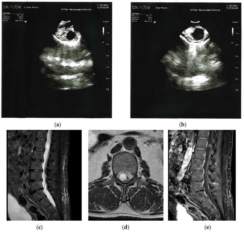 Figure 1
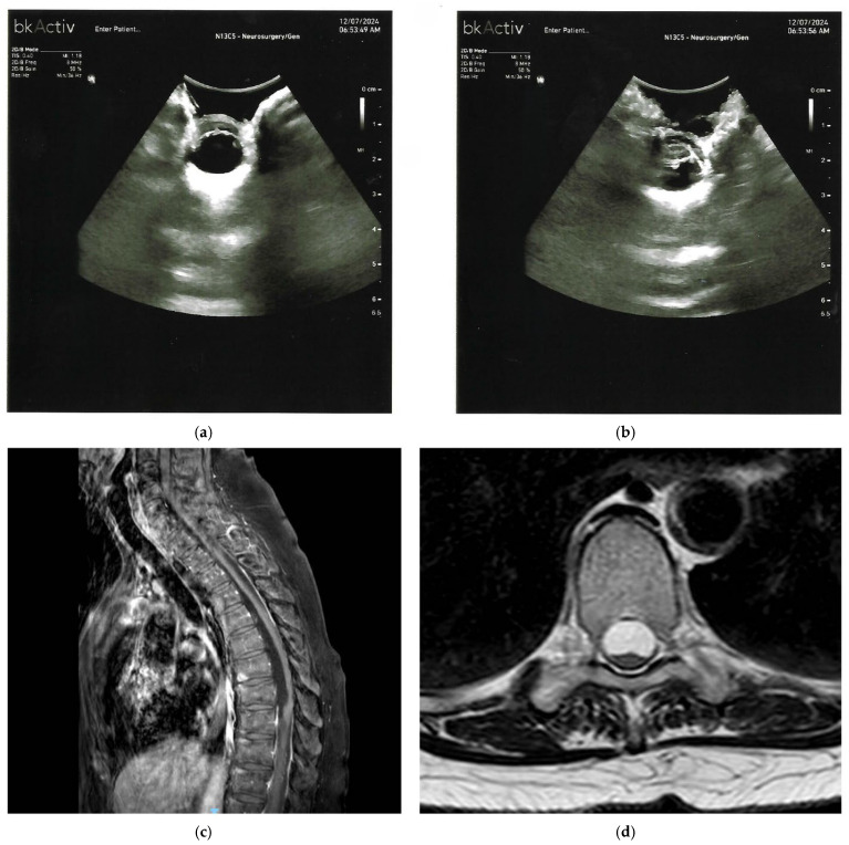 Figure 2
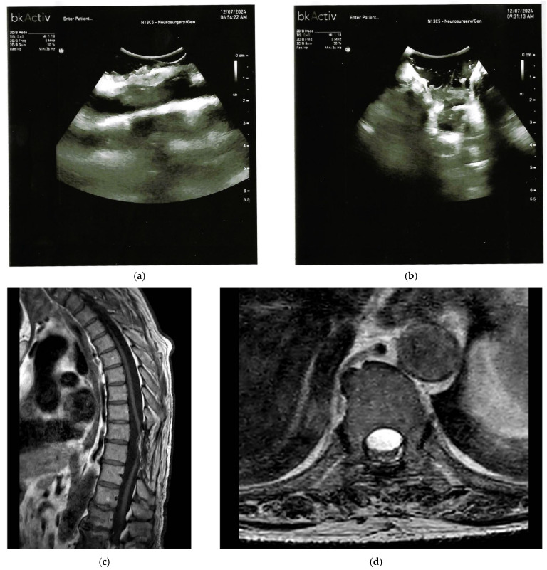 Figure 3
Peer Reviews
No public reviews on file for this paper yet. If you reviewed it on a platform where reviews are public (OpenReview, ICLR, NeurIPS, ICML), you can paste yours below so the community can read it here.
Videos
No videos yet. Explain this paper in a talk, walkthrough, or lecture? Add one.
Taxonomy
TopicsSpinal Dysraphism and Malformations · Glioma Diagnosis and Treatment · Spinal Hematomas and Complications