CT Morphometric Analysis of Ossification Centres in the Fetal Th12 Vertebra
Magdalena Grzonkowska, Michał Kułakowski, Zofia Dzięcioł-Anikiej, Agnieszka Rogalska, Beata Zwierko, Sara Kierońska-Siwak, Karol Elster, Stanisław Orkisz, Mariusz Baumgart

TL;DR
This study uses CT scans to track the growth of fetal Th12 vertebrae ossification centers, providing normative data for estimating fetal age and assessing spinal anomalies.
Contribution
The first comprehensive CT-based normative data for fetal Th12 vertebra ossification centers in the second and early third trimesters.
Findings
All morphometric parameters of the Th12 vertebral body and neural processes showed linear growth with gestational age (R2 = 0.94–0.98).
No significant sex-related or side-related differences were found, allowing for single normative growth curves.
The study provides reference values for prenatal and postnatal assessment of spinal anomalies at the thoracolumbar junction.
Abstract
Objectives: The present study aimed to determine the growth dynamics of the ossification centers of the twelfth thoracic vertebra in the human fetus, focusing on detailed linear, surface, and volumetric parameters of both the vertebral body and neural processes. Methods: The investigation was based on 55 human fetuses (27 males, 28 females) aged 17–30 weeks of gestation. High-resolution low-dose computed tomography, three-dimensional reconstruction, digital image analysis and appropriate statistical modeling were used to obtain detailed morphometric measurements. Results: All measured morphometric parameters of the Th12 vertebral body ossification center—transverse and sagittal diameters, cross-sectional area, and volume—increased linearly with gestational age (R2 = 0.94–0.97). A similar linear growth pattern was demonstrated for the length, width, cross-sectional area, and volume of…
Genes, proteins, chemicals, diseases, species, mutations and cell lines named across the full text — each resolved to its canonical identifier and authoritative record.
Click any figure to enlarge with its caption.
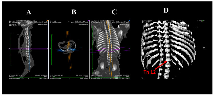 Figure 1
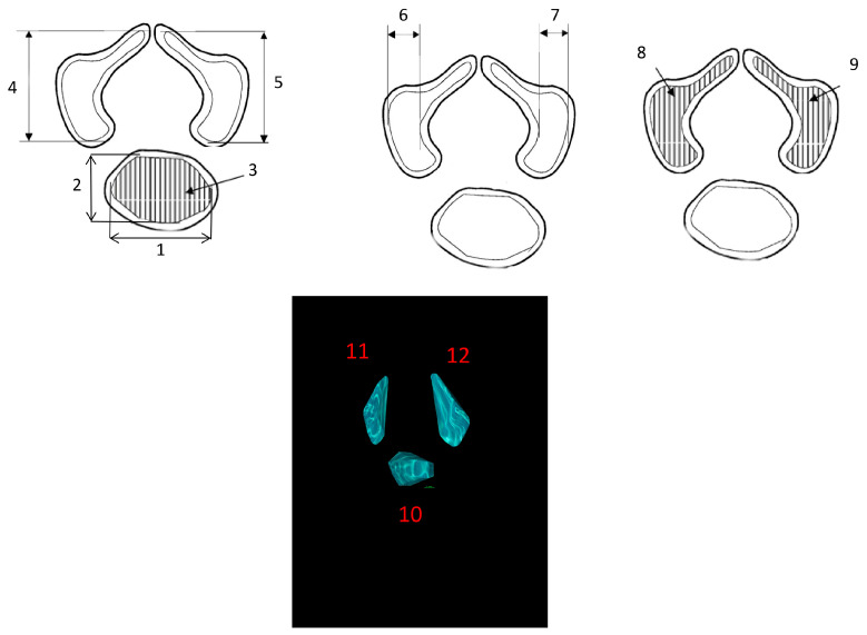 Figure 2
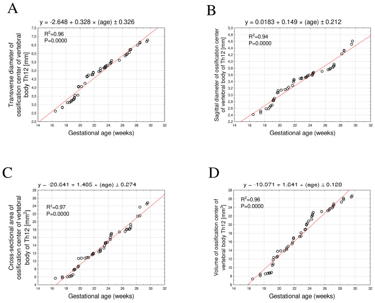 Figure 3
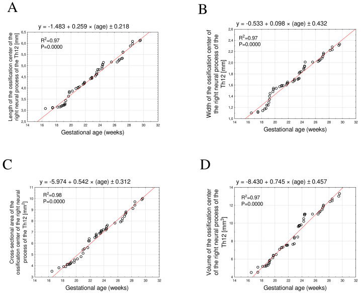 Figure 4
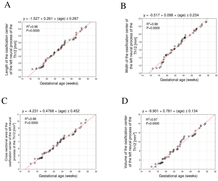 Figure 5
Peer Reviews
No public reviews on file for this paper yet. If you reviewed it on a platform where reviews are public (OpenReview, ICLR, NeurIPS, ICML), you can paste yours below so the community can read it here.
Videos
No videos yet. Explain this paper in a talk, walkthrough, or lecture? Add one.
Taxonomy
TopicsScoliosis diagnosis and treatment · Cervical and Thoracic Myelopathy · Spinal Fractures and Fixation Techniques