A Community Benchmark for the Automated Segmentation of Pediatric Neuroblastoma on Multi-Modal MRI: Design and Results of the SPPIN Challenge at MICCAI 2023
Myrthe A. D. Buser, Dominique C. Simons, Matthijs Fitski, Marc H. W. A. Wijnen, Annemieke S. Littooij, Annemiek H. ter Brugge, Iris N. Vos, Markus H. A. Janse, Mathijs de Boer, Rens ter Maat, Junya Sato, Shoji Kido, Satoshi Kondo, Satoshi Kasai, Marek Wodzinski, Henning Müller

TL;DR
This paper introduces a benchmark challenge for automatically segmenting pediatric neuroblastoma tumors in MRI scans to improve surgical planning.
Contribution
The paper presents the first segmentation challenge in extracranial pediatric oncology and evaluates deep learning methods for tumor segmentation.
Findings
Nine teams participated, with a wide variation in performance metrics like Dice score and HD95.
The top team used a pre-trained model and achieved a median Dice score of 0.82.
Pre-operative tumor segmentations showed significantly lower scores, suggesting limitations in current methods.
Abstract
Surgery plays a key role in treating neuroblastoma. To assist surgical planning, anatomical 3D models derived from the segmentation of anatomical structures on MRI scans are often used. Automation using deep learning can make segmentations less time-consuming and more reliable. We organized the Surgical Planning in PedIatric Neuroblastoma (SPPIN) challenge, to stimulate developments and benchmarking of automatic segmentation of neuroblastoma on MRI. SPPIN is the first segmentation challenge in extracranial pediatric oncology. Nine teams provided a valid submission. Evaluation was based on the Dice similarity coefficient (Dice score), the 95th percentile of the Hausdorff distance (HD95), and the volumetric similarity (VS). A combination of these scores determined the ranking of the teams. The spread in the median evaluation scores per team was large (Dice: 0.21–0.82; HD95: 63.31–7.69;…
Genes, proteins, chemicals, diseases, species, mutations and cell lines named across the full text — each resolved to its canonical identifier and authoritative record.
Click any figure to enlarge with its caption.
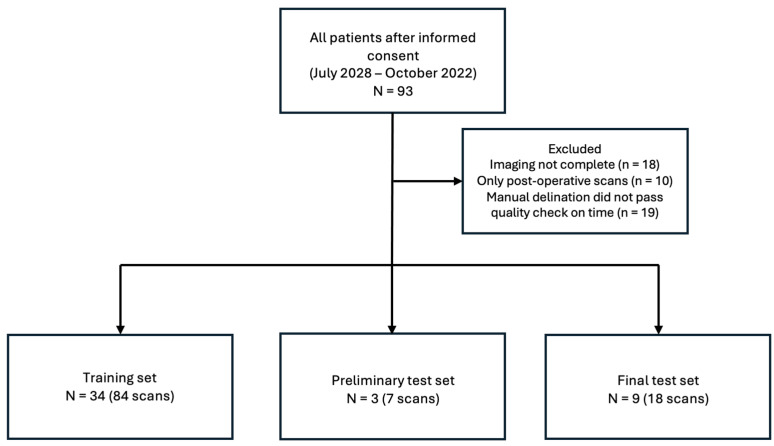 Figure 1
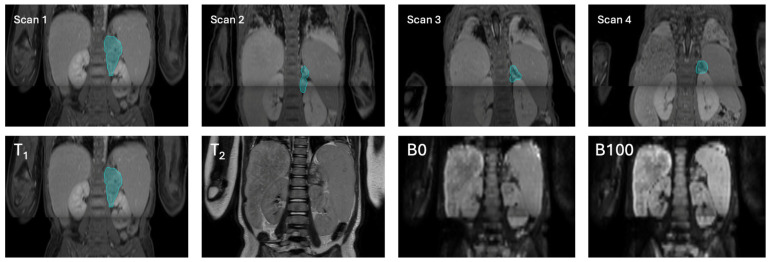 Figure 2
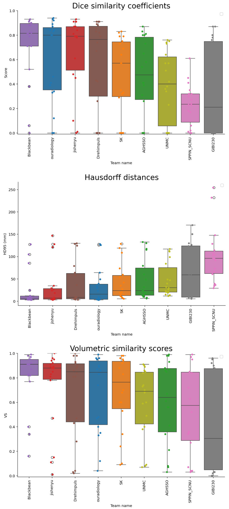 Figure 3
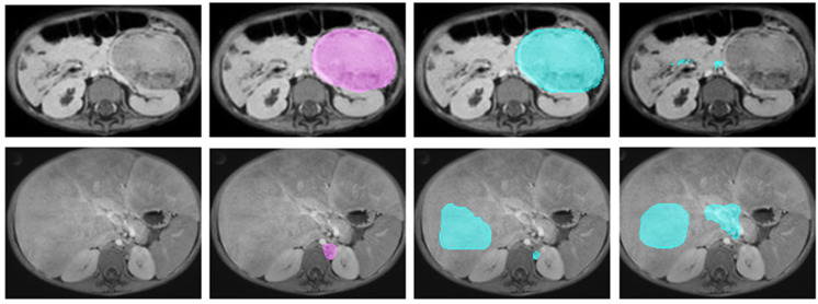 Figure 4
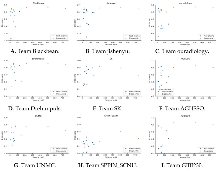 Figure 5
Peer Reviews
No public reviews on file for this paper yet. If you reviewed it on a platform where reviews are public (OpenReview, ICLR, NeurIPS, ICML), you can paste yours below so the community can read it here.
Videos
No videos yet. Explain this paper in a talk, walkthrough, or lecture? Add one.
Taxonomy
TopicsNeuroblastoma Research and Treatments · Glioma Diagnosis and Treatment · Medical Imaging and Analysis