Distinguishing benign from malignant thyroid nodules via virtual biopsy: a study on using quantitative parameters and classical radiomics features from dual-energy CT imaging
Jian He, Changyu Du, Mengting Hu, Jingyi Zhang, Qiye Cheng, Yijun Liu, Jianying Li, Jiageng Shen

TL;DR
This study explores using dual-energy CT imaging and radiomics models to distinguish between benign and malignant thyroid nodules more accurately than radiologists.
Contribution
The study introduces a fusion model combining DECT parameters and radiomics features for improved thyroid nodule classification.
Findings
Normalized iodine concentration (NIC) from DECT is an independent predictor of thyroid nodule malignancy.
The multi-image radiomics model achieved an AUC of 0.966, outperforming radiologists in predicting nodule nature.
The fusion model combining NIC and radiomics features showed strong diagnostic performance in decision curve analysis.
Abstract
To evaluate the predictive value of dual-energy computed tomography (DECT)-based quantitative parameters and radiomics models for the preoperative differentiation between benign and malignant thyroid nodules, and to compare their performance with radiologists’ interpretations. A retrospective analysis was conducted on 215 patients who underwent contrast-enhanced DECT of the thyroid, with pathological outcomes for thyroid nodules obtained. Patients were randomly assigned to training and testing groups in a 7:3 ratio. The images were evaluated by radiologists. Quantitative parameters derived from DECT were identified through univariate and multivariate logistic regression analyses to construct a DECT model. Radiomics features were extracted from the 40, 70, and 100 keV virtual monochromatic images, as well as iodine-based material-decomposition (IMD) images in the arterial phase (AP) and…
Genes, proteins, chemicals, diseases, species, mutations and cell lines named across the full text — each resolved to its canonical identifier and authoritative record.
Click any figure to enlarge with its caption.
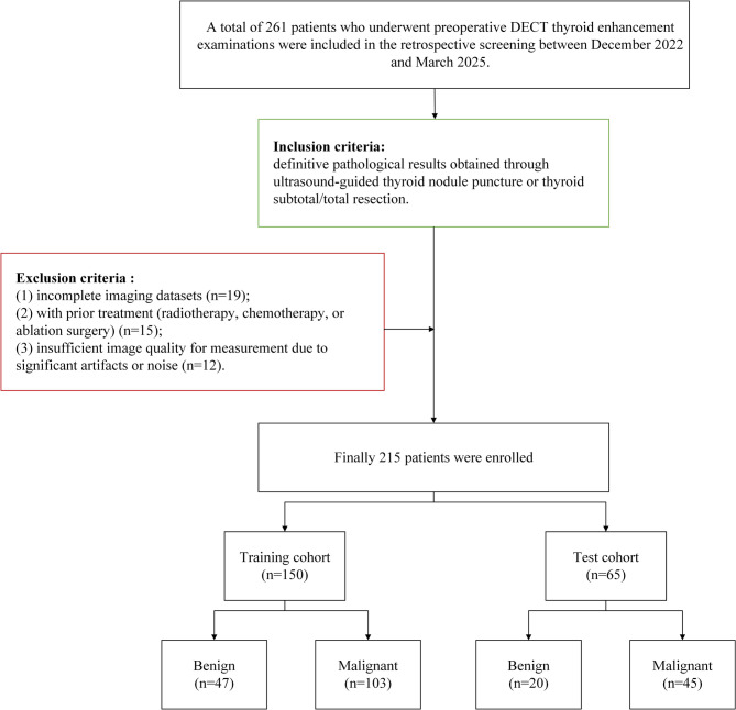 Figure 1
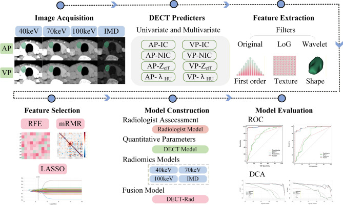 Figure 2
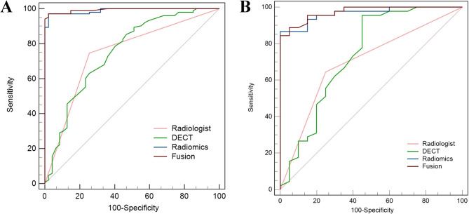 Figure 3
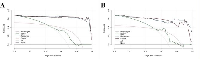 Figure 4
Peer Reviews
No public reviews on file for this paper yet. If you reviewed it on a platform where reviews are public (OpenReview, ICLR, NeurIPS, ICML), you can paste yours below so the community can read it here.
Videos
No videos yet. Explain this paper in a talk, walkthrough, or lecture? Add one.
Taxonomy
TopicsAdvanced X-ray and CT Imaging · Thyroid Cancer Diagnosis and Treatment · Radiomics and Machine Learning in Medical Imaging