High-resolution multimodal imaging reveals spatial and temporal heterogeneity of airway mucus plugging in mice with muco-obstructive lung disease
Claudia V. Benke, Julia Duerr, Annika Engel, Christian Dullin, Wolfram Stiller, Heinz Horstmann, Claudia Redenbach, Maximilian Ackermann, Hans-Ulrich Kauczor, Thomas Kuner, Mark O. Wielpütz, Marcus A. Mall, Willi L. Wagner

TL;DR
This study uses advanced imaging to show how mucus builds up in the lungs of mice with a lung disease, revealing differences across ages and structures.
Contribution
A novel multimodal imaging approach reveals spatiotemporal heterogeneity in mucus plugging across developmental stages in a mouse model of muco-obstructive lung disease.
Findings
Mucus scores in βENaC-tg mice were significantly higher than in wild-type mice at both juvenile and adult stages.
SRCT analysis showed a strong correlation between mucus area and contact ratios, indicating obstruction and adherence.
SEM imaging revealed detailed ultrastructure of mucus plugs in juvenile βENaC-tg mice.
Abstract
Mucus plugging is a hallmark of muco-obstructive lung diseases; however, the spatiotemporal dynamics of plug formation remain poorly understood. We used a multimodal high-resolution imaging approach to study mucus plugging in newborn (0–1 days), juvenile (13–16 days), and adult (49–59 days) βENaC-transgenic mice (βENaC-tg, n = 24), a model of muco-obstructive lung disease, in comparison to age-matched wild-type mice (n = 28). Micro-computed tomography (µCT) enabled whole lung mucus scoring on a scale from 0 (no obstruction) to 2 (> 50% obstruction) using an extended murine airway tree nomenclature. A subset of βENaC-tg lungs (5 juvenile, 5 adult) underwent localised synchrotron radiation-based computed tomography (SRCT) for segmentation-based calculation of mucus area and contact ratios as indicators of luminal obstruction and airway wall adherence. µCT-guided scanning electron…
Genes, proteins, chemicals, diseases, species, mutations and cell lines named across the full text — each resolved to its canonical identifier and authoritative record.
Click any figure to enlarge with its caption.
 Figure 1
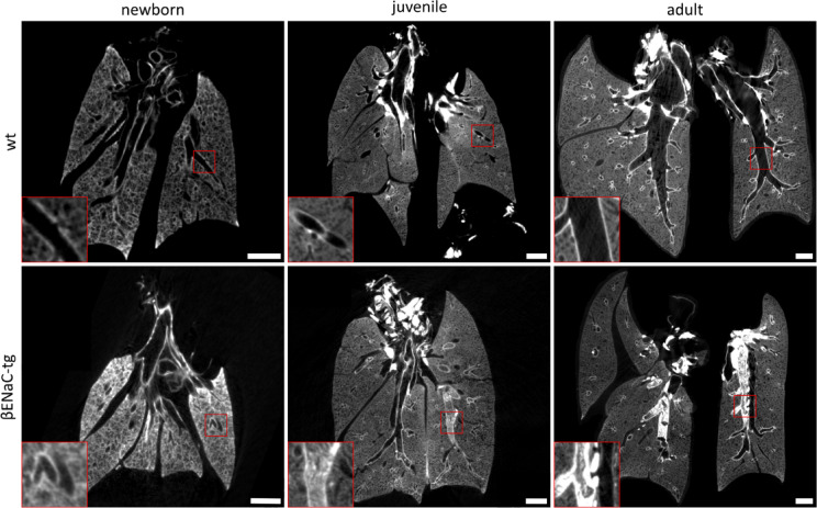 Figure 2
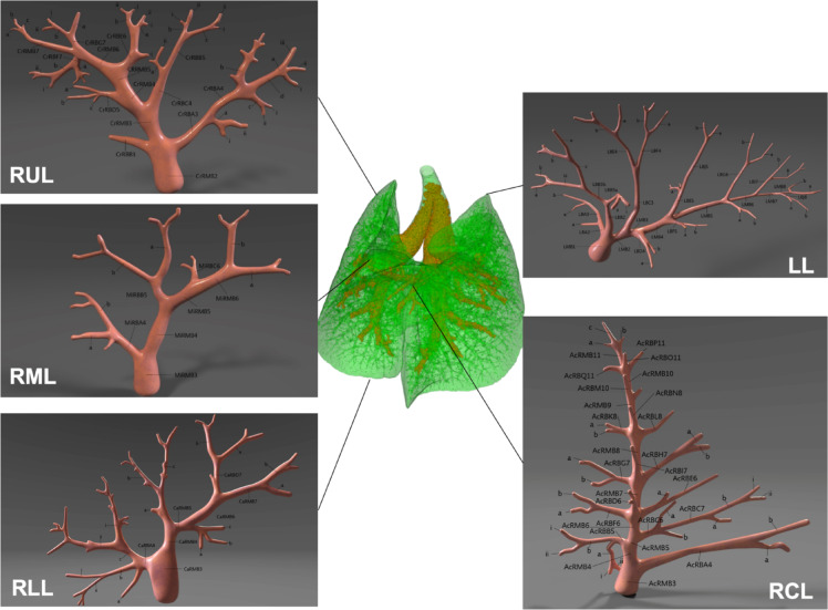 Figure 3
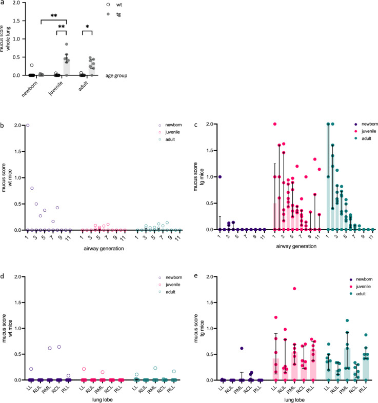 Figure 4
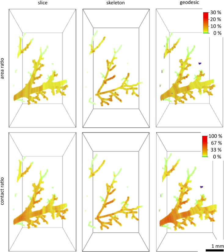 Figure 5
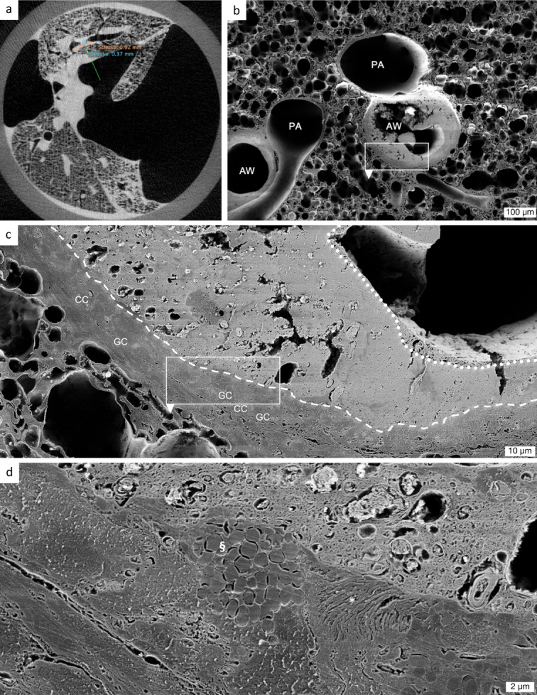 Figure 6
Peer Reviews
No public reviews on file for this paper yet. If you reviewed it on a platform where reviews are public (OpenReview, ICLR, NeurIPS, ICML), you can paste yours below so the community can read it here.
Videos
No videos yet. Explain this paper in a talk, walkthrough, or lecture? Add one.
Taxonomy
TopicsInterstitial Lung Diseases and Idiopathic Pulmonary Fibrosis · Inhalation and Respiratory Drug Delivery · Chronic Obstructive Pulmonary Disease (COPD) Research