Monitoring extracellular vesicle surface glyco-properties using fluorescent lectins and nanoparticle tracking analysis
Ninoslav Mitic, Filip Janjic, Jelena Danilovic-Lukovic, Sanja Goc, Tamara Jankovic, Miroslava Jankovic

TL;DR
This study compares methods to isolate extracellular vesicles and finds that certain techniques change their surface sugar patterns, which could impact their function.
Contribution
A new method using fluorescent lectins and nanoparticle tracking analysis is introduced to monitor extracellular vesicle surface glycosylation during isolation.
Findings
Lectin binding to extracellular vesicles changes with isolation methods like size exclusion chromatography and microfiltration.
Lectin-positive extracellular vesicles larger than 200 nm increase in samples isolated with size exclusion chromatography or microfiltration.
Ultracentrifugation alone preserves original surface glycosylation patterns better than combined methods.
Abstract
Extracellular vesicles are small particles released by all cell types. Different extracellular vesicle isolation methods are widely used, yet none achieve an optimal balance between yield, purity and structural integrity. This study aimed to establish a comparative approach for evaluating different extracellular vesicle preparations using nanoparticle tracking analysis. A simple one-step assay relying on fluorescence-based nanoparticle tracking analysis was used to evaluate lectin binding to extracellular vesicles as a measure of possible changes in their surface glycosylation during various isolation methods. Seminal extracellular vesicles were isolated from normozoospermic men using ultracentrifugation alone—UC-sEVs—or combined with size exclusion chromatography—UC-SEC-sEVs—or microfiltration—UC-MF-sEVs. They were analysed based on their size and lectin-binding properties using wheat…
Genes, proteins, chemicals, diseases, species, mutations and cell lines named across the full text — each resolved to its canonical identifier and authoritative record.
Click any figure to enlarge with its caption.
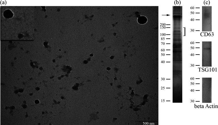 Figure 1
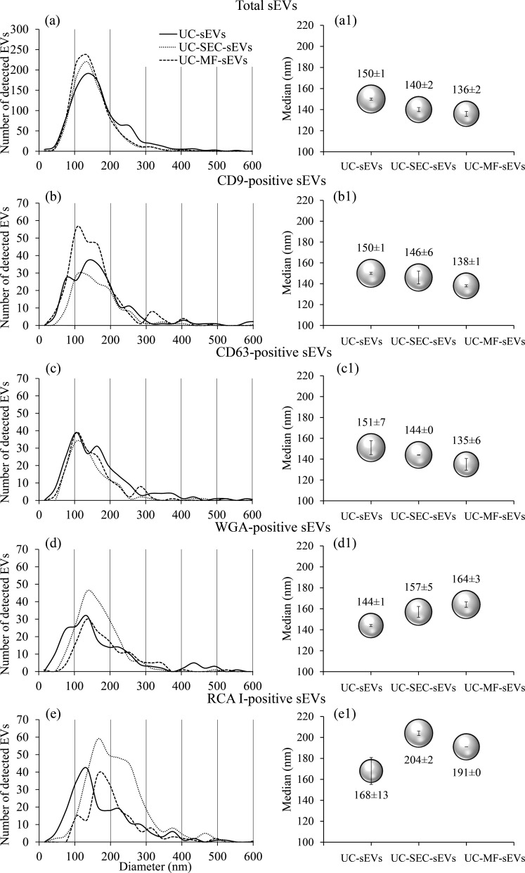 Figure 2
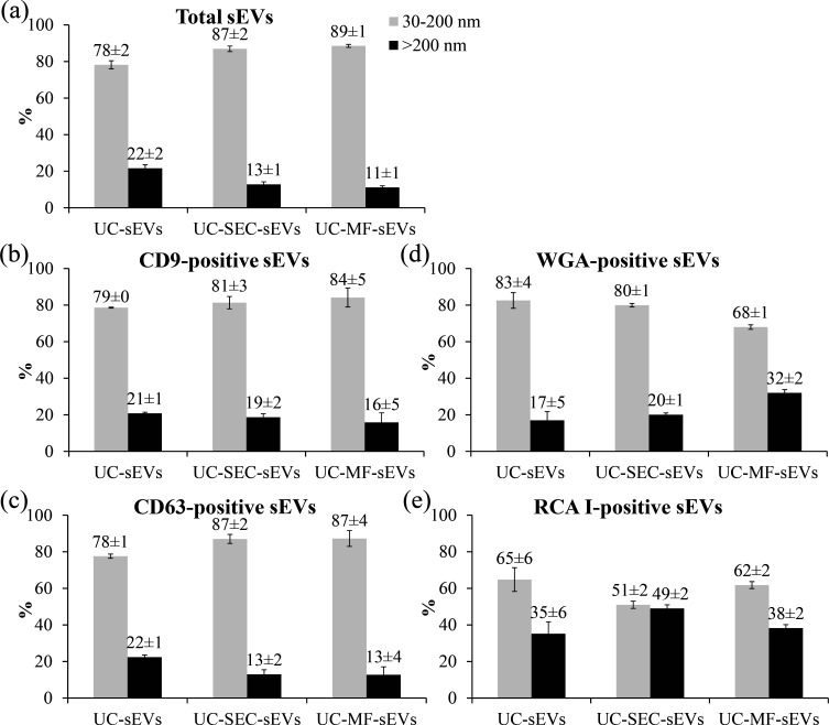 Figure 3
 Figure 4
Peer Reviews
No public reviews on file for this paper yet. If you reviewed it on a platform where reviews are public (OpenReview, ICLR, NeurIPS, ICML), you can paste yours below so the community can read it here.
Videos
No videos yet. Explain this paper in a talk, walkthrough, or lecture? Add one.
Taxonomy
TopicsExtracellular vesicles in disease · Bacterial Infections and Vaccines · interferon and immune responses