Pampiniform Venous Plexus Thrombosis Presenting As Testicular Pain: Report of a Rare Case
Pronami Borah, Dharavath Venkatesh, Debarati Majumder, Saanjhi Rawat

TL;DR
A rare case of pampiniform venous plexus thrombosis causing testicular pain is reported, highlighting the importance of Doppler ultrasound for diagnosis and avoiding unnecessary surgery.
Contribution
This paper presents a rare clinical case and emphasizes the diagnostic value of Doppler ultrasound in identifying pampiniform venous plexus thrombosis.
Findings
Spontaneous pampiniform venous plexus thrombosis can present as acute testicular pain.
Scrotal Doppler ultrasound effectively diagnosed the condition with absent venous flow.
Conservative treatment resolved symptoms and confirmed recovery on follow-up imaging.
Abstract
Spontaneous thrombosis of the pampiniform venous plexus is a rare vascular disorder that can manifest as acute testicular pain, often mimicking conditions such as epididymitis, orchitis, or testicular torsion. Due to its uncommon presentation, clinical diagnosis can be challenging, and a high level of suspicion is required. Scrotal Doppler ultrasonography serves as an effective, non-invasive diagnostic tool by demonstrating absent venous flow within the pampiniform plexus while confirming normal testicular perfusion. We present a case of spontaneous pampiniform venous plexus thrombosis diagnosed using Doppler ultrasound. The patient was successfully managed with conservative measures, including anticoagulation, analgesics, and brief bed rest, resulting in complete resolution of symptoms and normalization on follow-up imaging. This case underscores the importance of including pampiniform…
Genes, proteins, chemicals, diseases, species, mutations and cell lines named across the full text — each resolved to its canonical identifier and authoritative record.
Click any figure to enlarge with its caption.
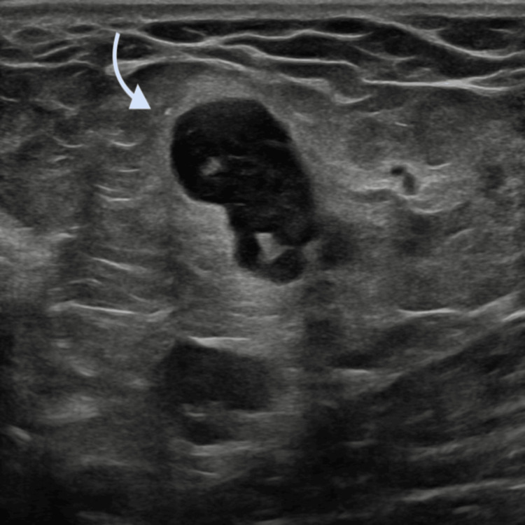 Figure 1
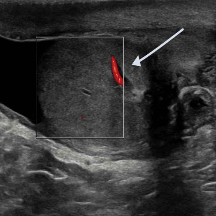 Figure 2
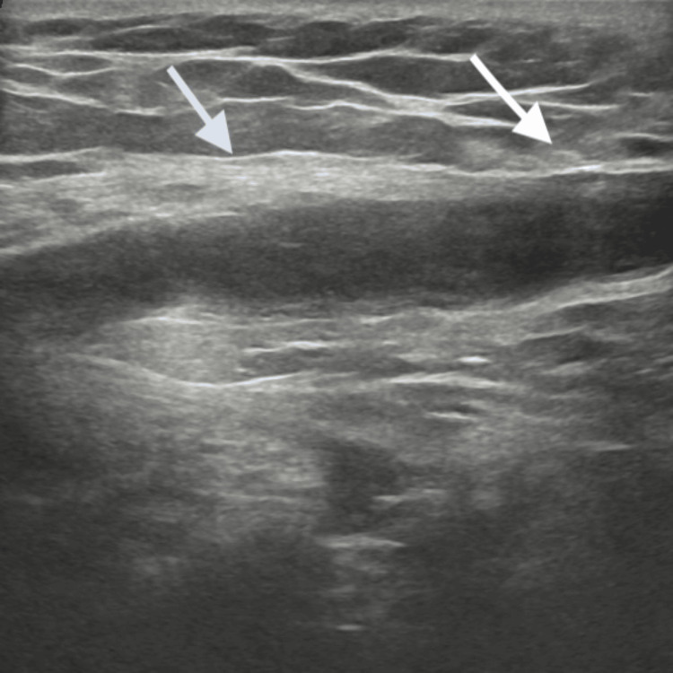 Figure 3
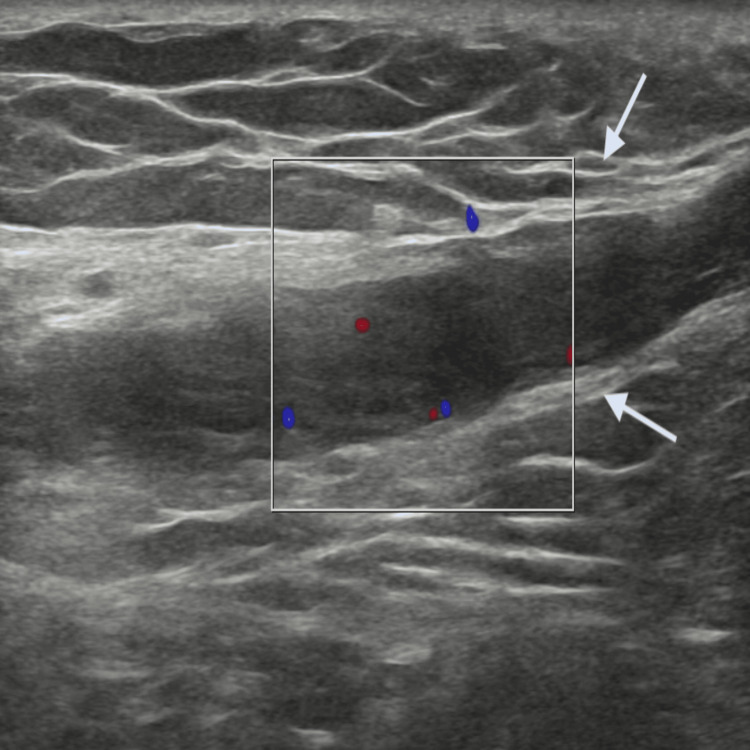 Figure 4
| Event | Timing | Details |
| Symptom onset | Day 0 | Sudden right testicular pain with mild swelling; no trauma or systemic symptoms |
| Initial evaluation & labs | Day 7 (on admission) | Comprehensive coagulation profile: PT, aPTT, INR—all normal; CBC and metabolic panel normal; no history of coagulopathies or systemic risk factors |
| Imaging (Doppler USG) | Day 7 | Dilated pampiniform vein, echogenic thrombus; absent venous flow; preserved arterial perfusion |
| Treatment initiated | Day 7 | Bed rest, analgesics, NSAIDs, anticoagulation |
| Resolution (follow-up) | 2 months after onset | Complete clinical and sonographic resolution of thrombosis |
Peer Reviews
No public reviews on file for this paper yet. If you reviewed it on a platform where reviews are public (OpenReview, ICLR, NeurIPS, ICML), you can paste yours below so the community can read it here.
Videos
No videos yet. Explain this paper in a talk, walkthrough, or lecture? Add one.
Taxonomy
TopicsVascular anomalies and interventions · Central Venous Catheters and Hemodialysis · Vascular Procedures and Complications
Introduction
Spontaneous thrombosis of the pampiniform venous plexus is extremely rare; fewer than 30 cases have been reported worldwide, making it a clinical curiosity that can easily be overlooked. Although it may occur in a wide age spectrum, its rarity and non-specific presentation often result in diagnostic uncertainty [1]. Patients typically present with acute scrotal pain, a clinical feature that overlaps with more common and urgent conditions such as testicular torsion, epididymo-orchitis, or incarcerated inguinal hernia [2,3]. Failure to recognize this entity may lead to unnecessary surgical exploration, which underscores the importance of timely and accurate diagnosis.
Scrotal Doppler ultrasonography remains the cornerstone of diagnosis. It enables real-time evaluation of pampiniform veins, revealing intraluminal echogenic thrombus, venous dilatation, absent or diminished venous flow, and preserved testicular arterial perfusion [4,5]. In certain cases, additional imaging modalities such as computed tomography (CT) or CT angiography may provide further anatomical details and help rule out other retroperitoneal or vascular pathologies. Nevertheless, Doppler ultrasound is favored in clinical practice due to its non-invasive nature, accessibility, and diagnostic reliability.
Management of pampiniform plexus thrombosis is generally conservative, consisting of analgesia, non-steroidal anti-inflammatory drugs, anticoagulation, and rest. Most patients show complete recovery without surgical intervention [6]. Given the diagnostic challenges and rarity of this condition, raising clinical awareness is essential, particularly in outpatient clinics and emergency departments. The present case highlights the diagnostic process, role of imaging, and successful conservative treatment of pampiniform plexus thrombosis, contributing to the existing body of literature and reinforcing its clinical relevance.
Case presentation
A 43-year-old male with no prior significant medical history presented to the emergency department with a seven-day history of right testicular pain. The pain had a sudden onset and progressively increased in severity, and was associated with mild swelling and discomfort in the right inguinal region. The patient denied fever, lower urinary tract symptoms, urethral discharge, trauma, abdominal pain, vomiting, constipation, or chronic cough. He had no history of hematuria, flank pain, or renal swelling.
A comprehensive coagulation study (prothrombin time (PT), activated partial thromboplastin time (aPTT), and international normalized ratio (INR)) was performed, with all results found to be within normal limits.
On physical examination, mild tenderness and induration were observed in the right inguinal region, which was consistent with enlarged inguinal lymph nodes (Figure 1). The right testis was tender but otherwise normal in size, shape, and consistency. Abdominal examination was unremarkable, with no palpable masses or flank tenderness.
Enlarged inguinal lymph nodes
Scrotal Doppler ultrasonography demonstrated a dilated vein within the right spermatic cord containing echogenic intraluminal material, findings consistent with thrombosis of the pampiniform venous plexus; testicular perfusion was preserved (Figure 2).
Color Doppler image demonstrating preserved testicular perfusion.
Ultrasound confirmed an enlarged pampiniform venous channel containing echogenic material consistent with thrombus (Figure 3).
Thrombosed vein within the pampiniform plexus in the spermatic cord.
Color Doppler of the pampiniform venous plexus demonstrated absence of vascular flow within the thrombosed vein on the right side (Figure 4).
Color Doppler ultrasound of the right pampiniform venous plexus demonstrating absence of vascular flow within the thrombosed vein
The patient was admitted and managed conservatively with bed rest, anti-inflammatory medications, analgesics, and a therapeutic course of anticoagulation. Symptomatic improvement was observed over 7-10 days. He was discharged in stable condition. Follow-up Doppler ultrasonography performed two months later showed complete resolution of the thrombus.
Table 1 presents the clinical timeline of the case from symptom onset to resolution.
Discussion
Pampiniform venous plexus thrombosis represents a rare cause of acute scrotal pain and is frequently underrecognized due to its clinical similarity with other scrotal pathologies [2,3]. Misdiagnoses have been reported, with patients initially suspected of having testicular torsion or incarcerated inguinal hernia, sometimes even undergoing surgical exploration before the correct diagnosis was established [4]. This reinforces the need for heightened clinical suspicion when evaluating patients with acute scrotal pain.
The underlying etiology of spontaneous thrombosis remains uncertain. Proposed mechanisms include venous stasis, hypercoagulable states, dehydration, local trauma, strenuous physical exertion, or increased venous pressure following vigorous sexual activity [5,6]. Associations with systemic diseases such as Buerger’s disease and other vasculopathies have also been postulated, though definitive causality remains unclear [7]. While most cases occur on the left side, likely due to anatomical factors such as the longer left testicular vein draining into the left renal vein at a right angle, right-sided cases-such as the one reported here-demonstrate that laterality should not exclude consideration of this diagnosis [8].
Doppler ultrasonography is both diagnostic and reassuring in this setting. The hallmark features include non-compressible, dilated pampiniform veins containing echogenic material consistent with thrombus and absence of venous flow, while intratesticular perfusion is preserved [4,5]. This distinction from testicular torsion, where arterial inflow is compromised, is crucial. In rare circumstances where ultrasound findings are equivocal, CT or MR venography may be warranted to confirm the diagnosis or exclude retroperitoneal and renal venous pathologies [1,9].
Treatment strategies are largely conservative. Most reports describe successful outcomes with a combination of analgesics, non-steroidal anti-inflammatory drugs, anticoagulation, and rest, leading to symptom resolution and radiological improvement [6,10]. Our patient demonstrated significant improvement within days and achieved complete resolution on follow-up imaging, consistent with prior observations. Surgical intervention is rarely required, though it may be considered in cases of diagnostic uncertainty, severe refractory pain, or complications [4]. Importantly, correct preoperative identification of this entity prevents unnecessary orchiectomy or varicocelectomy.
The clinical implications of this case extend to both emergency and outpatient settings, where physicians frequently encounter acute scrotal pain. Early consideration of pampiniform plexus thrombosis in the differential diagnosis may reduce the risk of mismanagement. Furthermore, the absence of standardized diagnostic and treatment guidelines reflects the need for larger case series and multicenter analyses. Future research should aim to define risk factors, establish diagnostic algorithms, and evaluate long-term outcomes to better inform clinical practice.
Conclusions
Spontaneous thrombosis of the pampiniform venous plexus is an exceedingly rare but clinically significant cause of acute scrotal pain. Its presentation closely mimics other urgent conditions such as testicular torsion and epididymo-orchitis, which often leads to misdiagnosis. Scrotal Doppler ultrasonography remains the diagnostic modality of choice, allowing differentiation from surgical emergencies by confirming venous thrombosis while preserving testicular perfusion. Conservative management with analgesics, anti-inflammatory agents, and anticoagulation is usually sufficient and highly effective. Increased awareness of this entity among clinicians, particularly in emergency and outpatient settings, can prevent unnecessary surgical exploration and optimize patient outcomes.
The reference list from the paper itself. Each links out to its DOI / PubMed record.
- 1Spontaneous pampiniform venous plexus thrombosis may cause chronic scrotal pain Investig Clin Urol Hernandez H Pineault K Sadeghi-Nejad H 420422652024 https://doi.org/10.4111/icu.2024009310.4111/icu.20240093 PMC 1123165938978222 · doi ↗ · pubmed ↗
- 2Thrombosed varicocele - a rare cause for acute scrotal pain: a case report BMC Urol Raghavendran M Venugopal A Kiran Kumar G 341820182973937710.1186/s 12894-018-0347-2PMC 5941692 · doi ↗ · pubmed ↗
- 3Testicular vein thrombosis mimicking epididymo-orchitis after suspected Covid-19 infection SAGE Open Med Case Rep Whiteley MS Abu-Bakr O Holdstock JM 20503139202110.1177/2050313 X 211022425 PMC 818216934158948 · doi ↗ · pubmed ↗
- 4Bilateral spontaneous thrombosis of the pampiniform plexus; a rare etiology of acute scrotal pain: a case report and review of literature Afr J Urol Kamel K Gassen S Mohamed M Bader BK 1418242018
- 5Right-sided varicocele thrombosis complicating an acute epididymo-orchitis: a case report and review of the literature Urol Case Rep Hamdouni W Bchir S Abidi M Boukhrissa N Ktari K Saad H 1017103920213422189810.1016/j.eucr.2021.101710 PMC 8243376 · doi ↗ · pubmed ↗
- 6Thrombosis of the pampiniform plexus: about a case report Urol Case Rep Ouanes Y Sellami A Chaker K Mokhtar B Ben Rhouma S Nouira Y 28292020182998878610.1016/j.eucr.2018.06.003PMC 6031866 · doi ↗ · pubmed ↗
- 7Spontaneous thrombosis of the pampiniform plexus Scand J Urol Nephrol Hashimoto L Vibeto B 252254402006 https://doi.org/10.1080/003655906005898231680927010.1080/00365590600589823 · doi ↗ · pubmed ↗
- 8Thrombosis of the spermatic vein in children J Urol Coolsaet B Weinberg R 290291219801202510.1016/s 0022-5347(17)55412-97401251 · doi ↗ · pubmed ↗