First application of carbon dioxide digital subtraction enterography for stricture evaluation in Crohn’s disease
Akihiro Maruyama, Kohei Takano, Junya Yamada, Hiroki Kato, Sakurako Isobe, Makoto Kobayashi

Abstract
Genes, proteins, chemicals, diseases, species, mutations and cell lines named across the full text — each resolved to its canonical identifier and authoritative record.
Click any figure to enlarge with its caption.
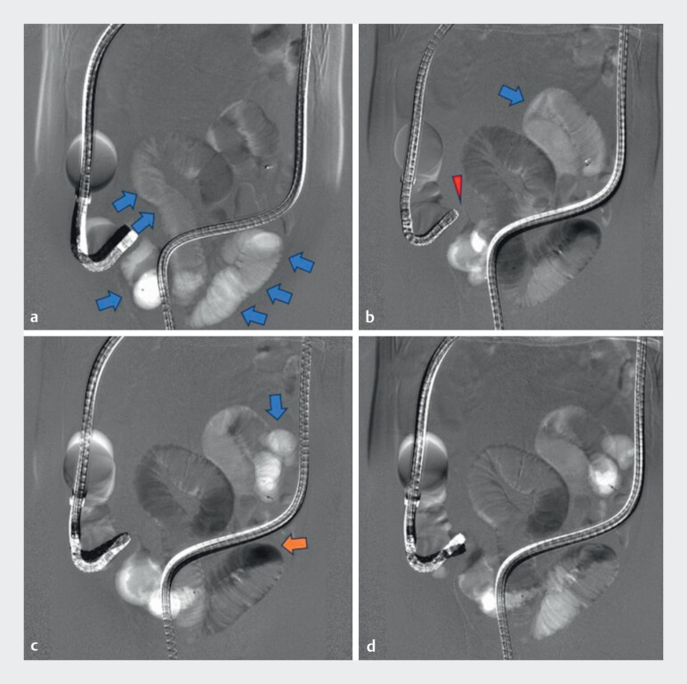 Fig. 1
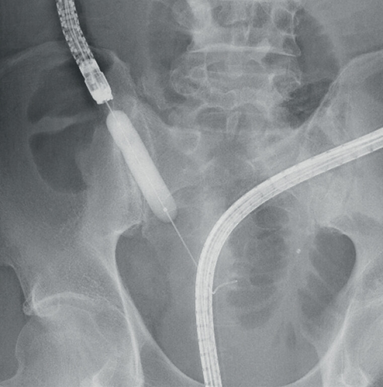 Fig. 2
Peer Reviews
No public reviews on file for this paper yet. If you reviewed it on a platform where reviews are public (OpenReview, ICLR, NeurIPS, ICML), you can paste yours below so the community can read it here.
Videos
No videos yet. Explain this paper in a talk, walkthrough, or lecture? Add one.
Taxonomy
TopicsInflammatory Bowel Disease · Gastrointestinal Bleeding Diagnosis and Treatment · Esophageal Cancer Research and Treatment
Digital subtraction imaging (DSI) is a radiological technique used to enhance the visualization of anatomical structures. It subtracts a pre-contrast image from a post-contrast image, thereby effectively eliminating background noise. This method is commonly used in angiography and interventional radiology 1 , and recent reports have also suggested its utility in gastrointestinal imaging 2 3 4 . We applied CO 2 digital subtraction enterography (CDDSE) as a novel approach during double-balloon endoscopy.
A 57-year-old man with a 10-year history of Crohn’s disease, notable for poor treatment adherence and irregular follow-up, presented with a 1-month history of recurrent nausea and progressive abdominal distension. Abdominal computed tomography demonstrated wall thickening at the terminal ileum. A transanal double-balloon endoscopy was performed using an EN-580T (FUJIFILM, Tokyo, Japan) equipped with a Cast Hood (TOP, Tokyo, Japan) at its tip. Conscious sedation was achieved with intravenous midazolam, and an antispasmodic agent, hyoscine butylbromide, was administered. DSI was conducted with the Ultimax-i DREX-U180 fluoroscopy system (Canon, Tokyo, Japan). CDDSE was then performed, which enabled evaluation of the stricture and the bowel up to the previously placed clip marking the maximal oral reach ( Video 1 and Fig. 1 ).
Panels ( a–d ) show serial CDDSE digital subtraction images. Compared with the pre-image, areas with greater CO 2 accumulation appear white (blue arrows), whereas those with less CO 2 appear black (orange arrows), reflecting CO 2 flow and enabling assessment of the intestine. The stricture is visualized as indicated by the red arrowhead.
Carbon dioxide digital subtraction enterography (CDDSE) performed during double-balloon endoscopy provided clear delineation of an ileal stricture in Crohn’s disease, demonstrating feasibility and procedural simplicity.Video 1
No adverse events occurred during the procedure. The total procedure time from endoscope insertion to balloon dilation was 31 minutes, and the cumulative duration of the two CDDSE acquisitions was 30 seconds. The total radiation dose was 27.3 mGy, of which 4.9 mGy was attributable to the two CDDSEs. Balloon dilation was subsequently performed ( Fig. 2 ), leading to prompt improvement of the patient’s clinical symptoms.
Balloon dilation was performed using a 15-mm balloon dilator, maintained for 1 minute.
As the technique requires only pressing the endoscopic insufflation button in synchrony with DSI acquisition, it enables clear delineation of intestinal strictures in a short time and with great procedural simplicity. Because CO 2 flows more readily than liquid contrast agents, CDDSE may enable visualization of longer intestinal segments within shorter acquisition times.
Endoscopy_UCTN_Code_TTT_1AP_2AD
The reference list from the paper itself. Each links out to its DOI / PubMed record.
- 1Modic MT Weinstein MA Chilcote WA Digital subtraction angiography of the intracranial vascular system: Comparative study in 55 patients AJR Am J Neuroradiol 1981252753410.2214/ajr.138.2.2997034508 · doi ↗ · pubmed ↗
- 2Maruyama A Kobayashi M Takeshima H Carbon dioxide digital subtraction enterography for route identification in post-Roux-en-Y biliary interventions Endoscopy 202557 E 153E 15410.1055/a-2523-288639933747 PMC 11813658 · doi ↗ · pubmed ↗
- 3Maruyama A Kobayashi M Takeshima H Digital subtraction imaging with carbon dioxide and liquid contrast in the biliary and pancreatic ducts Endoscopy 202557 E 388E 38940345251 10.1055/a-2589-1094 PMC 12064330 · doi ↗ · pubmed ↗
- 4Maruyama A Takano K Yamada J Digital subtraction pancreatography enhances ductal visualization in high grade pancreatic intraepithelial neoplasia Endoscopy 202557 E 1184 E 118510.1055/a-2718-484641161334 PMC 12571546 · doi ↗ · pubmed ↗