Macropinocytosis mediates neurotropism of Cryptococcus neoformans in a human organoid model of the blood-brain barrier
Amelia B. Bennett, Dylan M. Lanser, Kiem Vu, Amita R. Sahoo, Matthias Buck, Angie Gelli

TL;DR
This study shows how the fungus Cryptococcus neoformans enters brain cells using a process called macropinocytosis, which could help develop better treatments for brain infections.
Contribution
The paper identifies CD44 and EphA2 as key proteins involved in the fungal entry mechanism into brain endothelial cells via macropinocytosis.
Findings
Cryptococcus neoformans uses macropinocytosis to enter brain endothelial cells.
CD44 and EphA2 form a molecular complex that facilitates fungal entry.
Two predicted binding sites on EphA2 suggest cooperative signaling for macropinocytosis.
Abstract
The opportunistic and neuroinvasive fungus, Cryptococcus neoformans (Cn), causes a life-threatening brain infection that despite treatment can cause long-term cognitive deficiencies. Studies have shown that Cn can infiltrate the central nervous system (CNS) through a transcellular route across the brain endothelium, however the molecular process that drives brain endothelial cells to internalize Cn remains poorly defined. Here we examine the molecular interactions between fungal cells and the brain endothelium by utilizing a human 3D organoid model of the blood-brain barrier (BBB). We show that Cn exploits the process of macropinocytosis as the mechanism of endocytosis into brain endothelial cells by recruiting CD44 and EphA2 as a molecular complex. We identified two predicted binding sites on EphA2, suggesting that the two structurally distinct regions may provide a molecular basis for…
Genes, proteins, chemicals, diseases, species, mutations and cell lines named across the full text — each resolved to its canonical identifier and authoritative record.
Click any figure to enlarge with its caption.
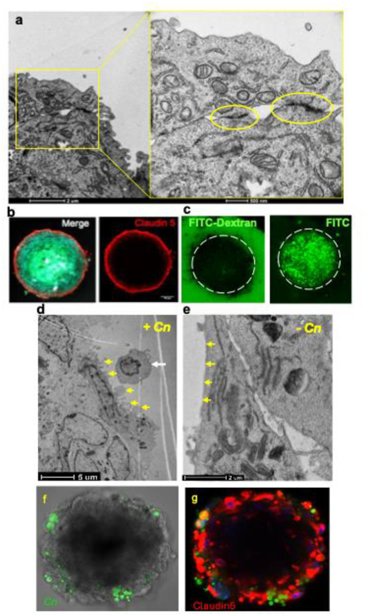 Figure 1
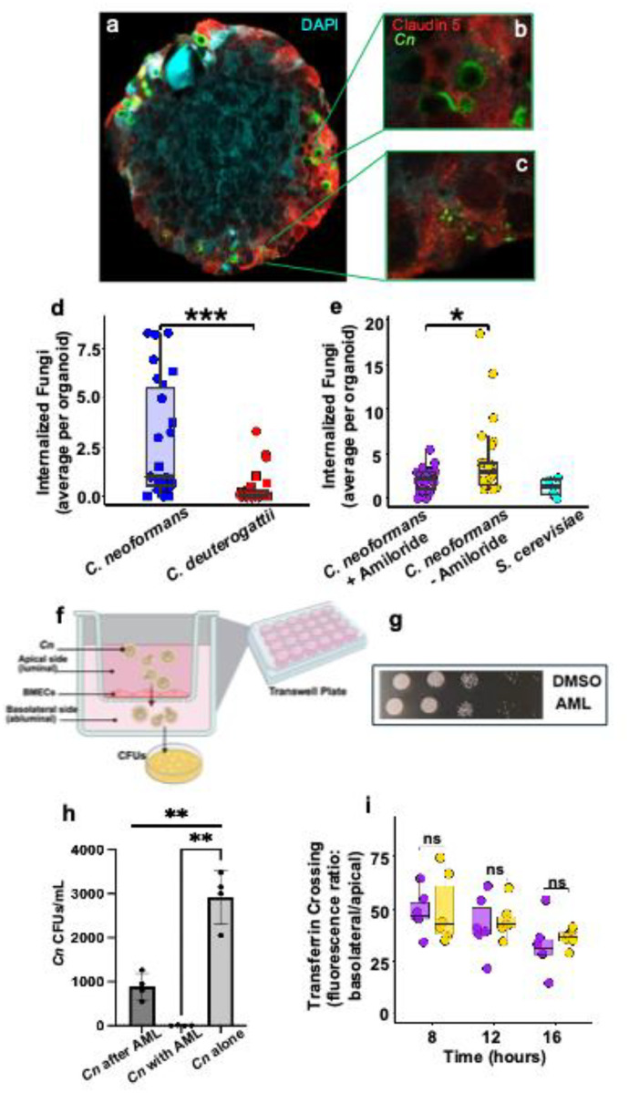 Figure 2
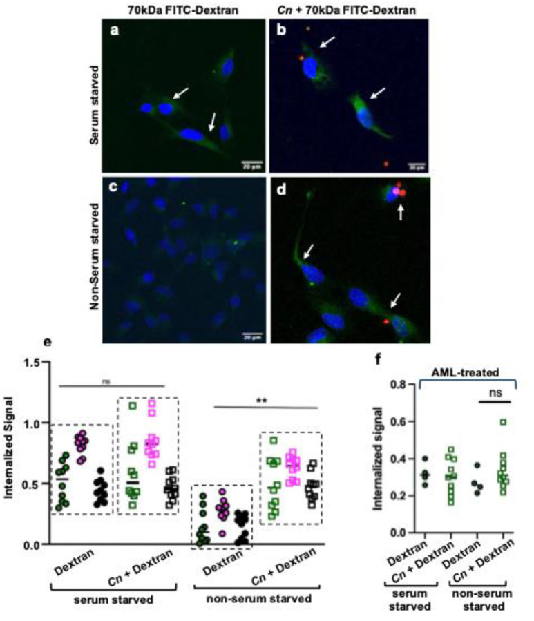 Figure 3
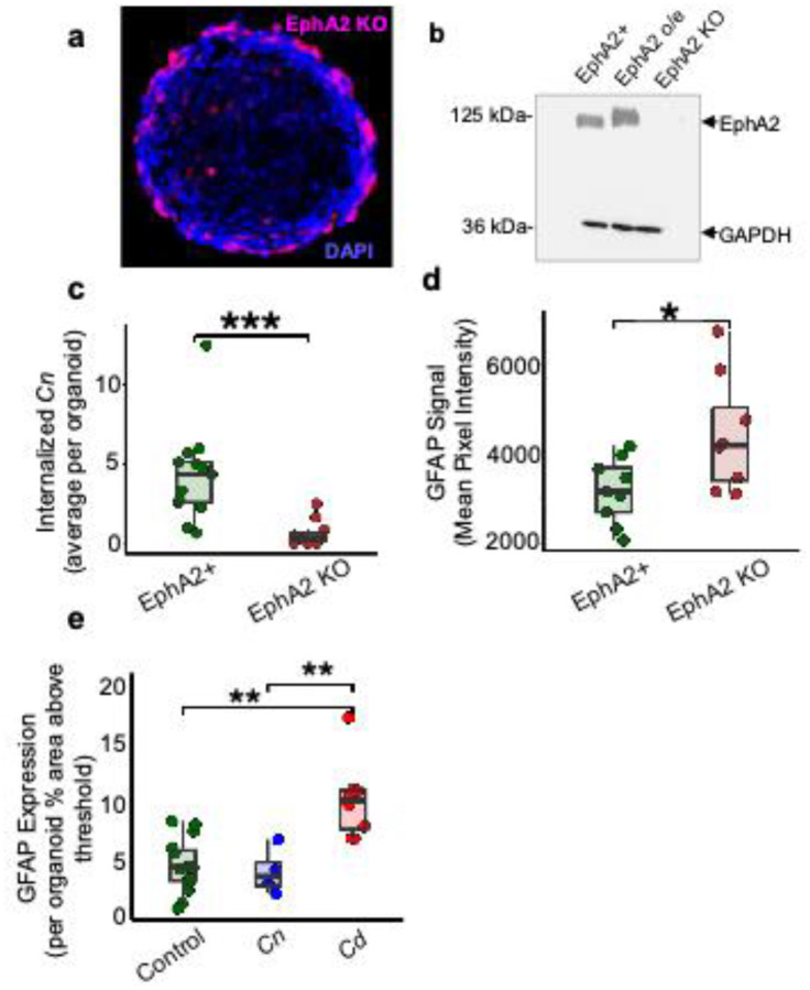 Figure 4
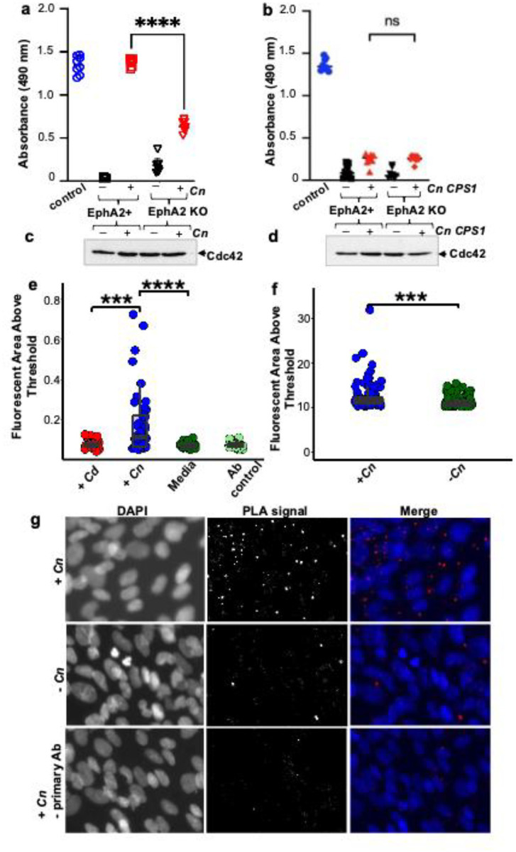 Figure 5
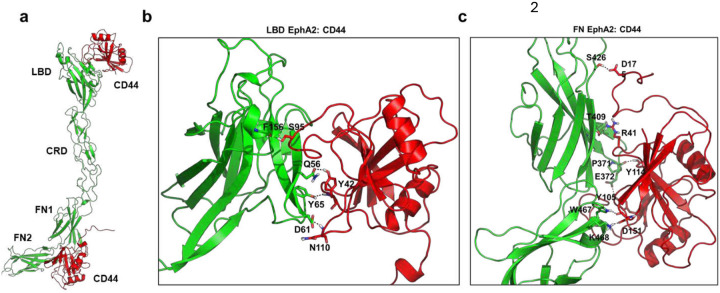 Figure 6
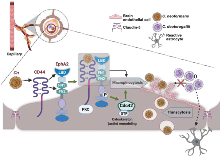 Figure 7
Peer Reviews
No public reviews on file for this paper yet. If you reviewed it on a platform where reviews are public (OpenReview, ICLR, NeurIPS, ICML), you can paste yours below so the community can read it here.
Videos
No videos yet. Explain this paper in a talk, walkthrough, or lecture? Add one.
Taxonomy
TopicsFungal Infections and Studies · Calcium signaling and nucleotide metabolism · Whipple's Disease and Interleukins
Introduction
In a healthy brain, components of circulating blood that could damage the central nervous system (CNS) are excluded by the blood-brain barrier (BBB). The tightly connected endothelial cells, the low rate of vesicular transport and the highly-selective permeability of the BBB collectively ensure a protective environment for the CNS.^1^ Despite this, Cryptococcus neoformans (Cn), a neurotropic fungal pathogen and leading cause of meningoencephalitis in immunosuppressed adults^2^, has developed adaptations that promote its entry^3^ into- and proliferation within the CNS.^4,5^ Several lines of evidence support multiple routes of CNS invasion, including: (i) “Trojan Horse,” in which Cn infiltrates the BBB via infected phagocytes^6–12^, (ii) paracellular crossing of free Cn via the disruption of the BBB endothelial cell junction proteins^12–15^, and (iii) direct fungal interaction with brain endothelial cells leading to endocytosis and transcytosis of free Cn.^16–22^ The molecular mechanistic details of these routes, the propensity of Cn using one route versus another and whether all three routes are used during infection remains largely unresolved.
Utilization of endothelial cells and exploitation of endocytosis to cross the BBB via a transcellular path is central to Cn’s ability to infiltrate and survive within the CNS. Several in vitro and in vivo studies have demonstrated transcytosis as the primary mechanism driving Cn across the BBB.^3,16,23,24^ Evidence includes observational studies that found cryptococci moving freely through the bloodstream and crossing the brain endothelium independently of phagocytes^14^, and quantitative analyses that established a direct association between brain endothelial cells and Cn in vivo.^16,23^ Collectively, these data support recruitment of a transcellular pathway for BBB crossing, however the molecular process by which brain endothelial cells internalize Cn remains poorly understood.
One form of endocytosis is macropinocytosis, an actin-driven endocytic process that internalizes extracellular cargo via large vesicles referred to as macropinosomes.^25–27^ Based on previously observed indicators of macropinocytosis such as actin-supported membrane ruffles in brain endothelial cells infected with Cn^16,21^, actin disruptors preventing BBB crossing^13,21,28^ and the lack of evidence for other processes, we questioned whether Cn might exploit macropinocytosis as the mechanism of entry to the CNS.^29^
To investigate this, we utilized a human organoid model of the BBB. Multicellular BBB organoids have been used as a reliable and predictive platform that recapitulate features of the native BBB.^30^ The present study examines the molecular interaction between Cn and the brain endothelium. We previously found that Cn induced the non-canonical, ligand-free activation of the ephrin tyrosine kinase receptor, EphA2, in human brain endothelial cells.^21^ While we demonstrated that transcytosis of Cn across the BBB relied on the phosphorylation-dependent activity of EphA2^21^, the mechanistic details of the endocytic process remained unresolved. It was also unclear whether EphA2 played a role in recruiting CD44, the glycoprotein receptor for hyaluronic acid, and a known contributor to the transcellular pathway co-opted by Cn to cross the brain endothelium.^31–33^
Results
Cellular organization and validation of a human blood-brain barrier (BBB) organoid model of neural cryptococcosis.
To examine whether macropinocytosis is the mechanism facilitating Cn internalization by the brain endothelium and transcytosis of Cn across the BBB, we constructed a 3D human organoid model of the BBB (Supp. Fig. 1a).^34,35^ To our knowledge, this is the first study that used a 3D BBB organoid model in the context of neurocryptococcosis. BBB organoids were generated by combining human immortalized brain endothelial cells (iBMECs) or primary brain endothelial cells, astrocytes and pericytes in a 1:1:1 ratio in commercially purchased low-adhesion 96-well plates specially designed to encourage the formation of unitary and uniformly sized (~200 μm) organoids (Akura microplates, InSphero, Brunswick, ME, USA, Supp. Fig. 1b and inset). Approximately 1000 cells of each neurovascular cell type were combined to generate organoids, which formed spontaneously within 2–3 days and remained viable and static for at least 2 weeks. Scaffold support was not required as each cell type spontaneously self-arranged into a recapitulation of the cellular organization of the BBB.^34^
Immunofluorescence was used to examine expression of cellular markers of BBB organoids that were fixed and cryo-sectioned (Supp. Fig. 2). Surface-localized brain endothelial cells spontaneously established tight junctions, indicated by Claudin-5 expression, a transmembrane protein that is key to the physical establishment of the BBB through prevention of paracellular leakage (Supp. Fig. 2c, 2d, merge). Astrocytes and pericytes concentrated in the core of the organoids (Supp. Fig. 2e, 2f). Under these conditions, the size of the organoids remained unchanged over 14 days of culture, suggesting that endothelial cells appeared to regulate the proliferation of the pericytes and astrocytes encapsulated within the organoids. As the data supported the expected orientation of the three cell types in the organoids, we next examined the integrity of the endothelial barrier.
Using transmission electron microscopy (TEM), we assessed the integrity of the organoid endothelium and observed structures indicative of tight junctions between endothelial cells (Fig. 1a). The inability of the FITC-dextran (70 kDa) macromolecule to cross brain endothelial cells and infiltrate the organoid interior demonstrated BBB impermeability and confirmed an intact endothelial barrier encapsulating the organoid (Fig. 1c). In contrast, FITC alone, which is a much smaller molecule, freely crossed the endothelial barrier as expected (Fig. 1c). BBB organoids showed strong expression of Claudin-5 co-localized with the outer layers of the organoid, further confirming an intact barrier and also illustrating the precise location of the endothelial cell layer along the surface of the organoid (Fig. 1c). TEM analysis of BBB spheroids exposed to a high inoculum of fungal (Cn) cells revealed extensive plasma membrane ruffling confined to the endothelial cells that was in close proximity to Cn (Fig. 1d, 1e), consistent with cytoskeleton remodeling and in agreement with similar morphological changes observed in previous studies.^13,16,28^ BBB organoids consisting of either immortalized or primary human brain endothelia cells (Fig. 1f, 1g) were similar in organization and function.
Treatment of the BBB with an inhibitor of macropinocytosis prevented Cn crossing of the BBB.
Next, we assessed whether the BBB organoids permitted Cn crossing to a degree that could be robustly analyzed. Cn, labeled in green with an antibody to the GXM of the polysaccharide capsule, appeared to penetrate the Claudin-5-labeled endothelial layer of BBB organoids (Fig. 2a–c). Numerous Cn were observed within endothelial cells and near DAPI-stained astrocytes or pericytes that were closest to the endothelial layer, but appeared to be largely excluded from the astrocyte core (Fig. 2a (inset), 2b). We also observed sloughing off of Cn capsule, indicated by green puncta (Fig. 2c), consistent with earlier studies that found remnants of the polysaccharide capsule with reactive astrocytes adjacent to cryptococcomas during infection.^36–38^ We found that Cn was readily internalized by BBB organoids, in contrast to the sibling species, Cryptococcus deuterogattii (Cd), and to the negative control, Saccharomyces cerevisiae (Sc) (Fig. 2d, 2e). Collectively, these results suggest that the BBB organoids are highly functional entities able to distinguish neuroinvasive pathogens from others, and could serve as a suitable model to examine the mode of Cn entry.
Macropinocytosis is uniquely susceptible to amiloride (AML), a chemical inhibitor of Na^+^/H^+^ exchange that inhibits macropinocytosis by lowering endomembrane pH and thereby inhibiting actin polymerization.^39^ Treating BBB organoids with AML prior to Cn exposure significantly reduced the number of fungal cells internalized, in contrast to the vehicle treated control (Fig. 2e; Supp. Fig. 3). Fungal cell viability was not impacted at the concentration of AML used in these assays (Fig. 2g). To confirm this result and by extension the BBB organoid-based model, we used a well-established 2D transwell-based model of the BBB^40,41^ to investigate Cn crossing of AML-treated cultured human brain microvascular endothelial cells (HBMECs)^41^ (Fig. 2f). In this model, HBMECs (similar to the cells used in the organoids), were grown on a transwell resulting in an intact, impermeable endothelial barrier that separated the bottom well (abluminal/basolateral) from the upper chamber (luminal/apical side).^21,40,42^ AML treatment was administered to the top chamber either before exposure to Cn or added to the fungal cell inoculum. The number of fungal cells crossing the endothelial barrier was reduced by ~3-fold with AML compared to the vehicle treatment, and the data were consistent with the results from the BBB organoids (Fig. 2h). Employing the same 2D transwell model of the BBB, the crossing of transferrin - used here as a previously validated negative control for macropinocytosis-dependent transcellular crossing^43^ - was examined 8 h, 12 h and 16 h post treatment with AML. The relative fluorescence unit (RFU) ratios of transferrin in the upper chamber versus the bottom well were not significantly different at all time points, suggesting that clathrin-mediated endocytosis of transferrin was not inhibited by AML (Fig. 2i). The AML-mediated inhibition of Cn internalization and crossing in both 2D and 3D in vitro BBB models was strongly indicative of macropinocytosis involvement.
Brain microvascular endothelial cells challenged with Cn induced the internalization of a macromolecule, further implicating macropinocytosis.
Serum starvation of cells can induce macropinocytosis thereby enabling cells to engulf extracellular cargo including macromolecules.^44,45^ To visualize the process of macropinocytosis in brain endothelial cells exposed to Cn, we examined the fate of a 70 kDa FITC-labeled dextran macromolecule (Fig. 3).^46^ Brain endothelial cells were either starved of serum or fed with medium containing serum (non-serum starved) for 24 h and subsequently exposed to Cn for 1 h in the presence of FITC-dextran. We observed a Cn-induced uptake of dextran in cells that was independent of serum starvation (Fig. 3d). Distinct peri-nuclear puncta (green), indicative of dextran uptake, observed in serum starved (Fig. 3a) and Cn-treated (Fig. 3b, 3d) were found in close proximity to cryptococci (red) (Fig. 3d). Quantification of the internalized dextran signal suggested that Cn induced significant uptake of the dextran macromolecule in non-serum starved cells to levels comparable to serum starvation in cells not exposed to Cn (Fig. 3e). Moreover, the uptake of FITC-dextran induced by Cn in non-serum starved cells was significantly diminished by AML-treatment, further implicating macropinocytosis (Fig. 3f).
Cn crossing of the BBB in the organoid model was contingent on EphA2 activity.
We next examined the contribution of the ephrin receptor tyrosine kinase, EphA2, to the neuroinvasion of Cn in BBB organoids that were genetically modified with the CRISPR/Cas9 system to knock out EphA2 in brain endothelial cells. The EphA2 knockout (KO) cells expressing Red Fluorescence Protein (RFP) exhibited the same peripheral localization as wild type (WT) endothelial cells (Fig. 4a). Western blot analysis of EphA2 KO cells confirmed the lack of EphA2 protein expression (Fig. 4b).
Organoids constructed with EphA2 KO endothelial cells were exposed to Cn for 24 h, after which the number of endocytosed fungal cells was quantified (Fig. 4c). Significantly fewer fungal cells were internalized by the organoids deficient in EphA2. Conversely, over-expression of EphA2 resulted in an increased rate of Cn crossing (Supp. Fig. 4). These results were consistent with our prior study using the 2D BBB transwell model showing that EphA2 is required for Cn to cross the brain endothelium.^21^ This also further demonstrated that the lack of EphA2 may not have compromised the integrity of the organoid endothelium to the extent that it permitted Cn crossing.
To assess whether organoid astrocytes were affected by the absence of EphA2 in endothelial cells, GFAP, a marker of astrocyte activation and indication of neuroinflammation, was quantified in intact organoids. The intensity of GFAP expression was significantly higher in organoids deficient in EphA2 when compared to wild type (EphA2+) organoids (Fig. 4d). Notably, GFAP expression increased significantly when organoids were exposed to C. deuterogattii (Cd) a fungal species known to cause disease primarily in immunocompetent individuals.^47,48^ This result was contrary to the levels of GFAP expression in organoids challenged with Cn, which were similar to control (Fig. 4e). These data are consistent with numerous studies that have reported a milder neuroinflammatory response associated with Cn brain burden compared to other neurotrophic pathogens, and further underscore the ability of BBB organoids to discern differences that the more commonly used 2D transwell BBB models overlook.^49,50^
Cn-induced activity of GTP-bound Cdc42 is dependent on both EphA2 and CD44.
Macropinocytosis, initiated by actin-polymerization and plasma membrane folding, produces cell surface ruffles that encapsulate extracellular cargo into macropinosomes. The entire process is regulated by signaling pathways, including small Rho GTPases such as Cdc42.^51^ To test the hypothesis that EphA2 promotes macropinocytosis of fungal cells by influencing actin remodeling through downstream signaling of Cdc42, we employed an activation assay to examine Cdc42 activity. GTP-bound (active) Cdc42 was detected in brain endothelial cells exposed to Cn with high levels of Cdc42 activity relative to unexposed cells (Fig. 5a). EphA2 KO endothelial cells that had been exposed to the same Cn inoculum had significantly reduced GTP-bound Cdc42 (Fig. 5a), suggesting that Cn stimulation of active Cdc42 was dependent on EphA2 activity. We confirmed by western blot analysis that Cdc42 protein expression levels were comparable in all endothelial cell lysates (Fig. 5c).
Next, we sought to examine whether adherence of Cn to brain endothelial cells via CD44 was required for the EphA2-mediated macropincytosis of Cn. Previous studies found that a hyaluronic acid-deficient strain of Cn, which lacks the gene encoding hyaluronic synthase (CPS1), failed to bind CD44, a transmembrane glycoprotein receptor for hyaluronic acid, and was defective at crossing the BBB.^31–33^ Employing the same Cdc42 activation assay as performed above, we detected significantly reduced levels of active GTP-bound Cdc42 in wild type (EphA2+) and EphA2 KO brain endothelial cells exposed to the CPS1 deletion strain of Cn (Fig. 5b). The lack of activity was not due to reduced or absent expression of Cdc42 in cell lysates as evidenced by western blot analysis (Fig. 5d). Collectively, the data suggest that Cn engages both CD44 and EphA2 to promote Cdc42-mediated signaling and thereby induce macropinocytosis.
Cn recruits CD44 and EphA2 as a complex in brain endothelial cells.
The dependence of Cn on both EphA2 and CD44 to adapt macropinocytosis and cross the BBB, led us to question whether EphA2 could form a molecular complex with CD44 in brain endothelial cells. To probe this possibility, we employed AlphaFold3 (AF3)^52^ to predict potential interaction interfaces between the EphA2 extracellular region and CD44. The structural predictions suggested two plausible binding sites for CD44: one located in the EphA2 ligand-binding domain (LBD) and a second in the fibronectin domains (FN1–FN2) positioned proximal to the plasma membrane (Fig. 6a). The predicted models yielded ptm scores of 0.49 and iptm scores of 0.13/0.11, indicating relatively low confidence in the inter-protein interface. Such low iptm values are not unexpected, as no structural evidence for EphA2–CD44 interactions currently exists in structural databases, making it challenging for AF3 to accurately capture the interface. Nevertheless, AlphaFold models can still serve as useful hypotheses for exploring possible binding modes. To test whether the predicted complexes could form stable assemblies, we carried out molecular dynamics (MD) simulations on the LBD:CD44 and FN:CD44 models independently. As a positive control, we simulated the EphA2 LBD bound to ephrinA5, using the crystal structure as the starting model.^53^ In all cases, the complexes remained stable throughout 200 ns of simulation in solution. The backbone RMSD profiles (Supplementary Fig. 5. a–c) exhibited minimal fluctuations, indicating that both CD44-binding modes are structurally compatible with EphA2 and can persist on the relevant timescale. We next clustered the simulation trajectories, using an RMSD cutoff of 5 Å for LBD:CD44 and FN:CD44 complexes, and 2 Å for the LBD:ephrinA5 control system. The dominant conformation of the LBD:CD44 complex (Fig. 6b) highlighted specific EphA2 residues, including Q56, Y65, D61, and F156, that formed stable contacts with CD44. Interestingly, the same EphA2 residue Q56 also mediates interactions with ephrinA5, together with R103, D53, and G39, in the LBD:ephrinA5 complex (Supplementary Fig. 5d). This overlap suggests that CD44 and ephrinA5 may compete for binding to the same functional interface of EphA2. By contrast, the dominant FN:CD44 conformation (Fig. 5c) revealed interactions involving both FN1 and FN2 domains, suggesting that CD44 can also engage EphA2 through a distinct, membrane-proximal site. To gain deeper mechanistic insights, we analyzed intermolecular contacts over the course of the simulations (Supplementary Fig. 6a-c). The interaction analysis confirmed that CD44 engages the LBD of EphA2 at an interface overlapping with ephrinA5 binding, while also being capable of forming a secondary, non-overlapping interaction with the FN domains. The ability of CD44 to bind EphA2 at two structurally distinct regions may provide a molecular basis for cooperative signaling in endothelial cells. In particular, competition with ephrin ligands at the LBD could modulate canonical EphA2 signaling, whereas FN-mediated interactions may stabilize CD44 association near the membrane to facilitate macropinocytic uptake.
To visualize the CD44 and EphA2 protein-protein complex, we used an in-situ fluorescence-based proximity ligation assay (PLA, DuoLink) in both primary and immortalized brain endothelial cells challenged with Cn. The detected fluorescent signal, evidenced by the red puncta in brain endothelial cells challenged with Cn, were indicative of a molecular interaction between EphA2 and CD44 (Fig. 5g, 5h). Quantification of the fluorescence signal detected in mouse primary brain endothelial cells (Fig. 5e) and in immortalized human brain endothelial cells (Fig. 5f) revealed stimulation of the EphA2-CD44 protein interaction by Cn in both cell types, which was not observed in brain endothelial cells exposed to Cd.
Discussion
A key innovation of the current study is the use of the 3D human BBB organoid model for the first time to elucidate mechanistic details by which Cn engages and crosses the BBB. A crucial feature of the organoid, vital to replicating the functionality of the BBB, is the direct contact of endothelial cells, pericytes and astrocytes, which collectively maintain the barrier integrity.^30^ Despite some limitations, human BBB organoids are a highly versatile and translational tool that overcome many of the constraints posed by 2D in vitro BBB models.^30^ By leveraging human BBB organoids in our studies, we determined that Cd and Cn elicit distinct responses from endothelial cells and astrocytes, which would have been difficult to discern in other BBB models. We also demonstrated that the BBB organoids are amenable to genetic manipulation. We engineered BBB organoids with endothelial cells that lacked EphA2 and detected a concomitant increase in GFAP+ astrocytes that likely acted in a compensatory manner to reinforce the barrier, as evidenced by the inability of Cn to cross the BBB.^54,55^ These results provide mechanistic insight into in vivo studies that found EphA2 deficiency preserved BBB integrity.^56,57^
In the present study we have made several compelling observations of the endocytic mechanism responsible for internalizing Cn and driving transcytosis across the brain endothelium. Despite the challenging nature of demonstrating the engulfment and transport of a pathogen by macropinocytosis, due to the non-specificity of this endocytic pathway, the data presented here strongly implicate macropinocytosis as the mechanism of entry. Exploitation of this process by Cn is of particular interest because the paucity of fluid phase endocytosis (i.e., macropinocytosis) is a distinguishing feature of the brain endothelium.^58–60^ At the same time, other pathogens have also adapted this mode of entry into non-phagocytic cells,^61–68^ which may have evolved from interactions either with innate immune cells that utilize macropinocytosis to survey and internalize the bulk extracellular milieu, or from single-celled eukaryotes that use macropinocytosis as a feeding-mechanism.^69^ Unlike other endocytic processes, macropinocytosis^70–73^ is not triggered by cargo binding or direct contact with large particles. Moreover, the larger size of macropinosomes (0.2–10 μm in diameter) would be the appropriate size for Cn (4–6 μm), unlike canonical endocytic pathways that typically produce vesicles less than 0.1 μm in diameter.^26,69^ While macropinocytosis is considered to be essentially nonselective (receptor-independent), recent evidence suggest that ligand-bound integrins and receptor tyrosine kinases may provide some degree of selectivity to this process.^74^
In a previous study, we demonstrated that transcytosis of Cn is mediated by a Cn- induced, ligand-independent activation of EphA2, potentially initiated by CD44, but the underlying mechanism had not been determined.^21^ Moreover, no studies have linked EphA2 and CD44 in the context of fungal neuroinvasion, although multiple lines of evidence have implicated their individual roles.^18,21,32,33^ In the present study, we found that CD44 and EphA2 are both critical for the successful entry and transcytosis of free Cn. Collectively our data support a model in which Cn has adapted macropinocytosis as the mechanism of entry to the CNS by recruiting both EphA2 and CD44 (Fig. 7). This is based on two key observations: (i) we detected a molecular interaction between EphA2 and CD44 in both human and primary mouse brain endothelial cells that is stimulated by Cn; and (ii) the absence of EphA2 or a failure to engage CD44 via hyaluronic acid-binding prevented the downstream activation of Cdc42, a key activator of macropinocytosis.^75,76^ Our model is further supported by the established independent roles of CD44 and EphA2 in Cn adherence to- and crossing of the brain endothelium, both of which require cytoskeleton remodeling, and activation of Rho-GTPase signaling cascades - activities attributed to both CD44 and EphA2.^77^ Our model suggests that the Cn-induced phosphorylation of EphA2 we previously reported as a required event for transcytosis^21^, is mediated by the association of EphA2 with CD44 upon binding Cn. This transactivation of EphA2 may involve PKC, a serine threonine kinase required for transcytosis of Cn^18^ and known to phosphorylate EphA2 independent of ligand binding (non-canonical signaling).^78,79^ The culmination of EphA2-CD44 complex formation would activate Cdc42-mediated signaling, and thereby trigger macropinocytosis. Our in vitro data is further supported by the in-silico prediction of a molecular interaction between EphA2 and CD44 such that CD44 may modulate canonical signaling of EphA2 via the LBD domain, while the FN-mediated interaction with CD44 may serve to stabilize the CD44-EphA2 complex near the cell surface and facilitate Cn entry.
Consistent with macropinocytosis as the mode of entry, we found that amiloride treatment prevented the internalization of Cn by brain endothelial cells in both the BBB organoid model and the monoculture transwell BBB model. Amiloride is known to inhibit macropinocytosis by reducing endomembrane pH and preventing Cdc42 signaling, which is required for the actin-polymerization/remodeling needed to form the membrane ruffles and maturation of macropinosomes.^39,76^ Our results further demonstrate that brain invasion by Cn does not damage tight junctions or proceed through lytic death of the brain endothelium, at least initially.^22^
Despite their advantages, certain tradeoffs associated with BBB organoids must be noted. While the BBB organoid model recapitulates the cell-to-cell contact required for BBB function and barrier integrity, it still cannot recapitulate all the relevant in vivo conditions, such as the directional shear forces from blood flow. Also, the organoid core appears to be solid, unlike the “less compact” organization of pericytes and astrocytes observed in brain tissue, which likely impedes the penetration of Cn deep into the organoid interior. Our organoid model does not yet include neurons, oligodendrocytes, and microglia, which are likely to respond to internalized fungal cells and further impact the integrity of the BBB.^4,5^ However, we emphasize that our organoid model readily captures other important interactions between cells of the neurovascular unit and cryptococci; we found that astrocytes were especially activated upon exposure to Cd, and yet Cd had lower crossing rates than Cn, meaning that the effect of Cd on astrocytes is much stronger than Cn given that relatively few astrocytes were in direct contact with Cd as compared to Cn. This suggests that secreted factors from Cd may be responsible for the astrocyte activity observed in our studies.
Astrocyte activation can occur as a result of BBB dysfunction, with tight junctions breaking down completely and allowing macromolecules (e.g., albumin) to come into direct contact with astrocytes, eliciting inflammation.^80,81^ However, if this had occurred in our study, it did not occur to the extent that permitted Cd to infiltrate between endothelial cells. This implies an A2 type astrocytic response, in which astrocytes act to reinforce the BBB^82–84^ and further suggests that Cn is entering the brain with the cooperation of brain endothelial cells, while Cd lacking this cooperation, elicits an astrocytic response, and is thwarted from crossing. However, further studies would be required to fully differentiate A1 and A2 astrocytes in the context of fungal neuroinvasion.^84^ That Cd has reduced BBB crossing relative to Cn in the BBB organoid model, is consistent with epidemiological studies demonstrating that Cd causes significantly fewer CNS infections compared to Cn.^85,86^ However, when Cd does infect the brain it often causes severe neurological complications likely due to a strong immune response that leads to inflammation.^87^
It is well known that capsule components of Cn downregulate the mammalian host immune response.^4,88^ The secreted Cn capsule component glucuronoxylomannan (GXM) in particular has been shown to influence the response of microglia to CNS-resident Cn, with cryptococcoma-proximal microglial cells adopting a non-phagocytic cell morphology near fungal cells producing a wild-type capsule.^4^ In the mouse model, GXM also reduces overall infiltration of microglia into cryptococcomas by directly reducing microglia motility, and skews the composition of circulating immune cells extravasating to sites of infection away from macrophages and B cells in favor of neutrophils and T-cells.^4^ The capsules of Cn and Cd differ in terms of capsule composition and average length of polysaccharides, with Cd expressing shorter GXM chains, and provoking a greater immune response in certain tissues,^49^ although Cd may have a certain advantage over Cn in immune evasion in the lung.^89^ Secreted capsule components that differ between Cn and Cd may therefore account for the differences in the astrocytic response observed in our BBB organoid model. Utilizing a human brain organoid model generated from human embryonic stem cells infected with Cn and Cd may provide further insight, as would expanding the human BBB organoid model to include additional cell types.^90^
Methods
Organoid model
Human brain microvascular endothelial cells (HBMEC/D3 endothelial cells; also referred to as immortalized BMECs or iBMECs) were a gift from Babette Weksler.^40^ These iBMECs were maintained in collagen-coated flasks with Endothelial Growth Media (EGM, Lonza) containing 0.25% FBS, 1 ng/μL Fibroblast Growth Factor, and Penicillin/Streptomycin. Human primary astrocytes and brain vascular pericytes were purchased from ScienCell (Cat. #1800 and Cat. #1020 respectively, Carlsbad, California). Astrocytes and pericytes were maintained in poly-L-lysine coated flasks with their respective growth media (ScienCell, Carlsbad, California). In some experiments, primary human brain microvascular endothelial cells (Cat. #1000, ScienCell), maintained in the supplier-provided media (Cat. #1001, ScienCell) were used. Organoids were formed as previously described.^34^ Briefly, astrocytes and pericytes were cultured separately in 2D cultures before being trypsinized in 0.025% Trypsin-EDTA, seeded at 1×10^3^ per well each in low-adhesion polymer-coated 96-well plates (InSphero, Brunswick, Maine) inclined at 30 degrees to foster the formation of single organoids in each well for 48 h (37°C, 5% CO_2_). Brain endothelial cells were then split and seeded at 1×10^3^ cells per well on top of the astrocytes and pericytes.
Baseline Permeability with FITC-dextran
Organoid permeability was established using low (FITC) versus high (FITC-dextran, 70 kDa) molecular weight fluid-phase fluorescent markers. FITC-dextran was diluted in EGM-2 media to a working concentration of 14.3 nM (10 μg/mL) and FITC to 500 nM (0.2 μg/mL) because it was empirically determined that these concentrations were approximately equivalent in relative fluorescence. Organoids were incubated in either FITC-dextran or FITC for 24 h, washed twice with warm PBS and once with media, then imaged on a confocal microscope at up to 100 μm depth in 8 μm slices. Average fluorescent signal in the FITC channel inside the spheroid at 100 μm was compared to assess permeability.
Fixation and sectioning of organoids
Following treatment, organoids were removed from 96-well plates using flame-polished glass aspirators and placed in conical tubes containing immunofluorescence buffer (IF buffer: 0.15 M NaCl, 5 mM EDTA, 20 mM HEPES pH 7.5) and washed thrice, with intervals to settle to the bottom of tubes between washes. PBS was replaced with 4% PFA (pH 7.4 in PBS) and fixed overnight at 4°C with agitation. Organoids were then washed in IF buffer thrice before being placed in a 30% sucrose solution for cryoprotection, which was left to diffuse into organoids for 3 h at 4°C. DAPI was included in the cryoprotectant solution to aid visibility in a subsequent step. Flame-polished glass aspirators in combination with a stereoscope were used to transfer organoids to embedding molds and subsequently flash-frozen in a dry-ice ethanol bath and stored at −80°C. OCT blocks were sectioned at 15 μm in a cryostat under near-UV illumination in order to distinguish DAPI-stained organoids against the white OCT background. In experiments not requiring antibody staining, slides were rehydrated in IF buffer and sealed with aqueous mountant beneath coverslips.
Live-cell membrane dye
An Invitrogen CellTracker fluorescent probe (Green, cat#: C7025) was used to stain pericytes prior to organoid formation to confirm the expected layering of cells in organoids. Staining procedure was done per the recommended manufacturer’s protocol. Briefly, the dye was dissolved in DMSO at 10 mM, then diluted in serum-free media to 15 μM. Cells were washed, trypsinized, pelleted at 200g for 3 min, and washed with warm PBS 3x. Cells were then incubated with dye-containing media for 30 min at 37°C in 5% CO_2_. Dyed cells were then washed twice with warm PBS before being combined with non-dyed cells as described above. Organoids were then fixed and sectioned as described previously before being imaged on a Leica TCS SP8 STED 3X confocal microscope with 50 nm laterally and 150 nm axially optical sectioning.
Immunofluorescence
After sectioning, slides with spheroid-containing sections were brought up to room temperature over 30 min, then rehydrated in IF buffer for 1 min. Slides were then transferred to antigen retrieval solution (10 mM Sodium Citrate Buffer, 0.05% Tween 20, pH 6.0) for 30 min at 60°C, washed twice with IF buffer, then permeabilized and blocked for 2 h at room temperature in 1% BSA, 0.1 Triton X-100 in IF buffer. Antibodies [rabbit anti-Claudin5 (abcam ab15106–1003), 1:250, and mouse anti-GFAP (Invitrogen MA5–12023), 1:2000] were diluted in 1% BSA-IF buffer. Slides were incubated with primary antibodies at 4°C overnight, with rotation. Secondary antibodies (goat-anti rabbit TR ab6719, goat-anti mouse Cy5 ab6563, all used at 1:1000) were prepared in 1% BSA-IF buffer. Slides were washed five times in IF buffer, then incubated at room temperature with secondary antibodies for 2 h with gentle agitation. Sections were washed five times and nuclear stained with DAPI or Hoechst.
TEM
Organoids were immersed in primary electron microscopy fixative (2.5% glutaraldehyde, 2% paraformaldehyde, 0.1 M sodium phosphate) for 3 h, rinsed with 0.1 M sodium phosphate, then fixed in secondary electron microscopy fixative (1% osmium tetroxide, 1.5 potassium ferrocyanide) for 1 h. Fixed organoids were washed thrice in cold deionized water, dehydrated in an ethanol series, then fixed in resin (Araldite 6005-Epon 812 mixture) overnight at 70°C. Resin blocks were then sectioned at 100 nm, transferred to copper grids, dried at 60°C for 30 min, and stained (4% uranyl acetate, 0.1 % lead citrate, 0.1 N sodium hydroxide). Imaging was performed with a FEI Talos L120C TEM 80kv.
Generation of EphA2 knockout (KO) cell line
CRISPR/Cas9 technology was used to generate the HCMEC/D3 EphA2 KO cell line lacking expression of EphA2. Two sets of plasmids were used in the CRISPR/Cas9 method - the EphA2 CRISPR/Cas9 KO plasmids and the EphA2 HDR plasmids, both of which were ordered from Santa Cruz Biotechnology. The EphA2 CRISPR/Cas9 KO plasmids (Cat# sc-400535-KO-2) consist of a pool of three plasmids each encoding the Cas9 nuclease and a EphA2-specific 20 nt guide RNA (gRNA) designed for high knockout efficiency. Sequences of the gRNA were derived from the GeCKO (v2) library and direct the Cas9 protein to induce a site-specific double strand break (DBS) in the genomic DNA. Co-transfection with the EphA2 HDR plasmids (Cat# sc-400535-HDR-2) were carried out to select for cells containing a successful Cas9-induced DSB. The HDR plasmids consist of a pool of 2–3 plasmids, each containing a homology-directed DNA repair template corresponding to the cut sites generated by the EphA2 CRISPR/Cas9 KO plasmids. Each HDR plasmid contains two 800bp homology arms designed to specifically bind to the genomic DNA surrounding the corresponding Cas9-induced DBS site, leading to insertion a puromycin resistance gene to enable selection of stable knockout cells and insertion an RFP (Red Fluorescent Protein) gene to visually verify transfection. The puromycin resistance and RFP genes are flanked by two LoxP sites to allow for further processing by Cre recombinase (Cat# sc-418923) if desired. Transfection steps were carried out according to the manufacturer’s instructions. Briefly, freshly passaged HCMEC/D3 cells were first grown on a collagen-coated 6-well culture dish to about 50–80% confluence. The sub-confluent monolayer was then co-transfected for 48 h with 3 μg of EphA2 CRISPR/Cas9 KO plasmids and 3 μg of the EphA2 HDR plasmids in combination with 5–10 μl of the UltraCruz transfection reagent (Cat# sc-395739). After 48hours of incubation, transfected cells were grown in media containing 2.5 μg/mL of puromycin to select for successful recombination events in which a stretch of the HDR plasmid spanning the puromycin resistance gene and an RFP gene were integrated into the genomic DNA, leading to loss of transcription and expression of EphA2. Puromycin-resistant and RFP-positive cells were further purified and enriched using FACs (UC Davis flow cytometry core).
Lentiviral Production and Transduction
To generate iBMECs overexpressing EphA2, lentiviral particles were first produced by transient transfection of HEK293T cells using a third-generation packaging system. Briefly, HEK293T cells were co-transfected with the transfer plasmid (pLenti-) and an optimized mix of packaging plasmids - pLP1, pLP2, and pLP/VSVG (ViraPower ^™^ Lentiviral packaging mix, thermofisher scientific) using Lipofectamine 2000 (Thermo Fisher Scientific) according to the manufacturer’s instructions. Viral supernatants were collected 48 and 72 h post-transfection, filtered through a 0.45 μm PVDF filter, and stored at −80°C. iBMECs were transduced with viral supernatant at an MOI of 5 and supplemented with 8 μg/mL polybrene (Sigma-Aldrich). After 48 h, the medium was replaced, and cells were allowed to recover. Transduced cells were selected using blasticidin (1.5 μg/mL) for 3 days before analyzing expression via western blot analysis (anti-V5 tag antibody, ab27671, abcam inc. dilution 1:1000).
BBB transcytosis assay
iBMECs were grown on collagen-coated transwells (Transwell-24 well plate, PET membrane, 8.0 μm pore size; Corning Inc., Lowell, MA, USA) as described previously.^40^ After differentiation of the cells, Cn at an MOI of 10 were added to the upper transwell chamber either in 1x EGM2 media to untreated cells (control treatment), in 1x EGM2 media to cells pretreated with 250 μm AML for 1 h (AML pretreatment) or in 1x EGM2 media with 250 μm AML to cells not pre-treated with AML (Cn and AML added together) to assess the effect of AML on the transcytosis of Cn. Cells were then incubated at 37°C, 5% CO_2_. At 12 h post-infection, the media in the lower chambers was collected and plated onto YPD agar plates to quantify the number of Cn that had crossed the iBMECs.
Organoid model (GFAP probing and quantification; C. neoformans vs C. deuterogattii; amiloride treatment assays)
Fungi (Cn, Cd, Sc strains, respectively - Cryptococcus neoformans H99, Cryptococcus deuterogattii VGII NIH-444, Saccharomyces cerevisiae W303) were grown overnight in 3 mL yeast peptone dextrose (YPD) liquid media at 30°C overnight. Second cultures were prepared 4 h prior to inoculation. Cultures were pelleted and washed in PBS 3x, diluted in PBS to 2×10^7^ cells/mL, then incubated in 500 μM CFDA-SE (10% DMSO) for 30 min at 37°C. The CFDA-SE-treated fungi were then washed and resuspended in cell culture media. Media over the organoids replaced with 100 μL of the fungal suspension, sufficient to completely bury organoids, which were returned to the incubator for 24–72 h, depending on the experiment. In experiments involving AML treatment, 250 μM AML or 0.25% DMSO (vehicle control) was added to the cell culture media along with fungi. Organoids were subsequently sectioned and imaged, as described above. BBB crossing was quantified in a semi-automated manner, with the edge of each organoid automatically delineated in ImageJ using a custom macro applied to the DAPI (cell nuclei) channel and redrawn on the FITC (fungi) channel (see supplementary information). This provided an unbiased boundary for manual counting of internalized versus adherent fungi. Fungal cells that had entered at least as far as the first layer of endothelial cells were considered as those that breached the organoid BBB.
Tight junction protein localization in organoids
BBB organoids were created with either regular or EphA2 KO hCMEC/D3 cells, treated with Cn for 48 h, and sectioned as described above. Sections were probed with rabbit anti-Claudin5 (1:1000, ab15106, abcam) primary antibodies and goat anti-rabbit Cy5 (1:1000, ab6565, abcam) secondary antibodies. To create a metric of protein localization within organoids, pixel intensity in the Claudin5 (Cy5) channel across the diameter of organoid sections was measured using the Plot Profile tool in ImageJ. Values were normalized to the maximum value within each profile. The coefficient of variation for each normalized profile was then calculated in order to measure signal localization.
2D FITC-dextran uptake
iBMECs were grown on 4 well chambered slides (Nunc^™^ Lab-Tek^™^ ll Chamber slide^™^ System, Thermo Fisher Scientific Inc., Waltham, MA) as previously described.^20,21^ Cells were either exposed to Cn (mCherry strain) at an MOI of 50 and 0.5 mg/mL of FITC-dextran or 0.5 mg/mL of FITC-dextran alone. Cells were incubated at 37°C, 5% CO_2_ for 1 h. Cells were immediately placed on ice, washed in 1x PBS (pH 7.4) and then washed in 0.1M NaCl solution and 0.1M Glycine solution (pH 3.00) 3x. After another 1x PBS wash, cells were fixed in 4% PFA for 10 min and prepared for confocal imaging (Leica, TCS SP8 STED 3x). For quantification of internalized dextran, an internalization index (dextran signal/area of the cell) was taken for 10 cells per treatment group using Image J software.
Cdc42 Activation Assay
WT and EphA2 KO iBMECs were grown to confluency on 6-well plates as previously described^21^ (6-well plates, Corning Inc., Lowell, MA, USA). After differentiation, iBMECs were exposed to Cn at an MOI of 10 on ice and then incubated at 37°C, 5% CO_2_ for 1 h. To measure Cdc42 activation, the G-LISA activation kit (Kit # BK127 Cytoskeleton Inc., Denver, CO) were used per manufacturer’s recommendations. This assay used Cdc42-GTP binding proteins linked to the wells of a 96-well plate. Active, GTP-bound Cdc42 in cell lysates bound to the wells while inactive GDP-bound forms were removed during wash steps. Bound GTP Cdc42 were detected by incubation with a Cdc42 specific antibody followed by a secondary antibody conjugated to HRP and a detection reagent. The signal was quantified by measuring absorbance at 490 nm using a microplate reader (Spectramax M5, Molecular Devices Inc., San Jose, CA).
Western Blot Analysis
Strains of C. neoformans (Cn WT and cps1Δ) were lysed with RIPA buffer (10X RiPA Buffer abcam Inc.) supplemented with a protease inhibitor (cOmplete mini, EDTA free cocktail protease inhibitor, Roche Inc.) and a phosphatase inhibitor (Roche PhosStop phosphatase inhibitor, Roche Inc.). The protein concentrations were measured by Bradford Assay (Quick Start^™^ Bradford Protein Assay; Bio- Rad Laboratories Inc.), and the samples were denatured and reduced in Laemmli buffer (Premixed 4X Laemmli protein sample Buffer; Bio-Rad Laboratories Inc.) and 2-mercaptoethanol (Bio-Rad laboratories Inc.), followed by boiling at 100°C for 5 min. SDS-PAGE electrophoresis was performed with 10% polyacrylamide gel at 80 V for 3 h (Mini-PROTEAN Tetra Cell; Bio-Rad laboratories Inc.). The proteins on the SDS PAGE gel were transferred to a nitrocellulose membrane (Nitrocellulose/Filter paper sandwiches 0.45μm, Bio-Rad Inc.) using the wet transfer method at 4°C (Mini Trans-Blot Cell; Bio- Rad Inc.), 30 V overnight for analyzing the EphA2 protein and 100 V for 2 h for analyzing the Cdc42 protein. The nitrocellulose membrane was stained with Ponceau Red (PONCEAU S High Purity; VWR Inc. 0.1% wt/vol with glacial acetic acid 5% vol/vol) to visualize polypeptide bands and then blocked with 5% milk (Blotting-Grade Blocker; Bio-Rad Inc.) for 1 h at room temperature. Membranes were then incubated with primary antibody (1:1000 dilution of Rb anti-Human EphA2; Cell signaling technology Inc. D4A2) (1:800 dilution of Ms anti-human Cdc42; cytoskeleton Inc. ACD04) at 4°C overnight. The membrane was washed several times with TBST buffer and then incubated in secondary antibody (1:10,000 dilution of Goat anti-Mouse IgG H&L HRP ab6789 and Goat Anti-Rabbit IgG H&L HRPab6721; Abcam Inc., Cambridge, MA, USA) at room temperature for 1 h. After washing with TBST, the chemiluminescent substrate solution for HRP (Supersignal West Pico Chemiluminescent substrate; Pierce Biotechnology, Rockford, IL, USA) was added to the membranes and X-Ray films (HyBlot ES Autoradiography Film; Denville Scientific Inc., Metuchen, NJ, USA) were used to detect the expression of EphA2 and Cdc42 proteins.
EphA2-CD44 Proximity Ligation Assay (PLA)
Brain endothelial cells (BECs) were seeded at 50% confluence in collagen-coated LabTek II chamber slides in EGM2 media containing 0.25% FBS and 1 ng/μL FGF (1X media). To promote the BBB phenotype, after the endothelial monolayer had reached confluency, the concentration of growth factors in media was reduced by half for 24 h, then reduced by half again for another 24 h before introduction of cryptococci (Cn or Cd). Cryptococci were grown at 30°C overnight in YPD, then secondary cultures were prepared from overnight cultures and grown 4 h prior to experiments to ensure that fungi were in logarithmic phase growth. The secondary cultures were washed thrice in PBS, then diluted to deliver 3.5×10^5^ cryptococci per 0.7 cm^2^ chamber (MOI of 5). Cryptococci and ECs were co-incubated at 37°C, 5% CO_2_ for 90 minutes. Control chambers that did not receive fungi were prepared as well. Cells were then washed with warm PBS and fixed with 4% PFA for 10 min at room temperature. A proximity-ligation assay (PLA) using the Duolink system (Sigma-Aldritch) was conducted following the supplier’s instructions (protocol source:
https://www.sigmaaldrich.com/deepweb/assets/sigmaaldrich/marketing/global/documents/154/882/introduction-duolink-pla-4383-ms.pdf). Briefly, after fixation, cells were blocked with Duolink blocking solution for 1 h at 37°C in a humidified chamber. Slides were then incubated with mouse anti-CD44 (Abcam ab254530) and rabbit anti-EphA2 (Abcam ab185156), diluted 1:500 and 1:50, respectively, in Duolink antibody diluent (Sigma-Aldrich), and incubated at 4°C overnight with agitation. Slides were washed twice with PBS before being incubated with anti-mouse PLUS and anti-rabbit MINUS probes diluted 1:5 in DuoLink antibody diluent and incubated for 1 h in a humidified chamber. Ligase (1:40 in ligation buffer) was then added to ligate PLUS and MINUS probes (up to 40 nm separation, per manufacturer specifications) and incubated for 30 min at 37°C. Amplification buffer containing the red detection reagent and polymerase (1:80) were incubated with slides for 100 min at 37°C. Control slides that did not receive primary antibodies were processed in parallel. After cover slips were added, slides were imaged using a fluorescence microscope. Quantification of fluorescence was performed in ImageJ.
Molecular Dynamics Simulations
We utilized Alphafold3^52^ (ref) to model the interactions of extracellular region (ECR) of EphA2 (A24-V537) with CD44 (Q21-S180). To analyze the protein-protein interface between EphA2-CD44, we started our md simulations with two systems: first one with the LBD (K27-L205) of EphA2 with CD44 and the second one with the FN domains (C325-E530) of EphA2 with CD44. As a control system we used the LBD of EphA2 with EphrinA5 from the crystal structure (PDB: 2X11). The FN:CD44 system and the LBD:CD44/EphrinA5 systems were encased in a box with dimensions measuring 110 × 110 × 110 Å^3^ and 90 × 90 × 90 Å^3^ respectively. The system was solvated using the TIP3P water model. To ensure neutrality, Na+ ions were added and also 150mM of NaCl was introduced using the CHARMM-GUI interface.^91^ Molecular dynamics simulations were performed using GROMACS 2023.3^92^ with the CHARMM36m force field.^93^ Production MD simulations were carried out in GROMACS using the leap-frog integrator with a 2 fs timestep and a total length of 200 ns. Non-bonded interactions were treated with the Verlet cutoff scheme, a 1.2 nm cutoff (force-switch for van der Waals), and PME for electrostatics. Temperature was maintained at 303 K using the velocity-rescale thermostat, and pressure at 1 bar with the C-rescale barostat. All bonds involving hydrogens were constrained with LINCS. A total of three replica simulations were conducted for each system. Trajectory analysis was conducted using the integrated modules within GROMACS. Subsequently, the data was visualized and plotted using Origin.
Statistical analysis
Data were analyzed for statistical significance with either GraphPad Prism 10 software or R v4.4.2. Statistical analysis of two data sets was performed using unpaired, two-sided parametric t-tests with Welch’s correction. Data sets greater than two were analyzed by one-way analysis of variance (ANOVA) with Tukey’s multiple comparisons test. (ns = not significant, *p<0.05, **p<0.01, ***p<0.001, ****p<0.0001).
Supplementary Files
This is a list of supplementary files associated with this preprint. Click to download.
The reference list from the paper itself. Each links out to its DOI / PubMed record.
- 1Kadry H., Noorani B. & Cucullo L. A blood-brain barrier overview on structure, function, impairment, and biomarkers of integrity. Fluids Barriers CNS 17, 69 (2020).33208141 10.1186/s 12987-020-00230-3PMC 7672931 · doi ↗ · pubmed ↗
- 2Sloan D.J. & Parris V. Cryptococcal meningitis: epidemiology and therapeutic options. Clin Epidemiol 6, 169–182 (2014).24872723 10.2147/CLEP.S 38850 PMC 4026566 · doi ↗ · pubmed ↗
- 3Zhou Y., The pathways and the mechanisms by which Cryptococcus enters the brain. Mycology 15, 345–359 (2024).39247889 10.1080/21501203.2023.2295409 PMC 11376299 · doi ↗ · pubmed ↗
- 4Enriquez V., Active Cryptococcus neoformans glucuronoxylomannan production prevents elimination of cryptococcal CNS infection in vivo. J Neuroinflammation 22, 61 (2025).40038673 10.1186/s 12974-025-03384-9PMC 11877788 · doi ↗ · pubmed ↗
- 5Lee S.C., Kress Y., Zhao M.L., Dickson D.W. & Casadevall A. Cryptococcus neoformans survive and replicate in human microglia. Lab Invest 73, 871–879 (1995).8558850 · pubmed ↗
- 6Santiago-Tirado F.H., Onken M.D., Cooper J.A., Klein R.S. & Doering T.L. Trojan Horse Transit Contributes to Blood-Brain Barrier Crossing of a Eukaryotic Pathogen. m Bio 8(2017).
- 7Charlier C., Evidence of a role for monocytes in dissemination and brain invasion by Cryptococcus neoformans. Infect Immun 77, 120–127 (2009).18936186 10.1128/IAI.01065-08PMC 2612285 · doi ↗ · pubmed ↗
- 8Kechichian T.B., Shea J. & Del Poeta M. Depletion of alveolar macrophages decreases the dissemination of a glucosylceramide-deficient mutant of Cryptococcus neoformans in immunodeficient mice. Infect Immun 75, 4792–4798 (2007).17664261 10.1128/IAI.00587-07PMC 2044542 · doi ↗ · pubmed ↗