Purkinje cell development and degeneration in the spastic Han-Wistar rat model of ataxia
Hanna Bellafard, Tanya Pelayo, Michael Aghopoo, Diba Sarhangnejad, Shayn Gaft, Luciana Werneck Zuccherato, Carlos Eduardo G. Amorim, Maria Elena Bellard

TL;DR
This study investigates how Purkinje cells degenerate in a rat model of hereditary ataxia, revealing early-onset motor deficits and genetic factors.
Contribution
The study identifies the timeline of Purkinje cell loss and potential genetic mutations in a rat model of hereditary ataxia.
Findings
Mutant spastic Han-Wistar rats show early-onset and progressive motor impairment.
Purkinje cell loss in the cerebellum correlates with motor deficits in the model.
Whole-genome sequencing identified amino acid-changing mutations consistent with autosomal recessive inheritance.
Abstract
Hereditary ataxia is a neurodegenerative disorder notable for its early onset, with symptoms appearing in patients as young as two years old. Although affected individuals exhibit severe motor deficits and early mortality rates, the timeline of Purkinje cell loss remains unclear. To address this gap, we used the spastic Han-Wistar rat model, which harbors an unknown homozygous recessive variant that causes Purkinje cell loss. Here, we aimed to determine the onset and temporal progression of Purkinje neuronal loss in the spastic Han-Wistar model. To achieve this, we employed immunohistochemistry, Hematoxylin and Eosin histology, and neuronal density quantification. Behavioral testing demonstrated early-onset, progressive motor impairment in mutant rats, which coincided with a gradual loss of Purkinje cells in the cerebellum. Additionally, guided by pedigree analysis from a previous study…
Genes, proteins, chemicals, diseases, species, mutations and cell lines named across the full text — each resolved to its canonical identifier and authoritative record.
Click any figure to enlarge with its caption.
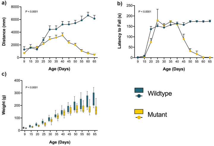 Figure 1
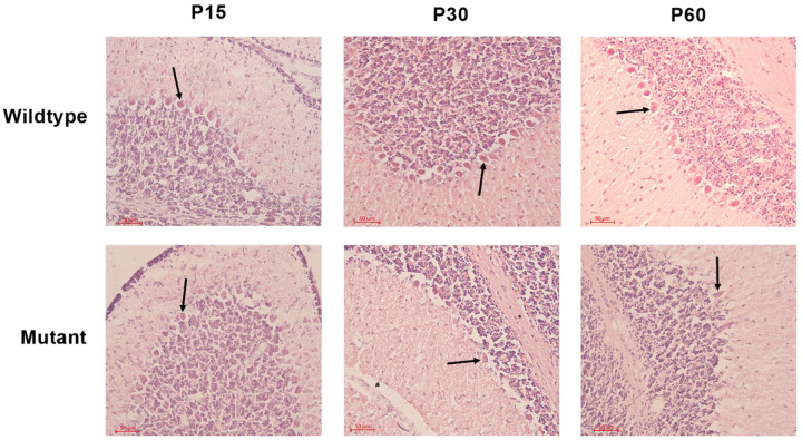 Figure 2
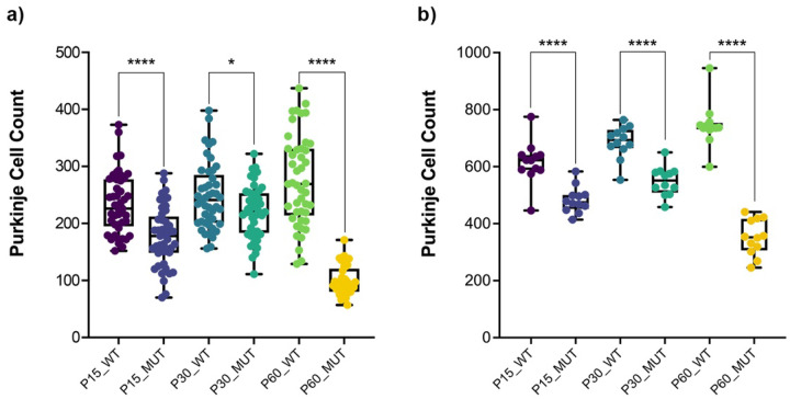 Figure 3
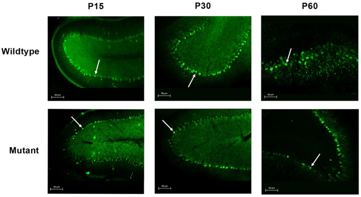 Figure 4
Peer Reviews
No public reviews on file for this paper yet. If you reviewed it on a platform where reviews are public (OpenReview, ICLR, NeurIPS, ICML), you can paste yours below so the community can read it here.
Videos
No videos yet. Explain this paper in a talk, walkthrough, or lecture? Add one.
Taxonomy
TopicsGenetic Neurodegenerative Diseases · Mitochondrial Function and Pathology · Neurological disorders and treatments
Introduction
Neurodegenerative diseases represent one of the most pressing challenges in modern medicine, affecting millions of individuals worldwide and imposing substantial burdens on healthcare systems and families^1^. Among these disorders, hereditary ataxias constitute a clinically and genetically heterogeneous group of conditions characterized by progressive cerebellar dysfunction, resulting in impaired coordination, balance, and motor control^2^. These disorders exhibit substantial phenotypic diversity, with onset ranging from early childhood to late adulthood and inheritance patterns spanning autosomal dominant, autosomal recessive, and X-linked transmission^3^.
The human cerebellum, though comprising only 10% of total brain volume, contains more neurons than the rest of the brain combined and serves as a critical hub for motor coordination and cognitive processing^4,5^. Purkinje cells function as the sole output neurons, integrating vast amounts of sensory information through their intricate dendritic arbors and providing inhibitory signals to the deep cerebellar nuclei^6,7^. These neurons are highly specialized yet particularly vulnerable to genetic mutations and environmental insults, with their degeneration serving as a hallmark of cerebellar ataxias^8,9^.
While the selective vulnerability of Purkinje cells in ataxia is well recognized, the temporal dynamics of their degeneration in relation to symptom onset remain unclear, which limits opportunities for early intervention strategies^10^. Furthermore, while numerous genetic loci have been associated with hereditary ataxias, approximately 75% of patients remain without a molecular diagnosis, highlighting the incomplete understanding of the genetic architecture underlying these disorders^11,12^.
Animal models have been invaluable for investigating the mechanisms of cerebellar degeneration and testing potential therapeutic interventions^13,14,15^. The spastic Han-Wistar (sHW) rat represents a proven model of hereditary ataxia, exhibiting progressive motor dysfunction, cerebellar degeneration, and early mortality by approximately 60 days of age (p60)^16^. Previous studies have documented significant Purkinje cell loss and structural abnormalities in the cerebellar cortex; yet, the genetic basis of this mutation remains unknown, and the precise timeline of neurodegeneration has not been systematically characterized^16–18^.
Here, we sought to provide a detailed temporal map of Purkinje cell loss in the sHW rat model, demonstrating that degeneration begins earlier (P15 vs. P21) and precedes the onset of motor symptoms. In addition, we present a comprehensive temporal investigation of Purkinje cell degeneration in the sHW rat model, combining behavioral assessments and histological analyses to address two fundamental questions: (1) What is the temporal progression of Purkinje cell loss relative to the onset of motor symptoms? and (2) What is the progression of motor impairments in mutant rats as measured by rotarod, open-field, and weight analysis? Through Immunohistochemistry (IHC), Hematoxylin and Eosin (H&E) staining, and quantitative neuronal density measurements, we demonstrate that Purkinje cell degeneration begins as early as P15, preceding the emergence of overt motor deficits. Additionally, we employed whole-genome sequencing and Sanger validation to identify candidate genes potentially responsible for the disorder. Our findings provide new insights into the pathological timeline of cerebellar degeneration and contribute to the growing understanding of genetic factors underlying hereditary ataxias.
Results
This study investigated the progression and timeline of Purkinje cell degeneration in the cerebellum of sHW mutant rats. We employed three experimental approaches: (i) behavioral assays including Open Field, Rotarod, and weight measurement; (ii) histological analyses using H&E staining and IHC; and (iii) whole-genome shotgun sequencing and Sanger sequencing to identify candidate genes underlying the trait.
Early-onset motor and systemic decline in ataxic rats
Locomotor activity, measured by the open-field test, was significantly reduced in mutant rats compared to wild-type controls. The total distance traveled declined progressively with age (Fig. 1a), reflecting reduced mobility. Wild-type rats traveled an average of 4,190 millimeters compared to 1,846 millimeters in spastic mutants, representing a 56% reduction in locomotor activity (two-way repeated measures ANOVA, p < 0.0001; mean difference = 2,344 units, 95% CI: 1,616–3,072). Motor coordination, assessed using the rotarod test, also declined over time in mutants (Fig. 1b). Wild-type rats remained on the rotarod for an average of 134.6 seconds compared to 73.97 seconds for spastic mutants, representing a 45% reduction in motor coordination performance (two-way repeated measures ANOVA, p < 0.0001; mean difference = 60.65 seconds, 95% CI: 42.24–79.07). Mutant animals exhibited early signs of motor impairment, falling before reaching the traditional target duration of 180 seconds^19^. Deficits became prominent by P35, when most mutants were unable to complete the trial, indicating impaired balance and coordination. Body weight measurements showed a similar progressive decline (Fig. 1c), with significant differences between wild-type and mutant. Weight loss coincided with the onset of motor impairment, suggesting a relationship between systemic health decline and reduced motor performance.
Disrupted morphology and reduced cell counts by H&E
To assess the morphology and physical appearance of Purkinje cells in the cerebellum, we utilized the H&E staining on the cerebellum of sHW mutants and wild-type rats at P15, P30, and P60. Initial observations revealed a noticeable reduction in the number of Purkinje neurons in mutant rats compared to their wild-type counterparts^16^.
In wild-type rats, Purkinje cells exhibited normal morphology, characterized by large, round nuclei (Fig. 2). The cell bodies were neatly aligned in a single layer within the Purkinje cell layer of the cerebellum. The dendritic arborizations were also visible, extending into the molecular layer. In contrast, Purkinje cells in the mutant sHW rats displayed signs of degeneration (Fig. 3a). There was a significant reduction in the number of Purkinje cells, with many neurons appearing shrunken and fragmented (Fig. 2). The alignment of Purkinje cells was disrupted. Quantitative analysis confirmed this reduction, with Purkinje cell loss ranging from 10.4% at P30 to 64.1% at P60, including a 24.6% decrease at P15 (Fig. 3a).
Calbindin staining confirms Purkinje cell degeneration
Purkinje cells were visualized using the Calbindin marker in cerebellar sections from wild-type and mutant rats at P15, P30, and P60 (Fig. 4). In wild-type animals, Purkinje cells formed a continuous monolayer with large, well-defined cell bodies and extensive dendritic arborization at all ages examined. Mutant animals had a significantly lower Purkinje cell count than age-matched wild-type controls at all time points, with reductions of 21.5% at P15, 20.0% at P30, and 52.8% at P60. Although the Purkinje cell count in mutants showed an increase from P15 to P30, it remained significantly less compared to wild-type and dropped further by P60 (Fig. 3b). These results parallel the neuronal loss observed in H&E-stained sections and provide cell-type-specific confirmation of Purkinje cell degeneration in the mutant cerebellum.
Genomic search for candidate genes underlying ataxia in Han-Wistar rats
Guided by previous pedigree analysis indicating autosomal recessive inheritance for this ataxia^17^, we used whole-genome shotgun sequencing (WGS) to search for candidate genes underlying this trait. WGS was performed on a trio from Family 1, consisting of the carrier mother, carrier father, and one affected offspring (Supplementary Fig. 1). Genome-wide average depth-of-coverage ranged from ~ 23x to ~ 25x.
Family 1 included three additional siblings, one affected based on phenotypic presentation, and two asymptomatic individuals whose carrier status was unknown at the time of sample collection. These additional siblings were later used for the validation of candidate genes through Sanger sequencing.
Following single-nucleotide variant (SNV) calling, we searched the data for variants following an autosomal recessive inheritance pattern as previously described for this trait^17^. In doing so, we assumed the trait is not caused by a compound heterozygous mutation, which is a common cause of autosomal recessive ataxia^20^. Functional annotation classified the variants into 17 high-impact candidates, 233 moderate-impact, and 24 low-impact, distributed across 273 genes (Supplementary Table 2). To refine this list, candidate prioritization was performed through a systematic review of publicly available resources, including peer-reviewed literature, the NCBI Gene database, the UCSC Genome Browser, and the Human Protein Atlas, to assess gene function, previously reported phenotypes, and tissue-specific expression profiles. This process identified ten genes of interest (Supplementary Table 1), one high-impact gene (Sbf2), six moderate-impact genes (Rhot2, Sh3bp4, Abi2, Cplane1, Stradb, Eif2ak2), and three low-impact genes (Pkd1l2, Ranbp9, Caprin2).
Based on predicted functional impact and biological relevance, the high-impact SNV candidate (chr1:175154510) in the Sbf2 gene was selected for validation in Family 1 using Sanger sequencing. The segregation pattern was consistent with an autosomal recessive model: both parents were heterozygous for the variant; both phenotypically affected offspring were homozygous for the alternate allele; and the two asymptomatic siblings were confirmed to be one heterozygous carrier and one homozygous for the wildtype reference genotype. To assess whether this variant was conserved across pedigrees, Sbf2 was subsequently evaluated in an independent family 2 (Supplementary Fig. 1). In this case, genotypes did not segregate with the disease phenotype, and the gene was thus excluded as the causal variant.
In parallel, copy number variant (CNV) analysis was performed on the WGS data. The frataxin (Fxn) gene emerged as a notable candidate due to its well-documented role in hereditary ataxia and extensive prior study. Fxn was therefore selected for validation in Family 1; however, Sanger sequencing results were inconclusive, and no definitive segregation pattern could be established.
Discussion
The spastic Han-Wistar rat is a well-established genetic model of hereditary ataxia, caused by a naturally inherited mutation that selectively targets Purkinje cells in the cerebellum. Previous studies suggested that Purkinje cell degeneration emerges around P21, coinciding with the onset of observable motor impairment^17,21^. Our study challenges this assumption by demonstrating that Purkinje cell loss begins earlier, at P15, well before overt behavioral symptoms become apparent. Such early vulnerability is consistent with observations in other ataxic mutants, such as Lurcher and pcd mice, where Purkinje cell loss occurs within the first postnatal weeks^22–24^. By refining the temporal onset in sHW rats, our findings address the long-standing question of when cerebellar pathology truly begins relative to symptom onset and provide a more precise disease timeline to guide biomarker discovery and pre-symptomatic intervention strategies.
Behavioral assessments in the sHW rat revealed progressive impairments in locomotion, coordination, and weight gain, with deficits becoming evident around P35. Importantly, these functional changes appeared weeks after histological evidence of Purkinje cell loss, indicating that substantial degeneration can occur before overt symptoms arise. This temporal dissociation has also been documented in spinocerebellar ataxias, where carriers often remain clinically silent despite ongoing neuronal loss^25–27^. A similar threshold-dependent effect is well established in other neurodegenerative disorders, such as Huntington’s disease, where compensatory networks delay clinical onset until a critical proportion of neurons is lost^28^. By extending this principle to the sHW rat, our findings emphasize its utility for modeling the pre-symptomatic phase of hereditary ataxia and for identifying biomarkers that could inform early intervention strategies.
The unresolved genetics of the sHW mutation further underscores the heterogeneity of hereditary ataxias. Our whole-genome sequencing analyses initially highlighted Sbf2 – the gene associated with Charcot-Marie-Tooth disease in humans – as a strong candidate, with segregation patterns consistent in one pedigree. However, follow-up testing in an independent family did not support this association, leading to its exclusion as a causal variant. Similarly, copy number variation analysis suggested a possible role for Fxn, a gene implicated in Friedreich’s ataxia in humans, but validation results were inconclusive. Notably, recent discoveries such as the identification of pathogenic GAA repeat expansions in the FGF14 gene, associated with SCA27B ataxia in humans, demonstrate that Next-Generation sequencing approaches, based on short sequencing reads, may fail to detect repeat expansion disorders, underscoring an additional challenge in resolving undiagnosed cases^29^. Together, these findings highlight the genetic complexity of hereditary ataxia and support the clinical observation that nearly 75% of patients remain without a molecular diagnosis, despite advances in sequencing^11,12,30^. Rather than representing a limitation, this process of iterative exclusion narrows the pool of candidates and mirrors the diagnostic pathway in human patients, where excluding false leads is often as critical as identifying the causative mutation^31^. By refining the genetic search space, our work provides a valuable resource for future studies with larger cohorts and more comprehensive sequencing approaches to pinpoint the underlying variant.
The identification of early Purkinje cell degeneration in the sHW rat also has important implications beyond genetics. Establishing a cellular timeline that precedes behavioral onset provides a foundation for identifying early biomarkers of disease progression. Such markers are crucial for designing interventions during the pre-symptomatic stage, when treatments are most likely to be effective. Moreover, clarifying the onset of degeneration aligns the sHW rat more closely with human ataxias, where cerebellar atrophy is detectable years before motor symptoms appear^25,27^.
Several limitations of this study should be acknowledged. The genetic analyses were conducted on small pedigrees, limiting the power to confirm segregation across multiple families. Larger, multi-generational cohorts will be essential to establish inheritance patterns more conclusively. In addition, although our histological data reveal degeneration by P15, finer sampling across earlier developmental ages will be necessary to determine whether Purkinje cell loss begins even sooner.
Conclusion
In conclusion, our study demonstrates that Purkinje cell degeneration in the sHW rat begins earlier than previously recognized, preceding the onset of motor deficits by several weeks. Behavioral decline closely follows cellular loss, reinforcing the central role of Purkinje neurons in ataxic pathology. In parallel, our genetic analyses excluded two high-priority candidate genes. We also present a curated list of 273 genes harboring mutations consistent with autosomal recessive inheritance, offering a valuable resource for ongoing efforts to identify the causal variant. Together, these findings establish a detailed temporal framework for cerebellar degeneration in this model and highlight its utility for investigating the early stages of hereditary ataxia and for testing pre-symptomatic therapeutic strategies.
Methods
Animals
Spastic Han-Wistar rats were obtained from the California State University, Northridge (CSUN) Vivarium breeding colony between the years 2023 and 2024. Both male and female rats were used, as no sex differences in phenotype have been reported in this model^17^. Animals were housed in groups of 2–4 and provided unrestricted access to standard rat chow and water. Housing was maintained on a 12 h light–dark cycle at 23–25°C. All procedures were conducted in accordance with CSUN’s Institutional Animal Care and Use Committee (IACUC) guidelines.
Breeding mutants
Upon weaning, animals were screened for symptoms of the mutant phenotype at approximately p15. Mutants were confirmed based on observable traits such as hyperactivity and severe whole-body tremors. Additionally, mutants tended to exhibit decreased body weight and weaker grip strength compared to their wild-type siblings.
Behavioral analysis
The behavioral component of this study aimed to evaluate motor coordination and activity levels in 12 mutant and 12 wild-type rats at various developmental stages. Animals aged 9 days old were selected for assessment. Firstly, they underwent the rotarod test. Each trial was conducted at 10 RPM for a maximum of 180 seconds and repeated three times per subject. Subsequently, the rats were placed in a 100 × 100 cm box made of black acrylonitrile butadiene styrene (ABS) plastic. This test consisted of placing the rodent in the described box three times, each time for 120 seconds, during which they were free to move about and explore their environment. The animals were given a 120-second break between each run. All actions within this chamber were captured with a Logitech Quick-Cam Pro 90000 (ffdshow codec 1:122 compression ratio, 15 fps) and recorded to a computer hard drive using VirtualDub. Video tracking and movement analysis were conducted using custom scripts written in Python with the OpenCV computer vision library (opencv.willowgarage.com). Lastly, the weight of each animal was recorded to monitor potential correlations between behavioral performance and physical development. These three tests were conducted every 5 days, starting when the rats were 9 days old and continuing until they reached 65 days of age.
Brain fixation and harvesting
At the designated age, the animals were euthanized, and the brains were extracted for analysis. This process involved exposing the animals to CO_2_ followed by a transcardiac perfusion. Animals were placed in a perfusing pan, and a midline incision through the sternum was made to expose the heart for upper body perfusion using transcardiac puncture. Using hemostats, the descending aorta was clamped to prevent whole-body perfusion. A 26-gauge needle was inserted into the left ventricle, the right atrium was then immediately incised, and a peristaltic pump was activated with a flow rate of 7 ml per minute. Animals were perfused with a 0.9% saline solution to clear most of the blood from the head and neck region. The paleness of the eyes, ears, and forelimbs visually confirmed clearance of blood, and the stiffness of the neck further confirmed clearance of blood. Afterward, 4% paraformaldehyde at pH 7.4 was used on the animal tissue until complete fixation was achieved. The perfusion is also confirmed by physical inspection of increased stiffness in the neck. The brain was then removed, placed in 4% paraformaldehyde, and stored at 4°C for future use.
Tissue sectioning and mounting by cryostat
Cerebellums of harvested brains were sectioned using a cryostat microtome, Leica CM1950, and mounted on a cryostat-mounting platform using tissue freezing media (Tissue-Tek OCT compound 4583). These brains were then sectioned at −20°C into 15μm sagittal cuts. After sectioning, the sections were promptly mounted onto superfrost slides and allowed to dry for 24–48 hours in a 4°C refrigerator to ensure proper adhesion before staining. For each animal, fifteen randomly selected tissue sections were used for H&E staining.
Hematoxylin and eosin staining
The morphology and structural integrity of Purkinje cells were evaluated using Harris’s hematoxylin and eosin (H&E) staining. Cerebellar sections from mutant rats and wild-type littermates were processed in parallel. Sections were rehydrated through graded ethanol (100% and 95%), immersed in hematoxylin for five dips (with staining duration adjusted to achieve optimal intensity), and rinsed in tap water. Slides were differentiated in 1% acid alcohol (1 mL HCl in 99 mL 95% ethanol), rinsed in 2% aqueous sodium bicarbonate to enhance nuclear staining, and counterstained with eosin Y for 20 s. Following dehydration in graded ethanol, slides were cleared in xylene and coverslipped using Permount. Purkinje cell morphology and evidence of neuronal degeneration were assessed in mutants at postnatal days 15, 30, and 60 using light microscopy.
Tissue embedding and vibratome sectioning
For IHC, cerebella were embedded in 4% agarose prepared in 1× phosphate-buffered saline (PBS). To prepare the agarose, 4 g of agarose powder was added to 100 mL 1× PBS, allowed to hydrate at room temperature, and then heated in a microwave oven for ~ 2 minutes with intermittent swirling every 30 s to prevent boiling and foaming, until fully dissolved. The solution was cooled to approximately 40°C before embedding to avoid heat damage to the tissue. Tissues were placed in appropriately sized disposable plastic molds, and molten agarose was gently poured to fully submerge the tissue, ensuring it was lifted slightly so that a thin agarose layer separated the tissue from the mold base. Once the agarose had solidified at room temperature, blocks were wrapped in a damp paper towel, sealed in plastic bags, and stored at 4°C for up to 24 hours before sectioning.
For vibratome sectioning, excess agarose surrounding the tissue was trimmed, and blocks were mounted with the desired orientation maintained. Sagittal sections of cerebellum were cut at 100 μm thickness using a vibratome (Leica, VT1000S) according to the manufacturer’s instructions. Sections for IHC were collected into wells containing 4% paraformaldehyde (PFA) for storage before staining.
Immunohistochemistry
Free-floating cerebellar sections were processed for IHC staining to visualize Purkinje neurons. Sections were transferred into new wells and blocked with 10% goat serum in phosphate-buffered saline (PBS) for 4 hours at 37°C to prevent nonspecific binding. Following blocking, the tissues were rinsed twice with 1× PBS and then incubated in 600 μL PBS containing the anti-Calbindin primary antibody (4 μL per well), a marker for Purkinje cells (1:500, Abcam, Cat# ab108404). Sections were incubated for 48 h at 37°C. After primary antibody incubation, sections were washed six times with PBS (30 min each) on a gentle shaker, adding PBS along the side of the well to minimize tissue disruption. Samples were then incubated overnight at 37°C with Alexa Fluor 488 conjugated anti-rabbit (4 μL/well) secondary antibodies. The following day, sections were washed six times with PBS (30 min each) using the same gentle washing method. Sections were mounted on slides, coverslipped, and imaged using a Zeiss fluorescence microscope equipped with appropriate filter sets for Alexa Fluor 488 (1:500, Invitrogen, Cat# A-11008). Images were acquired at consistent exposure times, saved as high-resolution digital files, and processed using AxioImage software (Zeiss).
Genetic mapping
A family trio was selected for whole-genome sequencing (WGS), comprising carrier parents, approximately one year of age, and an affected offspring. Both parents were heterozygous carriers of the spastic Han-Wistar allele, as they had previously produced progeny with and without the disease phenotype^17^. In the litter from which the trio was derived, an additional two affected and two unaffected offspring were identified at 60 days of age for further DNA extraction and Sanger confirmation of potential candidate genes. Pedigrees are shown in Supplementary Fig. 1. Total DNA was extracted from liver tissue samples obtained from mutants and wild-type rats using the DNeasy Blood & Tissue Kit (Qiagen, Germany), following the manufacturer’s protocol.
Raw sequencing reads were processed using the Illumina DRAGEN Germline Pipeline (v4.3.13), aligned to the Rattus norvegicus reference genome (Rnor_6.0). The DRAGEN pipeline performs key steps, including read mapping, sorting, duplicate marking, and germline variant calling. In each step, the default parameters from the DRAGEN pipeline were used. Quality control metrics were summarized using MultiQC. Variant Call Format (VCF) files generated from Germline analyses were annotated using the Ensembl Variant Effect Predictor (VEP, v113) with the Ensembl RN6.104 annotation set. VEP was used to determine the predicted functional consequences of each variant. CNV calling was performed using the previously generated BAM files and the DRAGEN Germline pipeline (v4.3.13). Segmentation was conducted using the Shifting Level Model.
Candidate gene analysis was performed by filtering both annotated SNV and CNV datasets to identify variants following an autosomal recessive inheritance pattern. For SNVs, variants where the father and mother exhibited heterozygous genotypes (0/1) while the proband displayed homozygous alternate genotypes (1/1) were extracted. For CNVs, only deletions following the same genotype pattern were considered, with additional copy number requirements of copy number = 1 in both parents and copy number = 0 in the proband. Functional impact prioritization was performed using VEP impact predictions, with variants classified as HIGH impact (transcript ablation, stop gained, stop loss, frameshift variants) and MODERATE impact (missense variants, in-frame indels) prioritized for downstream analysis. The resulting candidate gene list represented loci harboring putative pathogenic recessive variants that could explain the observed ataxia phenotype in the proband. All filtering and analysis steps were performed using bedtools^32^ and custom shell scripts.
Candidate gene validation
Primers were designed for Polymerase Chain Reaction (PCR) experiments based on candidate genes identified from the WGS results using Primer3 software (https://primer3.ut.ee/). PCR reactions were performed using 100 ng of genomic DNA in a reaction mixture containing 1× buffer, 4 mM MgCl_2_, 0.2 mM dNTPs, 0.4 μM forward and reverse primers, and 0.05 U/μL Taq DNA polymerase. Thermal cycling conditions were optimized as follows: an initial denaturation at 95°C for 2 min; 30 cycles of denaturation at 95°C for 30 s, annealing at 60°C for 30 s, and extension at 72°C for 1 min; and a final extension at 72°C for 7 min. The primers used were as follows: Sbf2 forward: 5′- ACATCTCAGCACCCGCTTCC-3′, reverse: 5′-TCACAGTAGCACGGAATTTGA-3′; Fxn forward: 5′-ATCTGATGCCCCTCTTCTGG-3′, reverse: 5′-TGGGAGTGAATTCAGGGCTT − 3’.
PCR products were purified using the ExoSAP-IT Express kit (Thermo Scientific, USA) according to the manufacturer’s protocol. Sequencing reactions were prepared in a final volume of 10 μL using the BigDye Terminator v3.1 Cycle Sequencing Kit (Thermo Scientific, USA), with 1 μL of purified PCR product and 1 μM of either forward or reverse primer. Thermal cycling for sequencing consisted of an initial denaturation at 96°C for 1 min, followed by 25 cycles of 96°C for 10 s, 50°C for 5 s, and 60°C for 45 s, with a ramp rate of 1°C/s. Sequencing products were purified using the BigDye XTerminator Purification Kit (Thermo Scientific, USA) and analyzed on a SeqStudio Genetic Analyzer (Thermo Scientific, USA). The resulting sequences were aligned to the Rattus norvegicus reference genome assembly Rnor_6.0 (rn6) using Geneious R9 software, and genotypes for the selected genes were annotated accordingly.
Data availability
Raw whole-genome sequencing data (FASTQ files) from the parent-offspring trio have been deposited in the NCBI Sequence Read Archive (SRA) under BioProject accession number [to be provided upon acceptance/submission]. The other datasets generated during the current study are available from the corresponding author upon reasonable request.
Supplementary Material
Supplementary Files
This is a list of supplementary files associated with this preprint. Click to download.
The reference list from the paper itself. Each links out to its DOI / PubMed record.
- 1Lamptey R. N. L. A review of the common neurodegenerative disorders: Current therapeutic approaches and the potential role of nanotherapeutics. Int. J. Mol. Sci. 23, 1851 (2022).35163773 10.3390/ijms 23031851 PMC 8837071 · doi ↗ · pubmed ↗
- 2Gorcenco S., Karremo C. & Puschmann A. Patients’ Perspective in Hereditary Ataxia. Cerebellum 23, 82–91 (2024).36525215 10.1007/s 12311-022-01505-1PMC 10864479 · doi ↗ · pubmed ↗
- 3Mokhtari D. Genetic investigation of patients with autosomal recessive ataxia and identification of two novel variants in the SQSTM 1 and SYNE 1 genes. Hum. Genome Var. 11, 35 (2024).39214971 10.1038/s 41439-024-00292-x PMC 11364807 · doi ↗ · pubmed ↗
- 4Van Essen D. C., Donahue C. J. & Glasser M. F. Development and evolution of cerebral and cerebellar cortex. Brain Behav. Evol. 91, 158–169 (2018).30099464 10.1159/000489943 PMC 6097530 · doi ↗ · pubmed ↗
- 5Magielse N., Heuer K., Toro R., Schutter D. J. L. G. & Valk S. L. A comparative perspective on the cerebello-cerebral system and its link to cognition. Cerebellum 22, 1293–1307 (2023).36417091 10.1007/s 12311-022-01495-0PMC 10657313 · doi ↗ · pubmed ↗
- 6Gruver K. M. Structured connectivity in the output of the cerebellar cortex. Nat. Commun. 15, 5563 (2024).38982047 10.1038/s 41467-024-49339-1PMC 11233638 · doi ↗ · pubmed ↗
- 7Witter L., Rudolph S., Pressler R. T., Lahlaf S. I. & Regehr W. G. Purkinje cell collaterals enable output signals from the cerebellar cortex to feed back to Purkinje cells and interneurons. Neuron 91, 312–319 (2016).27346533 10.1016/j.neuron.2016.05.037PMC 4969194 · doi ↗ · pubmed ↗
- 8Hekman K. E. & Gomez C. M. The autosomal dominant spinocerebellar ataxias: emerging mechanistic themes suggest pervasive Purkinje cell vulnerability. J. Neurol. Neurosurg. Psychiatry. 86, 554–561 (2015).25136055 10.1136/jnnp-2014-308421 PMC 6718294 · doi ↗ · pubmed ↗