Tannic Acid Shaped Microbiome Composition in Midguts and Rearing Microcosms of Aedes triseriatus (Say)
Shicheng Chen, Liang Cui, Bin Zuo, Jiangchao Zhao, Edward D. Walker

TL;DR
Tannic acid affects the gut microbiome of Aedes triseriatus mosquitoes, reducing diversity and altering bacterial composition, which could help in mosquito control.
Contribution
This study reveals how tannic acid alters mosquito gut microbiota and suggests its potential use in biocontrol strategies.
Findings
TA reduced microbial richness and diversity in mosquito guts but not in water or leaf samples.
TA increased Pseudomonadota and decreased Bacteroidota, with Pseudomonas linked to TA detoxification.
TA exposure enriched pathways related to metabolism and transport, indicating microbial adaptation to TA stress.
Abstract
Tannic acid (TA), a polyphenol derived from plants, often accumulates in water-holding containers where mosquitoes develop. Yet, its effects on mosquito gut microbiota remain poorly understood, representing an important knowledge gap. Because mosquito-associated microbiota are vital for host development, nutrition, and immunity, uncovering how TA shapes these microbial communities may yield new insights into mosquito biology and vector control strategies. In this study, we conducted a comparative analysis of bacterial communities in Aedes triseriatus midguts and rearing microcosms with or without TA supplement. Addition of TA at 0.35 mg/mL caused up to 50% larval Ae. triseriatus mortality, whereas combined supplementation with TA and kanamycin (100 μg/mL) increased mortality to 75% relative to controls. TA treatment significantly reduced microbial Chao 1 richness and Shannon diversity…
Genes, proteins, chemicals, diseases, species, mutations and cell lines named across the full text — each resolved to its canonical identifier and authoritative record.
Click any figure to enlarge with its caption.
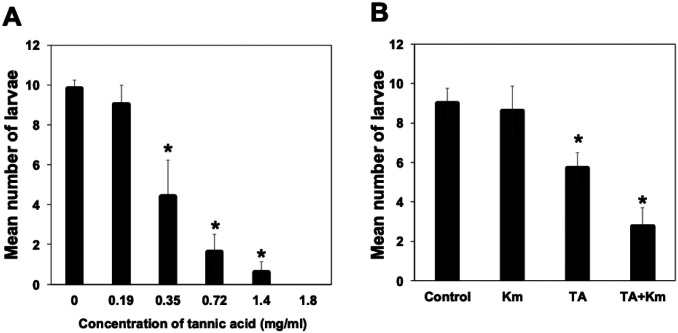 Figure 1
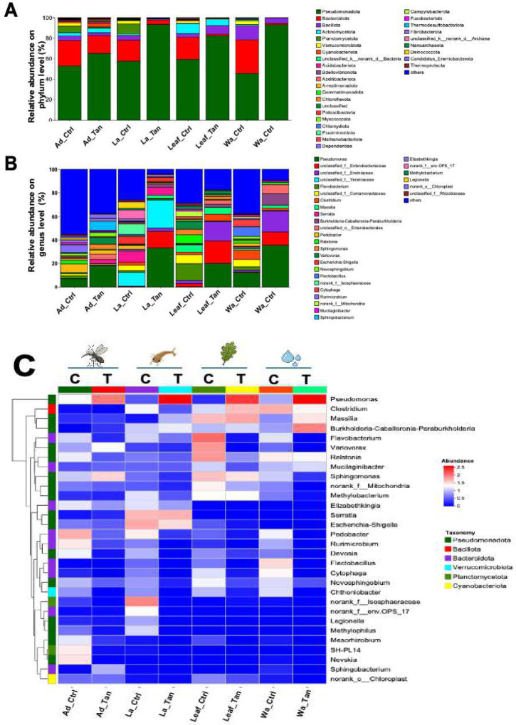 Figure 2
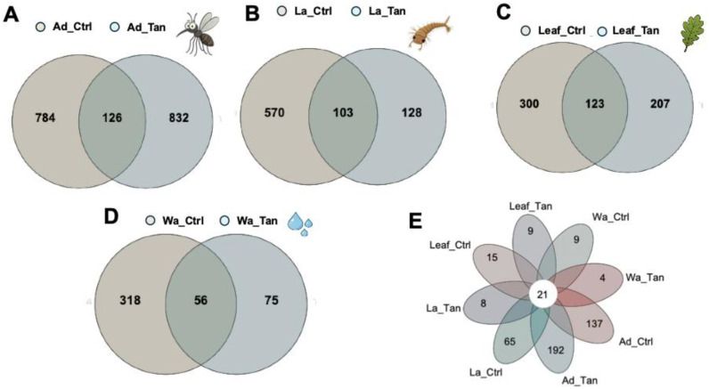 Figure 3
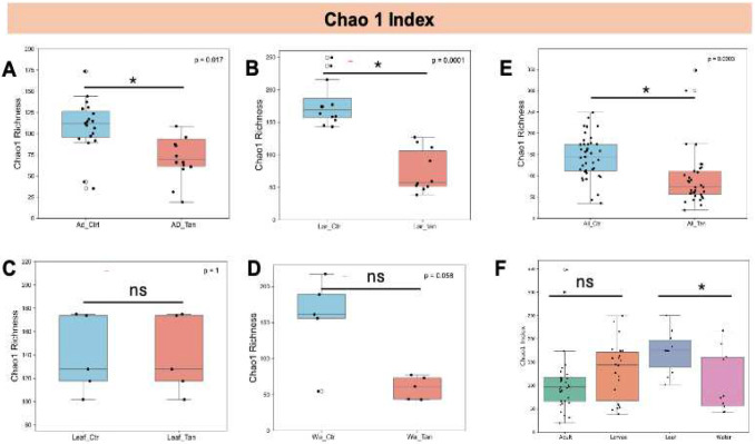 Figure 4
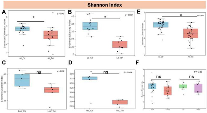 Figure 5
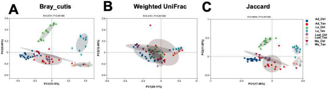 Figure 6
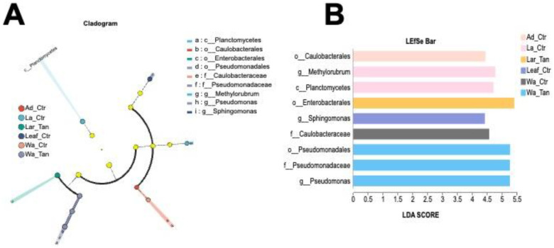 Figure 7
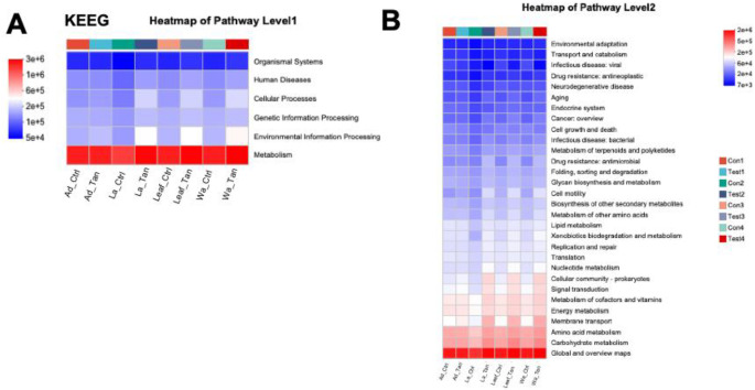 Figure 8
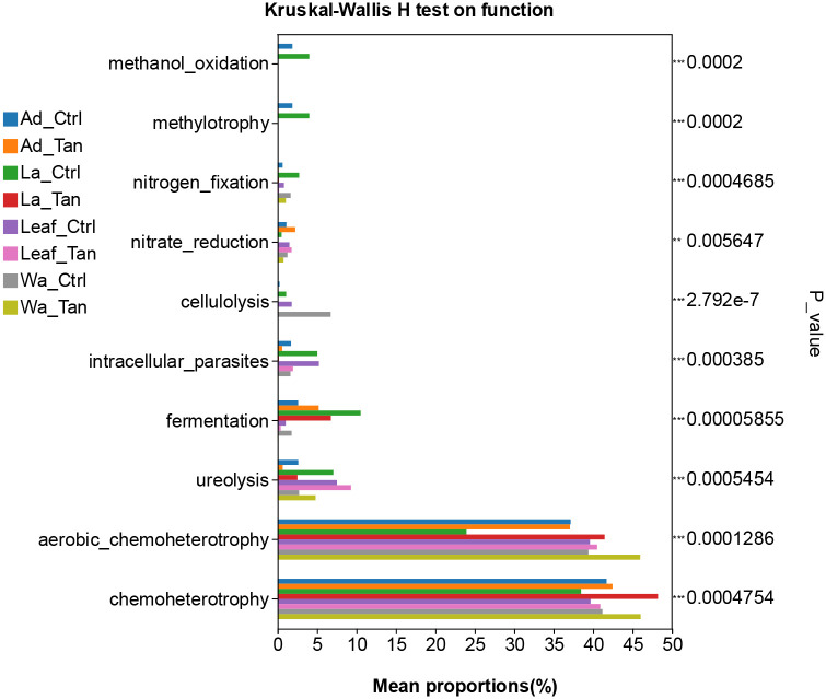 Figure 9
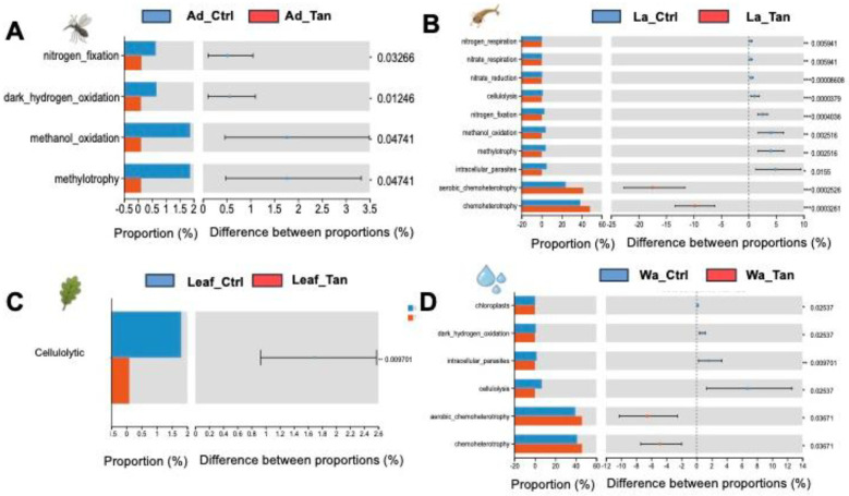 Figure 10
Peer Reviews
No public reviews on file for this paper yet. If you reviewed it on a platform where reviews are public (OpenReview, ICLR, NeurIPS, ICML), you can paste yours below so the community can read it here.
Videos
No videos yet. Explain this paper in a talk, walkthrough, or lecture? Add one.
Taxonomy
TopicsDengue and Mosquito Control Research · Insect symbiosis and bacterial influences · Mosquito-borne diseases and control
Introduction
Aedes triseriatus (Say) (Diptera: Culicidae), the primary vector of La Crosse virus in the Midwestern United States, breeds in natural habitats such as water-filled tree holes, as well as artificial containers like discarded tires [1]. These microhabitats are sustained by the accumulation of senescent leaves, plant detritus, and dead invertebrates, which drive microbial activity and support the base of the aquatic food web [2,3]. Although the water in these habitats is regularly replenished by rain water moving into tree holes as stemflow [2], the decomposition of leaf litter through chemical and biological processes alters its nutritional quality for mosquito larvae and other aquatic invertebrates [2]. Microbial metabolism in particular modifies water chemistry and releases various compounds, including tannic acid, which can accumulate to toxic levels and negatively impact mosquito larval development [4]. Despite its ecological relevance, the physiological and behavioral effects of leaf-derived compounds such as tannic acid on mosquito larvae remain poorly understood. Tannins in tree hole water inhibited growth of the western tree hole mosquito, Ae. sierrensis and caused larval mortality in a dose dependent manner [5,6]. Histological analyses have shown that midgut epithelium was damaged by TA or its derives, causing cellular degeneration - particularly affecting the clear cells of the anterior midgut before the dark cells of the posterior region [7]. Additionally, TA has been reported to interfere with the digestion of starches, lipids, and proteins, thereby disrupting nutrient absorption and leading to growth inhibition, as observed in Hyphantria cunea larvae [8].
Tannic acid (TA), a plant-derived polyphenol, exhibits broad-spectrum antimicrobial and anti-biofilm activities against various bacteria and fungi in vitro. It inhibited growth of both Gram-positive and Gram-negative bacteria by disrupting cell membranes, chelating essential metal ions, and interfering with enzymatic functions [9,10]. Tannic acid also effectively prevented biofilm formation by reducing microbial adhesion to surfaces, disrupting cell-to-cell signaling, and destabilizing extracellular polymeric substances [11,12]. These properties were observed in diverse microbial taxa, including Streptococcus, Bacillus, and fungal genera such as Candida [13–16], underscoring its ecological significance and potential applications in microbial community modulation and biofilm management [17]. In vivo studies in insects demonstrated that tannic acid disrupted gut microbial communities and interfere with digestive processes, contributing to reduced growth and survival [18]. For example, in Hyphantria cunea larvae, tannic acid ingestion caused significant damage to midgut epithelial cells and impaired the activities of key digestive enzymes, including amylase, lipase, and protease, resulting in poor nutrient absorption and growth retardation [8,18]. Additionally, tannic acid was shown to increase gut permeability in insects, potentially facilitating microbial translocation and immune activation [19]. These findings highlighted the dual role of tannic acid in directly affecting insect physiology and indirectly influencing development through microbiota modulation [18]. Given that mosquito larvae depended heavily on their gut microbiota for digestion and development, elevated levels of tannic acid possibly disrupted these microbial communities (either within the gut or the larval habitat), thus impeding larval development [20]. Furthermore, midgut epithelial damage may compromise the gut barrier, allowing microbial translocation into the hemocoel and potentially leading to sepsis. This hypothesis is supported by studies showing enhanced larvicidal efficacy of Bacillus thuringiensis when co-administered with tannic acid [21].
Several bacteria possess the ability to detoxify tannic acid through enzymatic degradation pathways, enabling them to survive in tannin-rich environments such as leaf litter or herbivore insect guts [22,23]. Key enzymes include tannase, which hydrolyzes tannic acid into gallic acid and glucose. Gallate decarboxylase then further metabolizes gallic acid [24]. Microbes such as Lactobacillus plantarum, Bacillus subtilis, Enterococcus faecalis, and Streptococcus gallolyticus have been shown to express these enzymes and tolerate tannic acid exposure [25–28]. This microbial detoxification not only benefits the bacteria themselves but may also alleviate tannin toxicity for host organisms, such as insects, by reducing oxidative stress and supporting gut homeostasis [17,18,22,24]. In this study, we examined how varying concentrations of tannic acid influenced larval mosquito survival and found that the removal of microbial communities further reduced larval survivorship, suggesting a protective role of the microbiota. We also employed the next-generation sequencing methods to characterize shifts in microbial communities in both larval habitats and mosquito midguts following tannic acid exposure. Our results support the potential application of tannic acid as a standalone or synergistic agent in mosquito control strategies. Furthermore, a deeper understanding of detoxification mechanisms in Aedes mosquitoes and their associated microbiota may reveal novel targets for disrupting vector populations and reducing disease transmission.
Materials and Methods
Mosquitoes and experimental design
Ae. triseriatus mosquito larvae were from a laboratory colony originating from larvae collected from bark-lined, pan tree holes in American beech (Fagus grandifolia Ehr.) on the Michigan State University campus and maintained under standard insectary conditions (27 °C, 12:12 h light:dark cycle, and 85% relative humidity) [29]. Upon adult emergence, mosquitoes were provided with a 10% sucrose solution ad libitum via a cotton wick. Adult Ae. triseriatus were blood-fed using bovine blood (Hemostat Lab, Dixon, CA, USA) delivered through an artificial membrane feeder. Two days post-blood meal, oviposition substrates (filter paper moistened with water and placed in Petri dishes) were introduced into rearing cages. Collected eggs were transferred to plastic containers filled with distilled water for hatching. First instar larvae were fed Tetramin tropical fish food flakes (Tetra, Blacksburg, VA, USA) ad libitum, followed by daily ad libitum feedings of Purina Cat Chow (Nestlé) for subsequent instars.
To evaluate the effects of tannic acid (Sigma-Aldrich, St Louis, MO) on mosquito development and microbiome, experimental microcosms were prepared with 1 g of senescent, American beech leaf litter sampled in autumn, dried, and placed into 80 mL of Milli-Q water, and 1 ml of microbial inoculum, consistent with our previous experiments [29,30]. Freshly collected, senescent beech leaves leach up to 8 mg of tannic acid equivalents per gram of dried leaf within the first 24 hours of immersion in water, thus establishing a background of tannin in the microcosm water [31]. Immersed leaves condition rapidly with microbial colonization [30,32]. To evaluate the dose-dependent effect of tannic acid, larvae were hatched from eggs and seeded into microcosms; ten second instar larvae were exposed to a range of TA concentrations (0.00, 0.19, 0.35, 0.72, 1.4, and 1.8 mg/mL). The number of surviving larvae was counted after 1 week. The effects of tannic acid and kanamycin on Ae. triseriatus larval survival were assessed by counting surviving larvae one week after treatment [31]. For these combinatorial treatments, four groups were established: (1) kanamycin-only group (100 μg/mL), (2) tannic acid-only group (0.35 mg/mL), (3) tannic acid + kanamycin group (0.35 mg/mL tannic acid + 100 μg/mL kanamycin), and (4) untreated control group. After six days of conditioning, microcosms received either 2 mL tannic acid solution plus 2 mL Milli-Q water (treatment) or 4 mL Milli-Q water only (control). Ten second instar Ae. triseriatus larvae were introduced into each microcosm at the time of treatment. Larval survival rate was recorded after seven days of exposure. The experiment was replicated five times, and the data were analyzed using one-way analysis of variance (ANOVA).
DNA Extraction, Library Construction, and 16S rRNA Sequencing
A subset of microcosms was randomly selected for microbial community analysis after a ten-day incubation. Fourth-instar larvae were collected for sampling. Upon adult emergence, mosquito midguts were immediately dissected and pooled. Before dissection, adults were surface sterilized by three rinses in 70% ethanol followed by one rinse in sterile water. Midguts were removed under sterile conditions using finely sharpened, number 5 watchmaker’s forceps (Daigger Scientific, New Jersey, USA), placed into 200 μL of sterile phosphate-buffered saline (PBS), and pooled from six individuals per sample. Each pooled set was homogenized with a sterile pestle. All dissections and DNA extractions were conducted in a laminar flow biosafety cabinet to avoid contamination. Water samples were collected by centrifuging at 15,000 rpm for 10 min at 4 °C; resulting pellets were stored at −70 °C until further use. Leaf materials after removing surface water were flash-frozen in liquid nitrogen, and ground with a sterile pestle. For larvae, at least 12 replicates were analyzed in the TA-treated group and 10 in the control group. For adults, 14 TA-treated and 19 control samples were processed. For water and leaf samples, five replicates were included for each treatment group.
Mosquito midgut tissues, pellets from water and leaf materials were resuspended in 200 μL of lysis buffer, and DNA was extracted using the DNeasy Blood & Tissue Kit (Qiagen) according to the manufacturer’s protocol. DNA concentration was quantified using Qubit^™^ dsDNA HS Assay Kits, and integrity was confirmed by PCR with primers 63F (CAGGCCTAACACATGCAAGTC) and 1387R (CGGAACATGTGWGGCGGG). Amplicon tagging and 16S rRNA gene sequencing were performed at the Research Technology Support Facility (RTSF) at Michigan State University. The V4 region of the bacterial 16S rRNA gene was amplified using primers 515f (GTG CCA GCM GCC GCG GTA A) and 806r (NNN NNN GGA CTA CHV GGG TWT CTA AT), with unique 6-bp error-correcting barcodes included in the reverse primer. Amplicons were purified, pooled, and sequenced using the Illumina MiSeq platform with a 2 × 250 bp paired-end format and 500-cycle v2 reagent kit. Base calling was performed with Illumina Real Time Analysis (RTA) v1.18.54, and output files were demultiplexed and converted to FASTQ format using Illumina Bcl2fastq v1.8.4.2.3.
Bioinformatics analysis and statistical analysis
To obtain high-quality sequences, bases at the head or tail with Phred quality scores below Q30 were trimmed, and sequences shorter than 100 bp were discarded. Raw sequence data were processed in QIIME2 (version 2024.2) [33], including sequencing and PCR error reduction, and denoising with DADA2 (filtering, dereplication, chimera removal, and merging of paired-end reads) to generate amplicon sequence variants (ASVs) for taxonomic analysis. ASVs representing less than 0.1% of the total sequences across all samples or annotated as chloroplast or mitochondrial contaminants were removed. Sequencing depth was normalized to the minimum read count among samples. Taxonomic classification was performed using a Naive Bayes classifier trained on the SILVA 138/16S rRNA database (https://www.arb-silva.de/). Alpha diversity was calculated in Mothur (version 1.30.2) (https://mothur.org/) using the Chao1 and Shannon indices based on randomly subsampled sequences from each sample. Beta diversity was assessed using PERMANOVA (999 permutations) based on Bray–Curtis distances, weighted UniFrac, and Jaccard dissimilarities, implemented in the vegan package (version 2.4–1) in R (version 3.3.1). Microbial biomarkers for each experimental group were identified using LEfSe (version 1.0) [34].
Microbial functions were predicted using PICRUSt2 (v2.2.0-b) [35], and functional differences between treatment groups were assessed using FAPROTAX (version 1.2.11) [36]. Predicted genes were annotated against the Enzyme Commission (EC) database (https://enzyme.expasy.org/), the Kyoto Encyclopedia of Genes and Genomes (KEGG) database [37–39], and the MetaCyc Metabolic Pathway Database (https://metacyc.org/). Group differences were analyzed in STAMP, with two-group comparisons performed using the Wilcoxon test followed by false discovery rate (FDR) correction. Statistical significance was defined as P < 0.05.
Sequence Data Accession Number
Raw paired-end reads per sample without barcode and primer bases were submitted to the Sequence Read Archive of the NCBI (PRJNA1320966).
Results
Effects of tannic acid and antibiotic treatment on larval mosquito
Larval Ae. triseriatus survival declined with increasing tannic acid concentrations, with 100% mortality observed at 1.8 mg/mL TA (Figure 1A) (ANOVA: F-test, df=4, P < 0.05). Larval survival was high and equivalent between controls and kanamycin alone but was lower with TA (at 35 mg/mL) and lowest with TA and kanamycin in combination (Figure 1B) (ANOVA: F-test, df=4, P < 0.05), indicating a synergistic negative effect on larval survivorship.
Effects of tannic acid treatment on bacterial composition in insect guts and microcosms
After filtering, denoising, merging, and chimera removal, 3,313,743 high-quality reads were obtained, with a median depth of 44,183 reads per sample. To minimize sequencing depth bias, read counts were normalized to the smallest sample size (1,042 reads) by random subsampling. Rarefaction curves (Figure S1) indicated that samples from rearing water, leaf surfaces, and mosquito midguts (larvae and adults) with or without TA addition did not fully reach asymptotic saturation, suggesting the presence of additional rare taxa. Nonetheless, all curves displayed the characteristic concave upward trend with increasing sequencing effort.
Taxonomic profiling assigned sequences to at least 38 bacterial phyla. The five most abundant were Pseudomonadota, Bacteroidota, Bacillota, Planctomycetota, and Actinomycetota (Figure 2A). Across all habitats (rearing water, leaf surfaces, larval midguts, and adult midguts) Pseudomonadota predominated (45.4–93.5%), regardless of TA treatment. Bacteroidota ranked second (1.7–33.0%). TA addition doubled the relative abundance of Pseudomonadota while sharply reducing Bacteroidota (from 33% to 1.5%), indicating strong TA-mediated inhibition of this bacterial group. Bacillota abundance declined in TA-treated water, Verrucomicrobia nearly disappeared, and Actinomycetota decreased. Similar patterns were observed on leaves and in midguts, except Bacillota increased on TA-treated leaves. Planctomycetota were more common in larvae and adults from non-TA treatments but remained rare in water and leaves.
Genus-level composition varied widely between treatments and sample types (Figure 2B). In control water, ~37.5% of reads were unclassified at the genus level (“others”); dominant genera included Pseudomonas, Flectobacillus, Clostridium, unclassified Comamonadaceae, and Cytophaga. TA-treated water showed a marked shift. Pseudomonas nearly doubled in abundance (35.6%) while Flectobacillus, Clostridium, and Cytophaga declined to negligible levels (Figure 2C). Conversely, unclassified Erwinaceae, unclassified Enterobacteriaceae, Burkholderia–Caballeronia–Paraburkholderia, and unclassified Enterobacteriales were enriched (Figure 2C). In control leaves, Pseudomonas was rare (0.4%); dominant genera included Flavobacterium, unclassified Comamonadaceae, unclassified Enterobacteriales, Ralstonia, and Variovorax. TA-treated leaves were dominated by Pseudomonas, unclassified Erwinaceae, and unclassified Enterobacteriaceae, resembling TA-treated water communities. In control larval midguts, 55.4% of reads were unclassified (“others”), with Pedobacter, Pseudomonas, Rurimicrobium, Elizabethkingia, and Ralstonia as key taxa. TA treatment doubled Pseudomonas abundance, while Elizabethkingia and Pedobacter remained similar to controls; Sphingobacterium, Serratia, and Variovorax were also common (Figure 2C). Adult midguts in controls were dominated by unclassified Yersiniaceae, Serratia, unclassified Isosphaeraceae, unclassified env.OPS_17, Escherichia–Shigella, and Legionella. In TA-treated adults, Pseudomonas, unclassified Enterobacteriales, and unclassified Yersiniaceae increased markedly (33.3%, 23.8%, and 14.0%, respectively).
Venn analysis showed that TA-treated and control samples shared only a limited number of ASVs (Figure 3). In adult guts, 1,742 ASVs were detected - 126 (7.2%) shared, 832 (47.8%) unique to TA-treated, and 784 (45.0%) unique to controls (Figure 3A). In larval guts, 801 ASVs were identified - 103 (12.9%) shared, 570 (71.2%) unique to controls, and 128 (16.0%) unique to TA-treated (Figure 3B). Leaves had 630 ASVs - 123 (19.5%) shared, 300 (47.6%) control-specific, and 207 (32.9%) TA-specific (Figure 3C). Water had 449 ASVs - 56 (12.5%) shared, 318 (70.8%) control-specific, and 75 (16.7%) TA-specific (Figure 3D). Across all habitats combined, only 21 ASVs (4.8%) were shared (Figure 3E).
Alpha and beta diversity affected by TA in the insect guts and microcosms
Building on our initial observations of microbial community composition, we next assessed whether alpha diversity was influenced by TA treatment (Figure 4). Bacterial richness, as measured by the Chao1 index, was significantly reduced in TA-treated adult guts compared to controls (p < 0.05) (Figure 4A). A similar decrease in richness was observed in larval samples following TA exposure (Figure 4B). In contrast, no significant richness differences were detected between TA-treated and untreated leaf samples (p > 0.05) (Figure 4C), nor in water samples (Figure 4D). However, when comparing all non-TA-treated samples (water, leaf, larvae, and adult grouped as controls) against all TA-treated samples (Figure 4E), a significant difference emerged (p < 0.05). Additionally, leaf samples generally exhibited higher Chao1 values than water samples, independent of treatment. When TA treatment was disregarded, no significant differences in richness were found between treated and untreated adults or larvae (p > 0.05) (Figure 4F).
Significant differences in species diversity, assessed via the Shannon index, were found between non-TA-treated controls and TA-treated adults (p < 0.05), with TA-treated larvae also showing reduced diversity relative to controls (Figure 5A and 5B). However, no significant diversity differences (p > 0.05) were observed between TA-treated and control leaves, nor between TA-treated and control water samples (Figure 5C and 5D). When ignoring TA treatment, Shannon diversity did not differ significantly (p > 0.05) among adult and larval mosquitoes or between leaf and water samples (Figure 5E and 5F).
For beta diversity analysis, we performed PCoA to assess similarities in microbial structures among TA treatment, rearing systems and development stages. PCoA analyses were based on Bray-Curtis distance, Weighted UniFrac and Jaccard, respectively (Figure 6). For Bray-Curtis distance analysis (Figure 6A), the dissimilarity revealed clear separation between TA-treated and control microbial communities (ANOSIM: R = 0.67, p = 0.001). The first two PCoA axes explained 25.05% of the total variance (PCoA1: 15.15%, PCoA2: 9.90%). Samples clustered primarily by TA treatment, with separation along axis 1 except for adult mosquito samples. Moreover, both Weighted UniFrac (ANOSIM: R = 0.42, p = 0.001) and Jaccard (ANOSIM: R = 0.68, p = 0.001) distance analyses supported significant structural differences between TA-treated and control microbiome (Figure 6B and 6C), agreeing with Bray-Curtis analysis. From the three different dissimilarity analyses, we also observed there were distinct gut microbial communities across developmental stages (larvae vs. adults) and in rearing systems (leaf vs. water) (Figure 6). Moreover, PERMANOVA analysis indicated that TA treatment (P = 0.001), rearing systems (P = 0.028), and developmental stages (P = 0.001) were all significant drivers of beta diversity (Table 1).
Linear discriminant analysis effect size (LEfSe) analysis
LEfSe analysis identified bacterial taxa differentially enriched across mosquito life stages and microcosms, spanning from phylum to genus level (Figure 7A and 7B). Only taxa with an LDA score greater than 2.0 were included. The cladogram revealed distinct phylogenetic patterns, highlighting three candidate markers in TA-treated water: order Pseudomonadales, family Pseudomonadaceae, and genus Pseudomonas. In contrast, family Caulobacteraceae was enriched exclusively in control adults, representing non-treated water. Two markers such as genus Methylorubrum and class Planctomycetes were enriched in control larvae, while genus Sphingomonas was a marker for TA-treated larvae. Additionally, family Caulobacteraceae served as a characteristic marker in leaf-reared control adults. No marker taxa were detected in treated leaf or treated adult groups.
Functional prediction analysis reveals potential differences in microbial community metabolic function.
PICRUSt2 predicted six major functional pathways (Level 1) based on KEGG (Figure 8A). These functional categories were enriched in metabolism, environmental information processing, genetic information processing, cellular processes, human diseases, and organismal systems (Figure 8A). Among all groups, metabolism was the predominant predicted function (72.91–76.5%), followed by environmental information processing (6.07–9.55%) and genetic information processing (4.80–6.50%). It is noted that more “environmental information processing” functions were predicted in the TA-treated samples. The most abundant pathways at Level 2 included carbohydrate metabolism, global and overview maps, amino acid metabolism, energy metabolism, and metabolism of cofactors and vitamins (Figure 8B). However, COG functional classification did not clearly resolve the predicted functional differences (Figure S2). Therefore, FAPROTAX was employed to refine functional predictions, focusing on processes such as plant cell wall degradation, nitrogen and carbon cycling, and nutrient assimilation, based on individual microbial profiles. This annotation identified 30 functional categories within the insect guts and rearing systems (supplemental Figure 3). The top five enriched pathways were chemoheterotrophy, aerobic chemoheterotrophy, fermentation, and ureolysis (Figure S3). We further analyzed the different predicted function enriched in treatment for further comparation (Figure 9 and Figure 10). Notably, cellulolysis and intracellular parasite-related functions were more abundant in water controls without TA compared to TA-treated water (Figure 9). The only significantly different pathway between TA-treated and untreated leaf surface samples was cellulolytic activity (Figure 10C). Furthermore, aromatic compound degradation and nitrate ammonification pathways were more prominent in water, leaf, and larval samples without TA. Conversely, “methanol oxidation” and “methylotrophy” were more abundant in larval and adult mosquitoes without TA, which likely benefits insect physiology (Figure 10A ang 10B). Genes involved in nitrogen metabolism were commonly detected in both larval and adult mosquitoes (Figure 10A and Figure 10B). Overall, TA-treated insects showed a reduced proportion of nitrogen fixation and methanol metabolism genes. However, several functions such as aerobic chemoheterotrophy showed higher abundance in TA-treated water and larval samples (Figure 10B and Figure 10D).
Discussion
Previous research on tannin-insect interactions largely focused on their impacts on the growth and physiology of herbivorous pests, with relatively little attention paid to aquatic insects [8,18,21,22]. In this study, we investigated the effects of TA on mosquito larval development, gut microbiota composition, and environmental microbial communities within experimental rearing microcosms. Our research centered on the feeding ecology of Ae. triseriatus larvae in container habitats, where early-life interactions with microbial communities can influence adult fitness and vector competence [40–43]. Larval Ae. triseriatus feed by filtering and browsing suspended particulate matter, detritus of plant leaves, and microbial biofilms found on submerged surfaces such as plant material and container walls [43]. In natural tree holes, microbial community composition was shaped by both leaf litter decomposition and larval mosquito activity [44]. However, little is known about how mosquito larvae tolerate or metabolize plant-derived polyphenol compounds such as tannins, which commonly accumulate in these environments [7]. The western tree hole mosquito was negatively affected by exposure to tannins, but any ameliorating effects of microorganisms on the effects were not investigated [5,6]. Overall, our findings demonstrated tannic acid influenced both mosquito survival and the structure of their associated microbial communities. The response of microbiota to elevated tannin acid concentrations suggested a potential microbial role in detoxification processes. These insights expand our understanding of how ecological and chemical factors shape mosquito–microbiome interactions and suggest that naturally occurring compounds such as tannins could be leveraged to manipulate mosquito-associated microbiota as part of integrated biocontrol strategies against mosquito populations and vector-borne diseases [18,21].
Our results showed that elevated TA concentrations significantly reduced larval survival rate. Although TA likely exerts its effects primarily by directly damaging the midgut epithelium of Aedes mosquitoes as reported previously [5,7], we cannot rule out additional pathways through which TA may influence larval physiology [11,18]. For example, tannic acid (TA) was shown to increase reactive oxygen species while decreasing antioxidant and detoxification enzyme activities in hosts [18]. In addition, dietary tannins were reported to change growth, behavior, and the gut microbiome of larval amphibians [45]. Because larval Aedes mosquitoes depend on microbial and detrital food resources that are frequently coated with plant-derived tannins, they are continuously exposed to these chemical stressors [30,32,43]. Such exposure may lower the nutritional quality of available food, disrupt gut microbial communities, and impose sublethal physiological costs that collectively slow larval growth and development. Another possible pathway for tannic acid–induced toxicity involves changes in water chemistry, especially reductions in dissolved oxygen (DO) levels, which may result from increased microbial respiration during the breakdown of tannin-rich organic matter [46]. Soaking American beech leaves for three days released tannins up to 120 mg/L, substantially lowering DO [46]. Reduced DO imposed metabolic stress, increased respiration costs, and impaired larval growth and survival [47]. Whether these mechanisms apply to mosquito larvae and other aquatic insects remains to be determined. Taken together, findings in this study and others indicate that tannins possibly affect mosquito survival through multiple, interacting pathways including direct physiological stress, altered food quality, gut microbiome disruption, and changes in water chemistry. It highlights their potential as natural compounds to modulate mosquito populations and inform integrated vector management strategies.
Exogenous toxicants are known to perturb insect gut microbiota, disrupting physiological homeostasis and impairing host growth and development [48]. Consistent with this, TA exposure significantly altered the richness, diversity, and taxonomic composition of gut microbiota in both larval rearing systems and the insects themselves, with TA-treated groups exhibiting reduced Chao1 richness and lower Shannon diversity indices compared to controls. PCoA analysis revealed clear differences in microbial community structures, highlighting a strong TA-driven influence on community composition. These shifts suggested that TA exposure selectively favored microbes capable of detoxification or otherwise supporting host health under chemical stress. Despite its antimicrobial properties, many bacteria exhibit tannin resistance [26,49–51]. Microbes have evolved diverse mechanisms for TA degradation in natural environments [51,52]. Notably, the relative abundance of Pseudomonas spp. consistently increased across insect midguts, rearing water, and leaf surfaces under sublethal TA exposure compared to controls. In agreement with this observation, Tan et al. reported a higher relative abundance of Pseudomonas in TA-treated larvae compared to untreated controls (37.43% vs. 33.59%) [18]. In Pseudomonas protegens Pf-5, TA exposure modulated iron homeostasis, upregulating genes for heme uptake and siderophore biosynthesis and transport, which may reduce reactive oxygen species generation and enhance bacterial survival [53]. Additionally, certain Pseudomonas species can metabolize tannic acid and its hydrolysis product gallic acid as sole carbon sources [54]. TA’s antimicrobial and protein-binding properties may inhibit tannin-sensitive bacteria, reducing competition and selectively enriching for tannin-tolerant taxa like Pseudomonas [50]. TA may alter the microenvironment within mosquito larval guts or associated habitats (e.g., pH, nutrient availability), indirectly favoring Pseudomonas proliferation [45]. As predominant mosquito commensals, Pseudomonas spp. played key roles in shaping larval gut communities and protecting hosts from toxic metals and insecticides [55], with disruptions to these populations linked to increased larval mortality and impaired development [56]. Collectively, these findings suggest that TA exposure drives a selective restructuring of mosquito gut microbiota, promoting tannin-tolerant taxa like Pseudomonas, which may provide detoxification functions and confer mutualistic benefits to the host.
Microbes within Burkholderia–Caballeronia-Paraburkholderia, unclassified Erwinaceae, and unclassified Enterobacteriaceae were also promoted by TA addition (Figure 2). Members of the Erwinaceae family, such as Pantoea and Erwinia, were commonly plant-associated bacteria [57,58]. Many plant-associated bacteria possessed tannin-tolerant or tannin-degrading enzymes including tannase (tannin acyl hydrolase) and polyphenol oxidases to survive in these phenolic-rich environments [27,51]. For instance, P. agglomerans was reported to tolerate polyphenols and degrade certain gallotannins [59]. Burkholderia exhibited a dramatic increase in relative abundance in water samples treated with TA compared to controls. It was also found in Aedes mosquito habitats [40,41]. This marked enrichment suggested a strong tolerance or adaptive response of Burkholderia to TA exposure, potentially mediated by specific genetic determinants that confer resistance or degradation capabilities. Serratia spp., another common commensal bacterium associated with mosquitoes [60], demonstrated a modest increase in relative abundance in TA-treated larval mosquitoes and persisted in adult mosquitoes. However, the physiological functions and ecological roles of Serratia in mosquito biology remain poorly characterized, warranting further investigation. The elevated abundance of Pseudomonas, Serratia, and Burkholderia collectively implies that these taxa may harbor intrinsic or acquired genes enabling resistance to the antimicrobial and protein-binding effects of TA, thereby facilitating their persistence and proliferation in TA-enriched environments [61,62].
In contrast, TA exposure resulted in a notable decrease in the relative abundance of Flavobacterium across both microcosms and mosquitoes [40,41]. The reduction of Flavobacterium is particularly significant, as this genus represents an important commensal gut bacterium in mosquitoes, commonly acquired and enriched from larval habitats [42]. Prior studies demonstrated that Flavobacterium played critical roles in mosquito nutrition and fitness [42]; its removal delayed larval growth, reduced adult longevity, and impaired overall mosquito fitness [63]. The antimicrobial properties of TA against this group highlight the potential ecological consequences of tannin presence in mosquito breeding sites, possibly influencing microbial community structure and, by extension, host health and development. Understanding these microbial dynamics is essential for elucidating the complex interactions between environmental chemicals, microbiota, and mosquito physiology.
LEfSe analysis identified Pseudomonadaceae and the genus Pseudomonas as markers enriched in TA-treated water, highlighting their role in tannic acid degradation or tolerance. In contrast, Caulobacteraceae were enriched only in non-treated water. It features a distinctive life cycle with motile swarmer and sessile stalked cells, enabling effective colonization of aquatic surfaces [64]. Widely distributed in various habitats, Caulobacteraceae contribute to nutrient cycling and organic matter decomposition [64,65], particularly in nutrient-poor environments such as water in native tree hole or disposed tires. In control larvae, two additional markers appeared: genus Methylorubrum and class Planctomycetes. Bacteria Methylorubrum are facultative methylotrophs that utilize single-carbon compounds like methanol, supporting carbon cycling and potentially host nutrition [66]. Planctomycetes are involved in nitrogen and carbon cycling and may help maintain gut microbial balance [67]. Sphingomonas species [68], common in various insect microbiomes including mosquitoes, were markers in TA-treated larvae. Known for degrading aromatic compounds, Sphingomonas likely aid in detoxifying tannin-rich environments [69]. They were also found in herbivorous insects, assisting in plant material digestion [70]. Lastly, Caulobacteraceae was a marker in leaf-reared control adults. LEfSe analysis did not discover significant marker taxa in two groups such as TA-treated leaf or treated adult groups. This usually means that those groups don’t have bacterial taxa with strong enough differential abundance (LDA score > 2.0) compared to others, or the microbial communities are more similar in these groups.
Functional predictions using PICRUSt2 revealed that carbohydrate and amino acid metabolism dominated microbial activity in mosquito guts and their habitats, while FAPROTAX identified key functions including chemoheterotrophy and fermentation, essential for nutrient cycling and organic matter breakdown. Cellulolytic activity and aromatic compound degradation were higher in untreated water and leaf samples, suggesting that TA inhibits microbes responsible for plant polymer and polyphenol degradation [71]. This shift likely alters microbial community composition and reduces the capacity to process tannin acid. Methanol oxidation and methylotrophy pathways were more abundant in untreated larvae and adults but reduced under TA treatment, potentially impacting host nutrition and metabolite production beneficial for mosquito development. Conversely, aerobic chemoheterotrophy increased in TA-treated water and larvae, indicating enrichment of microbes capable of tolerating or metabolizing tannic acid. Nitrogen metabolism genes were present across samples, but nitrogen fixation decreased in TA-treated insects, potentially limiting nitrogen availability critical for larval growth. Overall, TA reshaped mosquito-associated microbial functions by suppressing specific degradation pathways while promoting others, thereby influencing microbial detoxification capacity and nutrient support, which likely affected mosquito development and survival in tannin-rich environments.
Supplementary Material
1
The reference list from the paper itself. Each links out to its DOI / PubMed record.
- 1Grimstad P.R.; Walker E.D. Aedes Triseriatus (Diptera: Culicidae) and La Crosse Virus. IV. Nutritional Deprivation of Larvae Affects the Adult Barriers to Infection and Transmission. J Med Entomol 1991, 28, 378–386, doi:10.1093/jmedent/28.3.378.1875364 · doi ↗ · pubmed ↗
- 2Walker E.D.; Lawson D.L.; Merritt R.W.; Morgan W.T.; Klug M.J. Nutrient Dynamics, Bacterial Populations, and Mosquito Productivity in Tree Hole Ecosystems and Microcosms. Ecology 1991, 72, 1529–1546, doi:10.2307/1940953. · doi ↗
- 3Fish D.; Carpenter S.R. Leaf Litter and Larval Mosquito Dynamics in Tree-Hole Ecosystems. Ecology 1982, 63, 283–288, doi:10.2307/1938943. · doi ↗
- 4Rey D.; David J.-P.; Martins D.; Pautou M.-P.; Long A.; Marigo G.; Meyran J.-C. Role of Vegetable Tannins in Habitat Selection among Mosquito Communities from the Alpine Hydrosystems. Comptes Rendus de l’Académie des Sciences - Series III - Sciences de la Vie 2000, 323, 391–398, doi:10.1016/S 0764-4469(00)00136-0. · doi ↗
- 5Mercer D.R. Effect of Tannic Acid Concentration on Development of the Western Treehole Mosquito,Aedes Sierrensis (Diptera: Culicidae). J Chem Ecol 1993, 19, 1119–1127, doi:10.1007/BF 00987373.24249130 · doi ↗ · pubmed ↗
- 6Mercer D.R.; Anderson J.R. Tannins in Treehole Habitats and Their Effects on Aedes Sierrensis (Diptera: Culicidae) Production and Parasitism by Lambornella Clarki (Ciliophora: Tetrahymenidae). J Med Entomol 1994, 31, 159–167, doi:10.1093/jmedent/31.1.159.8158619 · doi ↗ · pubmed ↗
- 7Rey D.; Pautou M.-P.; Meyran J.-C. Histopathological Effects of Tannic Acid on the Midgut Epithelium of Some Aquatic Diptera Larvae. J Invertebr Pathol 1999, 73, 173–181, doi:10.1006/jipa.1998.4810.10066397 · doi ↗ · pubmed ↗
- 8Zhang A.; Li T.; Yuan L.; Tan M.; Jiang D.; Yan S. Digestive Characteristics of Hyphantria Cunea Larvae on Different Host Plants. Insects 2023, 14, 463, doi:10.3390/insects 14050463.37233091 PMC 10231093 · doi ↗ · pubmed ↗