Vasoactive neuropeptide dysregulation: A novel mechanism of microvascular dysfunction in vascular cognitive impairment
Willians Tambo, Keren Powell, Steven Wadolowski, Prashin Unadkat, Eric H. Chang, Christopher LeDoux, Daniel Sciubba, Ping Wang, Patricio Huerta, Chunyan Li

TL;DR
This study shows that vasoactive neuropeptide dysregulation causes microvascular issues that lead to cognitive decline in vascular cognitive impairment.
Contribution
The study identifies vasoactive neuropeptide dysregulation as a novel mechanism driving microvascular dysfunction in vascular cognitive impairment.
Findings
Microvascular vasoconstriction is the earliest and most persistent event in vascular cognitive impairment.
Dysregulation of vasoactive neuropeptides is the key driver of vascular and non-vascular dysfunction in chronic cerebral hypoperfusion.
Calcitonin gene-related peptide (CGRP) supplementation prevents vasoconstriction and improves cognitive function in chronic cerebral hypoperfusion.
Abstract
Neuropeptide dysregulation and microvascular injury are involved in pathogenesis of vascular cognitive impairment (VCI); however, the underlying etiology of this pathological axis remains unclear. We investigated pathological mediators across varying severities of VCI in a rat model of chronic cerebral hypoperfusion (CCH). Proteomic analysis guided the evaluation of neuropeptide and non‐neuropeptide markers associated with vascular and non‐vascular dysfunction, which were correlated with cognitive function to determine their role in VCI. Proteomic analysis revealed vasomotor dysfunction as the primary pathological pathway in VCI. Microvascular vasoconstriction was the earliest and most persistent event, initiating a cascade of both microvascular and non‐vascular dysfunction. Dysregulation of vasoactive neuropeptides was identified as the key driver of this process. Calcitonin…
Genes, proteins, chemicals, diseases, species, mutations and cell lines named across the full text — each resolved to its canonical identifier and authoritative record.
Click any figure to enlarge with its caption.
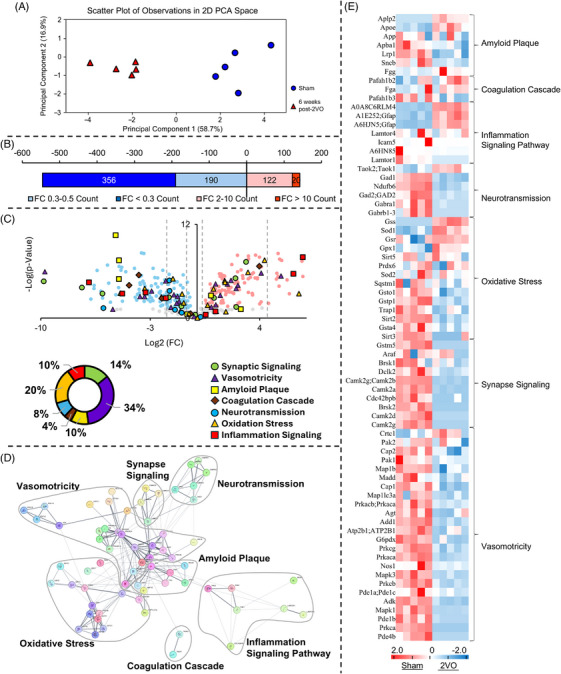 FIGURE 1
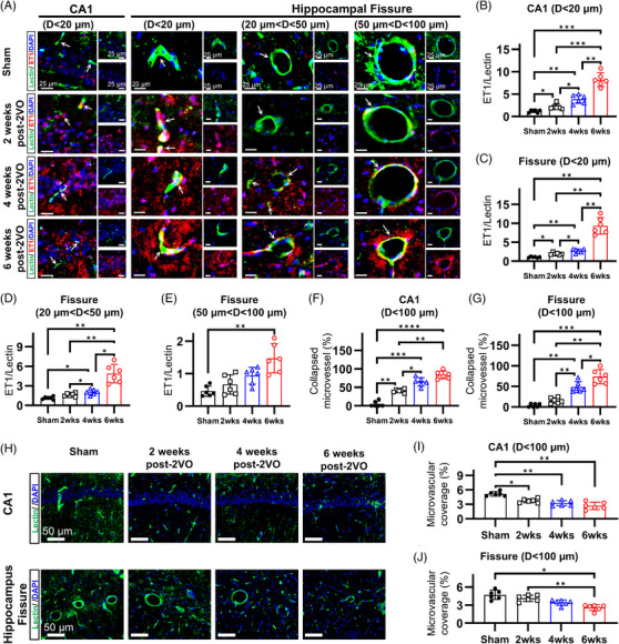 FIGURE 2
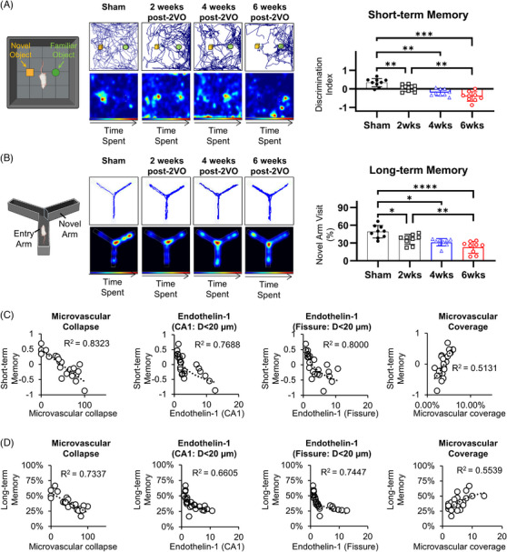 FIGURE 3
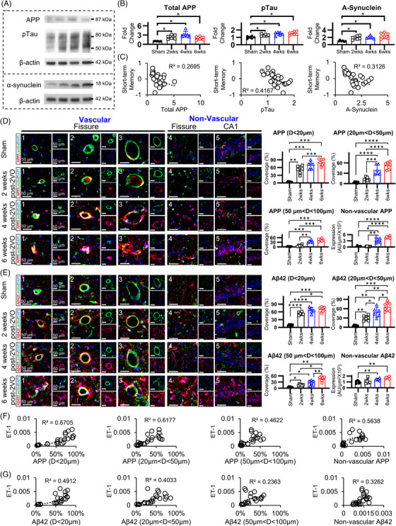 FIGURE 4
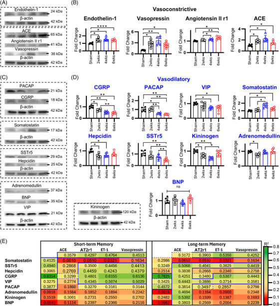 FIGURE 5
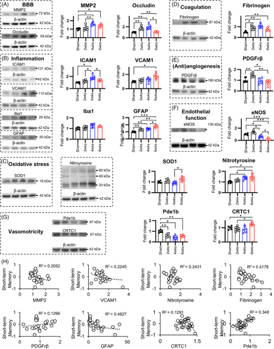 FIGURE 6
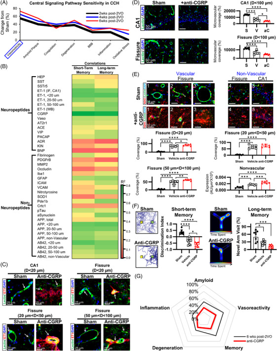 FIGURE 7
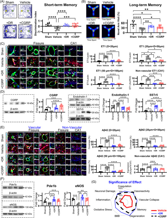 FIGURE 8
- —United States Army Medical Research Acquisition Activity (USAMRAA)
- —US Army Medical Research and Materiel Command (USAMRMC)
- —Zoll Foundation Award
- —The Feinstein Institutes for Medical Research
- —Advancing Women in Science and Medicine at the Feinstein Institutes for Medical Research
Peer Reviews
No public reviews on file for this paper yet. If you reviewed it on a platform where reviews are public (OpenReview, ICLR, NeurIPS, ICML), you can paste yours below so the community can read it here.
Videos
No videos yet. Explain this paper in a talk, walkthrough, or lecture? Add one.
Taxonomy
TopicsNeurological Disease Mechanisms and Treatments · Neurological Disorders and Treatments · Neuroscience and Neuropharmacology Research
INTRODUCTION
1
Vascular cognitive impairment (VCI) is the second most prevalent cause of cognitive decline, yet effective therapeutic strategies remain elusive.1 Chronic cerebral hypoperfusion (CCH) is a key contributor to the pathogenesis of VCI and other neurodegenerative diseases, such as Alzheimer's disease (AD). Despite numerous interventions targeting various pathophysiological processes in CCH, none have demonstrated clinically effective results, likely due to the disease's heterogeneity and varying severity.2, 3 Autonomic nervous system (ANS) dysfunction represents an early driver of CCH, manifesting as recurrent cerebral hypoperfusion episodes,4, 5, 6, 7 potentially mediated by ANS‐regulated neuropeptide dysregulation.8, 9 Clinically, ANS dysfunction correlates with the severity of cognitive decline and accelerates disease progression,7 thereby increasing the risk of dementia.6 These findings implicate neuropeptide dysregulation as a critical factor in both VCI and AD pathogenesis. Such dysregulation correlates with dementia onset and severity in AD,10, 11, 12 appears in clinical VCI cases, and has been proposed as a cognitive dysfunction biomarker across dementia subtypes.13, 14 Furthermore, preclinical studies have demonstrated that neuropeptide dysfunction contributes to cognitive decline in VCI,15, 16 and represents a promising therapeutic target.17 However, despite the essential roles that neuropeptides play in the pathological processes initiated by CCH,2, 13, 14, 18, 19, 20 it remains unclear whether neuropeptide dysregulation is the primary factor driving the pathological cascades associated with CCH. Moreover, while multiple neuropeptides have been investigated in the context of CCH,13, 17 the predominant neuropeptide and its specific pathomechanistic contributions have yet to be identified.
Microvascular dysfunction represents a central pathophysiological mechanism in CCH,21 with neuropeptides serving as critical mediators in its development.20 Therapeutic interventions targeting microvascular dysfunction demonstrate superior efficacy,2, 3 compared to approaches targeting non‐vascular cascades,21 offering disease‐modifying potential through attenuation of cerebrovascular injury progression.3 In clinical populations, reduced cerebral perfusion and associated microvascular dysfunction exhibit robust correlations with dementia severity and function as predictive indicators for progression from mild cognitive impairment to severe dementia.18, 19, 22, 23 This established relationship has stimulated investigation of therapeutic strategies focused on blood–brain barrier (BBB) restoration and microvascular angiogenesis promotion, yielding significant cognitive improvement outcomes.24, 25, 26, 27 However, despite these encouraging findings,14 the specific aspects of microvascular dysfunction that predominantly drive the progression of CCH remain unclear. Neuropeptide dysregulation disrupts multiple facets of microvascular function,13, 14, 20 precipitating microvascular impairment and cognitive deterioration in preclinical models.15, 16 This pathological process manifests with particular severity in regions characterized by low vascular density, notably the hippocampus, which represents a critical anatomical substrate for CCH‐associated cognitive decline.28 While current evidence establishes microvascular dysfunction as fundamental to CCH pathophysiology, significant knowledge gaps persist regarding the hierarchical importance of specific microvascular dysfunction mechanisms, their longitudinal persistence across disease progression, and the quantitative contribution of neuropeptide dysregulation to these pathological processes.
To elucidate the specific microvascular dysfunction mechanisms underlying CCH development and their potential relationship with neuropeptide dysregulation, we employed a rat model of CCH induced by bilateral common carotid artery occlusion (2VO). This model was chosen because it recapitulates key clinical features of vascular cognitive impairment and dementia (VCID), including the development of amyloid pathology.29 Pathological progression was evaluated at three distinct CCH stages: early (2 weeks post‐2VO), middle (4 weeks post‐2VO), and late (6 weeks post‐2VO). Initially, proteomic analysis was conducted during the late CCH stage to identify predominant pathomechanisms. Guided by the significant pathways identified in our proteomic analysis, we designed follow‐up experiments to assess both neuropeptide markers and non‐neuropeptide markers, with the latter further stratified into vascular and non‐vascular categories. To determine which markers demonstrated the strongest correlation with cognitive deterioration, marker expression was correlated with short‐term working memory and long‐term spatial memory assessments and established cognitive impairment indicators. Our findings indicate that vasoactive neuropeptide dysregulation constitutes a critical pathogenic mechanism in CCH pathology and VCI, functioning primarily through induction of microvascular constriction that promotes amyloid deposition. This mechanistic relationship was further substantiated through interventional studies employing both exogenous and endogenous calcitonin gene‐related peptide (CGRP) modulation.
METHODS
2
Experimental animals
2.1
Four cohorts of male Sprague–Dawley rats (Charles River Laboratories, USA) were utilized in this investigation, comprising a total of 159 animals. The first cohort consisted of 72 rats (225–250 g) for initial assessment of 2VO pathophysiological progression and identification of primary pathways involved in CCH pathology. The second cohort comprised 12 rats (225–250 g) to confirm acute changes at 3 days post‐2VO. The third cohort comprised 27 rats (225–250 g) to validate and begin characterizing the preliminarily identified primary pathway. The fourth cohort included 48 initially juvenile (100–125 g) rats for comparative 2VO, 2VO with intranasal CGRP therapy (2VO + CGRP), and 2VO with diving reflex therapy (2VO + DR) assessments. Within the fourth cohort, 12 rats underwent DR training into adulthood (200–250 g), while 36 rats from the same cohort aged concurrently for sham, 2VO‐vehicle, and 2VO + CGRP groups. For experimental consistency, 2VO induction was performed at equivalent ages across all experimental groups. Due to pre‐training requirements for the 2VO + DR group, only the 2VO + CGRP group was randomly assigned following 2VO induction.
All experimental procedures were conducted in accordance with the Guide for the Care and Use of Laboratory Animals30 following Institutional Animal Care and Use Committee approval.
Experimental design
2.2
A schematic of experimental protocols is depicted in Figure S1. Animals were systematically assigned to one of four experimental protocols. In all protocols, cognitive performance was assessed in every subject at predefined biweekly intervals prior to tissue collection. Based on preliminary estimates of inter‐subject variability, a minimum group size of n = 9 was determined to provide adequate statistical power for cognitive endpoints. After the final cognitive assessments, either hippocampal tissue was microdissected for biochemical analyses or animals underwent transcardial perfusion for subsequent histological evaluation.
RESEARCH IN CONTEXT
- Systematic review: The authors reviewed literature from PubMed and Google Scholar, as well as meeting abstracts and presentations. Neuropeptide dysregulation and microvascular injury are increasingly recognized for involvement in the pathogenesis of Vascular cognitive impairment and dementia (VCID); however, the underlying etiology of this pathological axis remains unclear. In the present study involving a rat model of VCID, we assessed the connection between chronic cerebral hypoperfusion‐induced neuropeptide dysregulation and microvascular dysfunction, and the development of cognitive decline.
- Interpretation: Our evidence shows that early, progressive vasoactive neuropeptide dysregulation drives microvascular constriction, constituting a pivotal mechanism in microvascular dysfunction and subsequent cognitive deterioration in chronic cerebral hypoperfusion. The data further elucidate the significant therapeutic efficacy of pharmacological and non‐pharmacological interventions directed at vasoactive neuropeptide pathways, which resulted in marked enhancement of cognitive function.
- Future directions: Our results indicate that aberrant vasoactive neuropeptide regulation constitutes a fundamental pathophysiological mechanism underlying the development and clinical manifestation of VCID. These findings have potentially substantial implications for the development of novel therapeutic strategies targeting this disorder, which represents the second most common etiology of cognitive deterioration.
Experiment 1 design
2.2.1
The initial cohort of 72 rats was randomized into six groups in a 2 × 3 factorial design: 2 weeks post‐2VO (early stage, n = 12), 4 weeks post‐2VO (mid stage, n = 12), 6 weeks post‐2VO (late stage, n = 12), and corresponding sham groups at 2, 4, and 6 weeks (each n = 12). CCH was induced in 2VO groups by bilateral common carotid artery occlusion, whereas sham animals underwent identical surgical handling without arterial occlusion. Standardized cognitive assessments were conducted at 2, 4, and 6 weeks post‐surgery. Animals (2VO and sham) were harvested in paired sets at each predefined survival time point. Histological outcomes were first compared across the three sham time points to confirm equivalence; upon verification, only the 6‐week sham group was retained as the reference for subsequent comparisons within this experiment.
Experiment 2 design
2.2.2
The second cohort comprised 12 animals randomly allocated to two groups: 3 days post‐2VO (acute stage, n = 6) and 3 days post‐sham (n = 6). In the 2VO group, CCH was induced by bilateral common carotid artery occlusion; sham animals underwent identical surgical handling without arterial occlusion. Behavioral testing was not performed in this cohort due to the documented variability and limited reliability of acute‐stage behavioral assessments in the 2VO model.
Experiment 3 design
2.2.3
The third cohort was randomized into three groups with a 6‐week survival: 2VO + fremanezumab (n = 9), 2VO + bosentan (n = 9), and sham (n = 9). CCH was induced in 2VO groups by bilateral common carotid artery occlusion; sham animals underwent identical surgical handling without occlusion. Fremanezumab (Ajovy; 30 mg/kg, intranasal; Teva Pharmaceuticals, Parsippany, NJ) was administered once weekly to inhibit CGRP, beginning 1 day prior to 2VO. Bosentan (Tracleer; 200 mg/kg/day, delivered in a flavored gel; Actelion Pharmaceuticals US, Inc., San Francisco, CA) was administered daily to inhibit endothelin‐1, starting 1 day before 2VO. Standardized cognitive assessments were performed at 6 weeks post‐2VO.
Experiment 4 design
2.2.4
The third cohort was randomized into four groups with a 6‐week survival: 2VO (n = 12), 2VO + DR (n = 12), 2VO + CGRP (n = 12), and sham (n = 12). Standardized cognitive assessments were conducted at 6 weeks post‐2VO. DR and CGRP served as endogenous and exogenous CGRP augmentation, respectively, and were administered 5 days per week throughout the 6‐week period. For exogenous augmentation, animals received intranasal CGRP daily (1 µg in 50 µL; Tocris Bioscience, Bristol, UK) divided equally between the nares, following established methodology.31 To enhance endogenous CGRP, we implemented a diving reflex (DR) protocol. A modified version of the McCulloch protocol32 was used to elicit DR in rodents, which has been validated for mammalian DR studies in mice and rats.33, 34, 35, 36 Animals were trained to voluntarily traverse an approximately 2.5 m underwater course over 3 weeks prior to 2VO. After 2VO, they completed 30‐min DR sessions consisting of seven sequential diving bouts, modeled after human DR paradigms.37 Five‐minute rest intervals were incorporated between bouts, as previously shown to reliably elevate CGRP during DR in rats.36
Rat model of chronic cerebral hypoperfusion
2.3
Male Sprague–Dawley rats underwent CCH induction via bilateral common carotid artery occlusion.38 Briefly, subjects were anesthetized with isoflurane and maintained at normothermia (37°C) using a thermal plate throughout the procedure. Following midline cervical incision, the left common carotid artery was isolated, carefully dissected from the vagus nerve, and permanently ligated. An identical procedure was subsequently performed on the right common carotid artery. Surgical incisions were closed using non‐absorbable nylon sutures, followed by subcutaneous buprenorphine administration for post‐operative analgesia. Animals were returned to their home cages and monitored for potential surgical complications. Sutures were removed at post‐operative day 10. During the survival period, animals underwent daily health monitoring. No mortality or morbidity requiring subject removal occurred as a consequence of CCH induction throughout the study duration.
Cognitive assessments
2.4
Cognitive assessments were conducted in a dedicated behavioral testing suite under standardized environmental conditions with consistent illumination and minimal extraneous auditory or visual stimuli. Test procedures adhered to established protocols and were sequentially administered from least to most stressful to minimize potential carryover effects and stress‐induced behavioral confounds. All behavioral sessions were digitally recorded and subsequently analyzed using automated tracking software (EthoVision XT, Noldus Information Technology, USA) to ensure consistent quantification of behavioral parameters. Following each assessment, testing apparatus and objects were thoroughly decontaminated using 70% ethanol followed by Peroxiguard disinfectant to eliminate residual olfactory cues that might influence subsequent subject performance. All behavioral evaluations were performed by two designated laboratory technicians who remained blinded to experimental group assignments throughout the study duration to prevent potential observer bias in test administration or data interpretation.
Novel object recognition test
2.4.1
Working recognition memory was assessed 24 h before euthanasia using the novel object recognition (NOR) test, which exploits rodents' innate exploratory preference for novel stimuli compared to familiar ones.39 Testing was conducted in a square open field arena (60 × 60 × 30 cm) constructed of opaque black acrylic to minimize external visual cues. The NOR paradigm consists of two sequential phases: habituation and testing. During the habituation phase, subjects were familiarized with an object. In the subsequent testing phase, a novel object was introduced alongside the familiar object, and interaction time with each object was quantified. Objects were matched for size and material composition to enhance test reliability. Recognition memory performance was quantified using a discrimination index (DI) calculated as DI = (TN–TF)/(TN + TF), where TN represents time spent exploring the novel object and TF represents time spent exploring the familiar object. This formula yields a normalized preference score ranging from −1.0 (exclusive exploration of the familiar object) to 1.0 (exclusive exploration of the novel object), with scores significantly above zero indicating intact recognition memory.
Y‐maze test
2.4.2
Long‐term spatial memory was assessed immediately before euthanasia in each animal using a validated two‐phase Y‐maze protocol.39 The testing apparatus consisted of a symmetrical Y‐shaped maze (MazeEngineers, USA) with three identical arms (40 cm length × 8 cm width × 15 cm height) positioned at equiangular intervals (120° separation) and constructed of blue, opaque polyvinyl chloride to ensure consistent visual cues while minimizing extraneous environmental influences. The assessment protocol comprised two sequential phases: spatial encoding and retrieval testing. During the initial encoding phase, subjects were placed at the terminus of the start arm and allowed 10 min of free exploration with one arm (novel arm) blocked by an opaque divider, thus restricting exploration to two arms (start arm and familiar arm). Following a 4‐h inter‐trial interval, the retrieval testing phase was conducted wherein subjects were reintroduced to the maze with all three arms accessible for a 5‐min exploration period. Arm entries were operationally defined as the center point of the animal crossing the threshold of an arm, as determined by the EthoVision XT program. Spatial memory performance was quantified primarily by calculating the percentage of novel arm exploration time (time in novel arm/total exploration time × 100). This methodology exploits rodents' innate preference for novel spatial environments; thus, subjects with intact long‐term spatial memory demonstrate preferential exploration of the previously inaccessible (novel) arm. Chance‐level performance in this paradigm represents approximately 33% exploration of each arm.
Brain tissue preparation for biochemical and histological characterization
2.5
Fresh brain hippocampus collection and cryopreservation
2.5.1
Subjects were deeply anesthetized via deep isoflurane anesthesia (5% in oxygen) until complete loss of pedal withdrawal reflex was confirmed. Cerebral tissue was rapidly extracted via decapitation to minimize ischemic degradation. Bilateral hippocampal formations were immediately microdissected on ice using microsurgical instrumentation. The excised hippocampal tissue was instantaneously flash‐frozen in liquid nitrogen to preserve protein integrity and prevent enzymatic degradation. Frozen specimens were subsequently pulverized to a homogeneous powder using a pre‐cooled mechanical tissue homogenizer under cryogenic conditions to maximize protein extraction efficiency. Processed tissue homogenates were maintained in sterile polypropylene cryovials at −80°C with stringent temperature monitoring and minimal freeze‐thaw cycling to preserve biomolecular integrity and prevent proteolytic degradation. A randomized subset of processed hippocampal specimens was allocated for comprehensive analytical characterization via two complementary approaches. First, samples underwent liquid chromatography‐tandem mass spectrometry (LC‐MS/MS) proteomic profiling under standardized analytical conditions. Second, parallel aliquots from the same specimens were subjected to targeted biochemical characterization of specific neuropeptide and non‐peptide biomarkers implicated in CCH pathomechanisms. This dual analytical approach enabled both hypothesis‐driven investigation of predetermined molecular targets and unbiased proteomic discovery of novel pathological mechanisms associated with experimental CCH.
Transcardial perfusion and cryosectioning for histomorphological analysis
2.5.2
Subjects were deeply anesthetized with isoflurane (5% in oxygen) until complete loss of pedal withdrawal reflex was confirmed. Transcardial perfusion was performed via left ventricular cannulation and right atrial incision with 200 mL cold phosphate‐buffered saline (PBS, pH 7.4, 4°C, Sigma‐Aldrich, USA) for vascular clearance, followed by 200 mL cold 4% paraformaldehyde (PFA, pH 7.4, 4°C, Sigma‐Aldrich, USA) in PBS for tissue fixation. Following perfusion, cerebral tissue was carefully extracted and post‐fixed in 4% PFA for 24 h at 4°C to ensure complete tissue fixation. Specimens were subsequently cryoprotected through sequential immersion in gradient sucrose solutions (10%, 20%, and 30% w/v in PBS) at 4°C until tissue saturation was achieved (confirmed by specimen submersion). The cryoprotected tissue was then embedded in a 1:3 mixture of 30% sucrose and Optimal Cutting Temperature (OCT, Thermo Fisher Scientific, USA) compound within disposable embedding molds and stored at −80°C prior to cryosectioning.
Serial coronal sections were obtained at 400 µm intervals throughout the rostrocaudal neuraxis using a cryostat microtome (CM1950, Leica Biosystems, Germany) maintained at −20°C. Adjacent sections of 18 µm thickness were mounted on Superfrost Plus adhesion microscope slides (Thermo Fisher Scientific, USA) for immunohistochemical analysis, while 14 µm sections were mounted on Polysine adhesion slides (Thermo Fisher Scientific, USA) for specialized histological procedures. Mounted sections were air‐dried for 30 min at room temperature and subsequently stored at −30°C in slide boxes containing desiccant to prevent frost formation prior to histological processing.
Proteomics analysis
2.6
Aliquots of pulverized hippocampal tissue from sham‐operated (n = 6) and 2VO‐vehicle (n = 6) subjects underwent comprehensive proteomic analysis via liquid chromatography‐tandem mass spectrometry at Creative Proteomics (Shirley, NY, USA). Computational data processing was performed using a multi‐platform bioinformatics workflow integrating R (MaxQuant v2.6.7.0, missMDA v1.19) and Python (Matplotlib v3.10.0) analytical environments. Raw spectral data underwent standardized preprocessing, including peak detection, peptide identification, and protein quantification. The resultant protein abundance dataset was subjected to log‐2 transformation followed by median normalization to minimize technical variability. Missing proteomic values were addressed through a hierarchical imputation strategy based on data sparsity patterns: singular value decomposition (SVD; imputation threshold: 50%, principal components: k = 5) for proteins with moderate missingness, quantile regression imputation (QRI) for proteins with substantial missingness (n = 4 or 5 missing values), and minimum value imputation for proteins with complete absence in one experimental group (n = 6 missing values). Initial principal component analysis (PCA) revealed one statistical outlier per experimental group (Hotelling's T ^2^ test, p < 0.01), which was excluded from subsequent differential expression analyses to enhance statistical robustness. Differentially expressed proteins were identified using dual selection criteria comprising fold‐change thresholds (> 2.0 or < 0.5) combined with statistical significance (Student's t‐test with Benjamini–Hochberg correction, p < 0.05). Further stratification designated fold changes of >10 and <0.3 as highly significant alterations. Visual representation of the proteomic landscape was accomplished through volcano plots and hierarchically clustered heat maps generated using Python Matplotlib and Seaborn libraries. Protein interaction networks and functional enrichment analyses were constructed using Cytoscape Software (v3.9.0) with STRING database integration. These preliminary proteomic analyses subsequently guided targeted biochemical investigations of specific protein candidates and relevant molecular pathways.
Quantitative analysis of neuropeptide and classical protein markers via Western blot
2.7
Pulverized hippocampal tissue specimens were homogenized in ice‐cold RIPA lysis buffer (150 mM NaCl, 1.0% NP‐40, 0.5% sodium deoxycholate, 0.1% sodium dodecyl sulfate (SDS), 50 mM Tris, pH 8.0; Thermo‐Scientific, USA) supplemented with protease and phosphatase inhibitor cocktail (Halt, Thermo‐Scientific, USA) at a 1:100 dilution. Mechanical tissue disruption was performed using a bead mill homogenizer (1.4 mm ceramic beads, 5 m/s, 4°C) with three 20‐s cycles separated by 30‐s cooling intervals to prevent protein degradation. Homogenates were subsequently centrifuged at 16,000 g for 5 min at 4°C to pellet cellular debris and insoluble fractions. Supernatants containing solubilized proteins were harvested and total protein concentration was determined using the bicinchoninic acid (BCA) protein assay kit (Pierce, Thermo‐Fisher, USA) with bovine serum albumin standards (0.125–2 mg/mL). Equivalent protein quantities (40 µg/lane) were denatured in Laemmli buffer (5 min, 95°C) and resolved by sodium dodecyl sulfate‐polyacrylamide gel electrophoresis on 4%–20% gradient Mini‐PROTEAN TGX precast gels (Bio‐Rad, USA) at 80 V for 15 min and 150 V for 40 min. Molecular weight determination was facilitated by concurrent electrophoresis of prestained protein standards (10–250 kDa). Separated proteins were electrophoretically transferred onto methanol‐activated polyvinylidene difluoride membranes (0.2 µm pore size) using semi‐dry transfer methodology (Trans‐Blot Turbo, Bio‐Rad, USA) at 25 V for 30 min. Membranes were blocked with 5% non‐fat milk in Tris‐buffered saline containing 0.1% Tween‐20 (TBS‐T) for 1 h at ambient temperature to minimize non‐specific binding and subsequently incubated with primary antibodies (Table S1) overnight at 4°C with gentle rocking. Following three 10‐min TBST washes, membranes were incubated with appropriate horseradish peroxidase‐conjugated secondary antibodies for 1 h at ambient temperature. Immunoreactive bands were visualized by enhanced chemiluminescence using SuperSignal West Dura Extended Duration Substrate (Thermo Fisher Scientific, USA) and digitally captured on a ChemiDoc MP Imaging System (Bio‐Rad, USA) with optimized exposure parameters. Densitometric analysis was performed using ImageJ software (NIH, version 1.53c) to quantify relative protein expression. β‐Actin (1:25,000; Sigma, USA) served as an endogenous loading control for normalization of target protein signals. Western blot data are presented as fold change relative to sham controls.
Quantitative spatial analysis of biomarker distribution via immunofluorescent histochemistry
2.8
Tissue sections mounted on Polysine adhesion slides underwent immunofluorescent labeling according to established protocols.40 Following rehydration, sections were subjected to three 5‐min washes with TBS‐T (pH 7.6, Sigma, USA) and subsequently blocked with 10% normal goat serum (Abcam, USA) supplemented with 1% bovine serum albumin (Fraction V, Sigma, USA) for 1 h at ambient temperature to minimize non‐specific binding. Primary antibody incubation (Table S2) was performed in humidified chambers at 4°C for 16–18 h. Following three TBS‐T rinses, sections were incubated with appropriate fluorophore‐conjugated secondary antibodies (1:400 dilution) for 1 h at ambient temperature with light protection. Nuclear counterstaining was performed using 4′,6‐diamidino‐2‐phenylindole (DAPI, 1:2000, Thermo Fisher Scientific, USA) for 10 min, followed by three additional TBS‐T washes. Sections were mounted with Vectashield Antifade mounting medium (Vector Laboratories, USA) and coverslipped. Fluorescent micrographs were acquired using an EVOS M7000 imaging system (Thermo Fisher Scientific, USA) equipped with an appropriate filter at 20× magnification. Exposure parameters were standardized within experimental series to ensure valid comparative analyses. Quantitative image analysis was performed using ImageJ software (version 1.53c, National Institutes of Health, Bethesda, USA) with consistent thresholding parameters applied across all specimens.
Microvessel density was assessed by calculating lectin‐positive area as a percentage of total analyzed area (0.25 mm^2^) within anatomically defined regions, including CA1 pyramidal layer and hippocampal fissure. Immunoreactivity for endothelin‐1, Iba1, glial fibrillary acidic protein (GFAP), amyloid precursor protein (APP), and Aβ42 was quantified semi‐automatically using standardized region‐of‐interest selection and background subtraction algorithms.41 Signal intensities were quantified with pixel values ranging from 0 (minimum) to 255 (maximum). Raw immunofluorescence data for microglial (Iba1), astrocytic (GFAP), and vasoactive (endothelin‐1) markers are presented as reciprocal intensity (arbitrary units, AU). Endothelin‐1/lectin signal ratio was calculated as an indicator of vasoconstrictive propensity within the microvasculature. Microvascular morphology was assessed by measuring vessel diameter from lectin‐positive structures, while endothelin‐1 expression was quantified exclusively on vascular elements. Differential analysis of APP and Aβ42 localization (vascular vs. parenchymal) was accomplished via co‐localization analysis with lectin immunoreactivity, with vascular characterized according to outer vessel diameter. Neuronal viability was evaluated using NeuN immunoreactivity, with morphological assessment conducted by two independent observers blinded to experimental conditions. Results are expressed as percentage of neurons exhibiting aberrant morphology (nuclear pyknosis, cytoplasmic shrinkage, or dendrite fragmentation) relative to total neuronal count per field.
Quantification of microvascular constriction and collapse
2.9
To evaluate overall microvascular constriction and collapse, brain sections (18 µm thickness) were subjected to standard hematoxylin and eosin (H&E) staining protocol. Stained sections were visualized using an EVOS M7000 imaging system (Thermo Fisher Scientific, USA) at 20× magnification with consistent illumination and exposure parameters. Digital micrographs of anatomically defined regions were systematically acquired, focusing on the CA1 stratum pyramidale and hippocampal fissure. Parenchymal microvessels (<100 µm diameter) were identified based on characteristic morphological features, including endothelial lining, smooth muscle layer, and identifiable luminal space. Total vessel counts were manually enumerated throughout each anatomical region by two independent observers blinded to experimental conditions, with agreement by consensus for discrepant classifications. A minimum of 100 vessels per region were analyzed across multiple tissue sections to ensure adequate sampling. Vascular collapse was rigorously determined using dual complementary criteria and expressed as percentage of total arteriole number within each region of interest. The primary criterion for identifying vascular collapse was the presence of perivascular space between internal and external elastic laminae, visualized as distinctive separation between the vascular wall layers. The secondary criterion was minimum 30% reduction in luminal diameter compared to morphologically normal vessels within the same anatomical region. This dual‐criteria approach was implemented to minimize potential misclassification, as tissue processing‐induced deformation artifacts could potentially manifest as artificial separation between vascular laminae, thus confounding accurate assessment when relying solely on perivascular space as a collapse indicator.42 Vessels meeting both criteria were classified as collapsed, while those meeting only one criterion underwent additional morphological evaluation before final classification. The percentage of collapsed microvessels was calculated using the formula (number of collapsed vessels/total vessel count) × 100.
Statistical analysis
2.10
Sample size determination was conducted a priori using statistical power analysis. Specifically, power calculations were performed using a two‐sided 95% confidence interval for a single mean with a power (1‐β) of 0.80, based on effect sizes observed in previous investigations employing comparable methodologies. This analysis determined that n = 6 animals per experimental group was sufficient to detect statistically significant differences in Western blot analyses and immunofluorescent examinations. For behavioral assessments, n = 9 animals per group was calculated as necessary to detect significant differences in novel object recognition and Y‐maze task performance.
All statistical analyses were conducted using GraphPad Prism software (version 9, GraphPad Software Inc., USA). Data are presented as mean ± standard deviation (SD) unless otherwise specified, with statistical significance established at p < 0.05. Prior to parametric testing, the normality of data distribution was verified using both Shapiro–Wilk and Kolmogorov–Smirnov tests. For datasets meeting normality assumptions, between‐group differences in cognitive performance, neuropeptide expression, and vascular/non‐vascular parameters were assessed using one‐way analysis of variance (ANOVA) followed by Tukey's post‐hoc test to correct for multiple comparisons.
For longitudinal behavioral and biomarker assessments, repeated‐measures ANOVA was employed to evaluate time‐dependent changes across experimental groups. In instances where normality assumptions were violated, non‐parametric equivalents (Kruskal–Wallis test followed by Dunn's multiple comparisons) were utilized. Correlation analyses were performed between behavioral metrics (novel object recognition and Y‐maze performance) and molecular parameters (neuropeptide expression levels, vascular integrity markers, and non‐vascular indicators) using Pearson's correlation coefficient for normally distributed data or Spearman's rank correlation for non‐parametric datasets. To characterize both linear and non‐linear associations, we fitted and compared four regression models: linear, logarithmic, quadratic (second‐order polynomial), and cubic (third‐order polynomial). These correlations were visualized as either scatter plots with regression lines or composite heat maps to illustrate relationship patterns across multiple variables simultaneously. For clarity, figures present the regression model with the highest correlation coefficient, while coefficients for all evaluated models are provided to enable comprehensive comparison.
RESULTS
3
Global proteomic profiling reveals distinct expression patterns in the hippocampus following CCH
3.1
To determine the potential pathways involved in CCH pathophysiology, samples from late‐stage CCH (6 weeks post‐2VO) were analyzed by proteomic methods. Principal component analysis was conducted to evaluate sample relationships and identify primary sources of variation in the proteomic dataset. PCA revealed clear segregation between chronic CCH and sham groups (n = 5) (Figure 1A). Differential expression analysis identified 688 significantly altered proteins from a total of 2388 proteins. Of these, 546 proteins were upregulated (≥2‐fold change) and 142 were downregulated (≤0.3‐fold change) (Figure 1B). The volcano plot illustrates the distribution of protein expression differences between groups, with the logarithm base 2 of fold change on the horizontal axis and the negative logarithmic‐transformed p‐value on the vertical axis. Functional categorization of differentially expressed proteins revealed seven major protein classes: synaptic signaling (14%), vasomotricity function (34%), amyloid plaque formation (10%), coagulation cascade (4%), neurotransmission (8%), oxidative stress (20%), and inflammation signaling pathway (10%) (Figure 1C). Protein–protein interaction (PPI) network analysis using STRING revealed extensive interconnectivity among proteins mediating vasomotor function, oxidative stress response, and amyloid‐associated signaling cascades. Notably, synaptic signaling components demonstrated significant interactions exclusively with the vasomotor regulatory network, with neuronal nitric oxide synthase (NOS1) serving as a central hub in this interaction (Figure 1D). Conversely, proteins involved in coagulation processes, inflammatory signaling, and neurotransmission displayed minimal interaction connectivity following CCH. Heat map visualization of differentially expressed proteins categorized by functional domains revealed significant alterations in essential regulatory molecules, particularly those mediating vasomotor responses. The substantial enrichment of vasomotor‐associated proteins, coupled with their remarkably consistent directional changes in CCH condition (Figure 1E), indicates a coordinated dysregulation of vascular tone modulatory pathways under pathological conditions. Collectively, these results indicate that vasomotricity‐related proteins exhibited the most pronounced alterations, suggesting significant modifications in critical protein interactions and signaling pathways during CCH progression.
Comprehensive proteomic analysis reveals differential molecular signatures in hippocampal tissue following chronic cerebral hypoperfusion. (A) PCA revealing statistically significant separation (p < 0.05) between sham (blue) and CCH (red) experimental cohorts. (B) Quantitative distribution analysis of significantly modulated proteins (fold change thresholds: ≤0.3 and ≥2). (C) Volcano plot representation of differentially expressed proteins, comprising 546 significantly upregulated and 142 significantly downregulated proteins (fold change ≥2 or ≤0.3; p < 0.05). Categorical distribution diagram illustrating the percentage of differentially expressed proteins across functional domains: synaptic signaling, vasomotricity, amyloid plaque formation, coagulation cascade, neurotransmission, oxidative stress, and inflammatory signaling. (D) Protein–protein interaction network analysis of 71 differentially expressed proteins (nodes) organized into functional clusters. (E) Hierarchical clustering heatmap demonstrating differential expression patterns of 71 proteins between experimental groups (p < 0.05). 2VO, bilateral common carotid artery occlusion; CCH, chronic cerebral hypoperfusion; PCA, principal component analysis.
CCH induces progressive microvascular deterioration in discrete hippocampal subfields
3.2
Vasoconstrictive responses and consequent morphological remodeling of the cerebral microvasculature were quantitatively assessed across three distinct stages of CCH: early (2 weeks post‐2VO), middle (4 weeks post‐2VO), and late (6 weeks post‐2VO). Microvascular dynamics in the CA1 region and hippocampal fissure were evaluated through concurrent analysis of endothelin‐1 immunoreactivity and lectin‐based vascular labeling (Figures 2A and S2A). Quantitative assessment revealed progressive intensification of perivascular endothelin‐1 expression following CCH, indicating escalating vasoconstrictive signaling (Figure 2B–E). This increase was initially detectable at 2 weeks post‐2VO and continued to amplify throughout the 6‐week experimental timeline in both hippocampal regions. Notably, endothelin‐1 upregulation demonstrated pronounced vessel‐caliber specificity. Microvasculature within the CA1 region and smaller vessels (<20 µm diameter) within the hippocampal fissure exhibited the most substantial endothelin‐1 expression at 2 weeks post‐2VO, with continued progressive increases thereafter (Figure 2B, C). No significant differences in endothelin‐1 immunoreactivity were detected across the 2‐, 4‐, and 6‐week sham groups in vessels of any caliber (Figure S2B), so the 6‐week sham cohort was adopted as the control for all subsequent analyses. To rule out artifacts from histological processing, endothelin‐1 findings in the 2‐week 2VO samples were triple‐confirmed by immunofluorescent staining for platelet‐derived growth factor receptor beta (PDGFRβ) and alpha smooth muscle actin (αSMA), markers of pericyte‐ and smooth muscle cell‐mediated constriction, respectively (Figure S3A,B). In agreement with the endothelin‐1 results, microvessels in the CA1 region and hippocampal fissure exhibited the highest PDGFRβ and SMA expression. Both markers displayed vessel‐caliber specificity, most prominently for PDGFRβ. These convergent observations indicate a vessel‐size‐dependent susceptibility of the hippocampal microvasculature to vasoconstrictive mechanisms during chronic cerebral hypoperfusion progression.
*Chronic cerebral hypoperfusion induces progressive microvascular deterioration in hippocampal subfields. Temporal assessment of microvascular constriction and degeneration in the CA1 and hippocampal fissure of CCH‐induced rats at 2, 4, and 6 weeks post‐2VO via immunofluorescence analysis. (A) Immunofluorescent visualization of ET‐1/lectin co‐localization within the CA1 region and hippocampal fissure demonstrating progressive microvascular constriction. (B–E) Quantitative analysis of ET‐1/lectin signal ratio showing continuous temporal elevation throughout the 6‐week experimental timeframe, with vessel size‐dependent vulnerability. (F, G) Morphometric analysis of vascular collapse demonstrating significant stepwise progression at 2, 4, and 6 weeks post‐2VO compared to sham controls within both CA1 and hippocampal fissure regions. (H) Immunofluorescent visualization of ET‐1 within the CA1 region and hippocampal fissure demonstrating progressive microvascular degeneration. (I, J) Quantitative assessment of lectin‐positive signal within the CA1 and hippocampal fissure showing progressive diminution, indicative of ongoing vascular degeneration. 2VO, bilateral common carotid artery occlusion; CCH, chronic cerebral hypoperfusion; D, diameter; DAPI, 4′,6‐diamidino‐2‐phenylindole; ET‐1, endothelin‐1 (*p < 0.05, **p < 0.01, ***p < 0.001, ***p < 0.0001).
The functional consequences of enhanced endothelin‐1 signaling were evident in the progressive microvascular constriction/collapse observed within the hippocampus (Figure S4). Morphometric analysis revealed significant increases in the proportion of constricted vessels beginning at 2 weeks post‐2VO, culminating in 83.5% of vessels exhibiting constriction in the CA1 region by 6 weeks (Figure 2F). Similar patterns of progressive vasoconstriction were observed within the hippocampal fissure (Figure 2G). Concurrent with these vasoconstrictive changes, we documented progressive diminution of lectin‐positive vascular signals, indicating substantial microvascular degeneration (Figure 2H). This rarefaction resulted in 59.2% reduction of vascular coverage in the CA1 region (Figure 2I) and 50% reduction in the hippocampal fissure by 6 weeks post‐2VO (Figure 2J). Notably, although endothelin‐1‐driven vasoconstrictive signaling and microvascular narrowing were already pronounced in both the CA1 region and hippocampal fissure by 2 weeks post‐2VO, histological signs of degeneration in the fissure did not appear until 6 weeks post‐2VO. This temporal dissociation indicates that microvascular constriction precedes overt vascular degeneration. To confirm this sequence, we performed dual endothelin‐1 and lectin staining on animals at the acute (3‐day) post‐2VO time point (Figure S5). At 3 days post‐2VO, hippocampal microvessels showed strong endothelin‐1 expression but retained full lectin labeling, demonstrating that constriction occurs in the absence of detectable degeneration at the acute stage.
These findings demonstrate that CCH induces temporally progressive, vessel‐caliber‐specific microvascular constriction, with the smallest vessels exhibiting the earliest and most pronounced vasoconstrictive alterations across the early, middle, and late stages of cerebral hypoperfusion. Furthermore, these vasoconstrictive changes precede and potentially contribute to subsequent microvascular degeneration. The vessel‐size specificity and temporal progression of these alterations suggest that microvascular constriction represents a primary pathophysiological mechanism rather than a secondary consequence of CCH, potentially serving as a key initiating factor in the cascade of pathological events leading to hippocampal dysfunction.
Microvascular constriction demonstrates strong temporal correlation with progressive cognitive deterioration in CCH
3.3
To characterize the cognitive sequelae of CCH, we evaluated both short‐term working recognition memory and long‐term spatial memory utilizing novel object recognition and Y‐maze paradigms, respectively. Our findings demonstrated that CCH induces progressive deterioration of short‐term working recognition memory, as evidenced by steadily declining discrimination indices across the experimental timeline (sham: 0.34 ± 0.22, 2 weeks post‐2VO: −0.019 ± 0.25, 4 weeks post‐2VO: −0.14 ± 0.23, 6 weeks post‐2VO: −0.36 ± 0.28, p < 0.05) (Figure 3A). The progressive shift toward negative values reflects a loss of preference for novel over familiar objects and mirrors the long‐term recognition deficits previously reported in CCH models.15, 16 Concurrently, we observed analogous deterioration in long‐term spatial memory function, demonstrated by progressive reduction in novel arm preference during Y‐maze assessment (sham: 46.4% ± 14%, 2 weeks post‐2VO: 36.9% ± 8%, 4 weeks post‐2VO: 34.5% ± 9%, 6 weeks post‐2VO: 27.2% ± 11%, p < 0.05) (Figure 3B). These decrements were characterized by diminishing preferential exploration of novel arms within the maze apparatus.
*CCH‐induced cognitive impairment demonstrates significant correlation with microvascular pathology, particularly microvascular collapse. (A) NOR assessment demonstrating temporal progression of short‐term working reference memory deficits at 2, 4, and 6 weeks post‐2VO. (B) Y‐maze spatial navigation paradigm revealing progressive deterioration of long‐term spatial memory function across the experimental timeline (2, 4, and 6 weeks post‐2VO intervention). (C) Regression analysis demonstrating relationship between microvascular pathological parameters (collapse and vascular degeneration) and short‐term memory deficits. Short‐term memory dysfunction exhibited significant correlation with microvascular collapse, with secondary correlation to vascular degeneration in hippocampal formations. (D) Regression analysis between microvascular pathological parameters (collapse and vascular degeneration) and long‐term memory deficits. Long‐term memory dysfunction displayed significant correlation with microvascular collapse, with secondary correlation to vascular degeneration in hippocampal formations. 2VO, bilateral common carotid artery occlusion; CCH, chronic cerebral hypoperfusion; NOR, novel object recognition test (*p < 0.05, **p < 0.01, ***p < 0.001, ***p < 0.0001).
To determine the mechanistic relationship between cerebrovascular pathology and cognitive dysfunction, we conducted comprehensive correlation analyses examining correlations between various parameters of microvascular injury and cognitive performance metrics. Notably, microvascular constriction/collapse in the CA1 region demonstrated the strongest correlation with short‐term working recognition memory deficits (R ^2^ = 0.8328 [linear]), establishing a robust relationship between structural microvascular integrity and cognitive function (Figure 3C). Strong logarithmic correlations were observed between short‐term memory impairment and endothelin‐1 expression (CA1: R ^2^ = 0.7688, hippocampal fissure: R ^2^ = 0.8000), a marker of vasoconstriction, as well as moderate polynomial correlation with vascular degeneration indices (R ^2^ = 0.5131) (Figures 3C and S6). Similar associations were identified between microvascular parameters and long‐term spatial memory performance, although correlation coefficients were generally lower than those observed for short‐term memory outcomes (Figures 3D and S6). This differential correlation suggests potential domain‐specific vulnerability to microvascular dysfunction, with short‐term recognition memory processes demonstrating greater sensitivity to cerebrovascular compromise.
These findings collectively demonstrate that CCH induces progressive cognitive deterioration, with particularly pronounced effects on short‐term working memory function. The strong correlations between cognitive performance metrics and parameters of microvascular dysfunction—especially microvascular collapse and endothelin‐1‐mediated vasoconstriction—provide compelling evidence for the primacy of cerebrovascular pathology in CCH‐induced cognitive impairment. These data establish microvascular constriction/collapse as a critical determinant in both the initial manifestation and subsequent progression of cognitive decline in the context of CCH.
Vascular amyloidogenic protein expression, but not non‐vascular or total levels, correlates with cognitive decline in CCH
3.4
To establish the primacy of microvascular pathology in cognitive deterioration during CCH, we analyzed temporal changes in amyloid‐associated protein markers previously implicated in VCI and AD.13 Hippocampal tissue analysis revealed that CCH induces progressive increases in phosphorylated tau (pTau) and α‐synuclein throughout the experimental timeline (Figure 4A, B). In contrast, APP demonstrated non‐linear kinetics, with expression increasing until 4 weeks post‐2VO, followed by subsequent reduction at 6 weeks (Figure 4A, B). Correlation analyses examining the relationship between identified protein markers and cognitive performance metrics revealed modest associations for both memory domains assessed. Short‐term memory performance demonstrated determination coefficients (R ^2^) ranging from 0.2695 to 0.4167 (Figure 4C), while long‐term memory assessments yielded R ^2^ values between 0.3561 and 0.54089 (Figures S7A–C and S8). The relatively limited explanatory power of these correlations suggests that whole‐tissue abundance of these protein biomarkers may not constitute the principal mechanistic driver of cognitive dysfunction in this model system.
*Vascular‐associated, but not total, amyloidogenic protein expression correlates with cognitive impairment in chronic cerebral hypoperfusion. Temporal assessment of amyloidogenic proteins (APP, pTau, and α‐synuclein) in CCH‐induced rats at 2, 4, and 6 weeks post‐2VO utilizing Western blot analysis and immunofluorescence techniques. (A, B) Densitometric analysis demonstrating progressive upregulation of APP (maximal at 4 weeks), pTau, and α‐synuclein protein expression compared to sham controls. (C) Regression analyses between total amyloidogenic protein expression and short‐term memory performance following CCH. Total APP, pTau, and α‐synuclein expression demonstrate poor correlation with short‐term memory deficits in CCH. (D, E) Differential quantification of vascular‐associated and parenchymal APP and Aβ42 accumulation within the CA1 region and hippocampal fissure. Vascular‐associated APP and Aβ42 deposition exhibits vessel size‐dependent progression, with primary involvement of microvessels followed by larger caliber vessels. Significant parenchymal APP or Aβ42 accumulation manifests only after 4 weeks post‐intervention, subsequent to vascular deposition. (F, G) Correlation analyses between APP/Aβ42 expression and ET1 immunoreactivity. Correlation coefficients demonstrate an inverse relationship with vessel caliber, with highest correlation in microvessels and decreased correlation in non‐vascular regions. 2VO, bilateral common carotid artery occlusion; Aβ42, amyloid β42; APP, amyloid precursor protein; CCH, chronic cerebral hypoperfusion; ET‐1, endothelin‐1; pTau, phosphorylated tau (*p < 0.05, **p < 0.01, ***p < 0.001, ***p < 0.0001.)
Given these findings, we conducted detailed spatial analysis of amyloid distribution to determine whether vascular‐specific amyloid deposition might demonstrate stronger associations with cognitive outcomes. Notably, immunohistochemical analysis revealed predominant accumulation of both APP and Aβ42 within vascular structures rather than non‐vascular hippocampal regions (Figures 4D, E and S7D). Vessel size‐specific analysis demonstrated distinct temporal patterns of amyloid accumulation, with microvasculature (<20 µm diameter) exhibiting the earliest increases in both APP and Aβ42, followed by progressive involvement of medium (20–50 µm) and large (>50 µm) vessels (Figures 4D, E and S8D). A temporospatial evaluation of APP distribution in large‐caliber vessels following 2VO revealed a progressive shift in localization: at 2 weeks, APP was chiefly intraluminal with minimal perivascular clustering; by 4 weeks, it formed pronounced perivascular aggregates; and by 6 weeks, APP had infiltrated the surrounding parenchymal tissue. Aβ42 exhibited a temporospatial progression similar to that of APP, although overall immunoreactivity was lower. Crucially, non‐vascular amyloid deposits in the parenchyma and CA1 region appeared only after a substantial vascular amyloid burden had formed, indicating a lag in parenchymal deposition relative to vascular accumulation. Consistent with our endothelin‐1 data, no significant differences were found among the 2‐, 4‐, and 6‐week sham groups (Figure S7), confirming stable baseline measures over time and validating the use of the 6‐week sham cohort for all computational analyses.
To quantitatively assess the relationship between vascular amyloid deposition and microvascular dysfunction, correlation analyses were performed between amyloid markers and vessel constriction. Vascular APP accumulation strongly correlated with microvascular constriction‐associated marker endothelin‐1, particularly in the smallest microvessels (R ^2^ = 0.670), with progressively lower correlations in medium microvessels (R ^2^ = 0.618), large microvessels (R ^2^ = 0.462), and non‐vascular regions (R ^2^ = 0.564) (Figure 4F). Similarly, vascular Aβ42 demonstrated the strongest correlation with microvascular constriction in small vessels (R ^2^ = 0.491), followed by medium vessels (R ^2^ = 0.403), large vessels (R ^2^ = 0.236), and non‐vascular regions (R ^2^ = 0.326) (Figure 4G).
When examining correlations with cognitive outcomes, vascular amyloid deposition showed substantially stronger correlations compared to total tissue amyloid expression. Vascular APP demonstrated robust correlations with cognitive performance (R ^2^ = 0.4450–0.6163), as did vascular Aβ42 (R ^2^ = 0.4266–0.6262). By comparison, non‐vascular APP showed only moderate associations (R ^2^ = 0.459–0.525), and non‐vascular Aβ42 correlations were relatively weak (R ^2^ = 0.393–0.419) (Figures S8E,F and S9).
These findings establish that vascular‐specific amyloid deposition, particularly within the smallest constricted vessels, demonstrates the strongest correlations with both microvascular dysfunction and cognitive impairment. This spatial relationship provides compelling evidence that microvascular constriction precedes and potentially drives amyloid pathology, confirming the hypothesis that microvascular dysfunction plays a primary and causal role in CCH pathogenesis and resultant cognitive deterioration.
Pathogenic significance of early‐onset and persistent vasoactive neuropeptide dysregulation in CCH
3.5
To delineate the molecular drivers of microvascular dysfunction in CCH, we performed a comprehensive quantification of vasoactive neuropeptides previously implicated in CCH and other neurodegenerative pathologies (Figure 5A–D).13, 14 Based on their predominant effects on vascular tone, these peptides were grouped into two categories: vasoconstrictors (endothelin‐1, vasopressin, angiotensin II receptor type 1 [AGTIIr1], and angiotensin‐converting enzyme [ACE]) and vasodilators (CGRP, pituitary adenylate cyclase‐activating polypeptide [PACAP], vasoactive intestinal peptide [VIP], somatostatin [SST] and its receptor SSTr5, hepcidin, high‐molecular‐weight kininogen, adrenomedullin, and brain natriuretic peptide [BNP]).
*Chronic cerebral hypoperfusion drives progressive dysregulation of vasoactive neuropeptides. Temporal profiling of neuropeptide expression in 2VO‐induced CCH rats at 2, 4, and 6 weeks post‐2VO by quantitative Western blot analysis. (A, B) Densitometry shows significant upregulation of vasoconstrictive neuropeptides, highlighted by stepwise increases in endothelin‐1 and a marked rise in vasopressin. (C, D) Quantitative analyses reveal CCH‐associated downregulation of vasodilatory neuropeptides. CGRP exhibits a progressive decline, with more modest, incremental decreases observed for PACAP, VIP, and SSTr5. Hepcidin shows a sustained moderate reduction, whereas kininogen decreases transiently. Among nine vasodilatory neuropeptides assessed, only adrenomedullin and BNP do not show sustained downregulation, underscoring the breadth of CCH‐induced vasoactive imbalance. (E) Comparative visualization of changes in absolute neuropeptide levels versus neuropeptide ratios. Absolute levels indicate predominant dysregulation of the most potent vasodilator and vasoconstrictor measured, CGRP and endothelin‐1, respectively. Ratio analyses demonstrate a shift in neuropeptide homeostasis toward a milieu unfavorable to vasomotor function. 2VO, bilateral common carotid artery occlusion; ACE, angiotensin‐converting enzyme; BNP, brain natriuretic peptide; CCH, chronic cerebral hypoperfusion; CGRP, calcitonin gene‐related peptide; PACAP, pituitary adenylate cyclase‐activating polypeptide; SSTr5, somatostatin receptor 5; VIP, vasoactive intestinal peptide (*p < 0.05, **p < 0.01, ***p < 0.001, ***p < 0.0001).
Longitudinal profiling of vasoactive neuropeptides after induction of 2VO revealed a coordinated shift toward a pro‐constrictive milieu (Figure 5A–D). Among vasoconstrictors, endothelin‐1 and vasopressin levels rose steadily from 2 through 6 weeks post‐2VO. AGTIIr1 was significantly elevated only at the 6‐week time point, whereas ACE increased at 2 and 4 weeks before declining toward baseline by week 6. In contrast, most vasodilatory peptides were suppressed over time: CGRP remained downregulated throughout the entire 6‐week period; SST showed a transient increase at 4 weeks that reversed by week 6; and both hepcidin and SSTr5 were persistently reduced starting at 2 weeks. High‐molecular‐weight kininogen followed a biphasic pattern, decreasing at 2 and 4 weeks then rebounding at 6 weeks. Adrenomedullin experienced only a brief dip at 2 weeks with full recovery by week 4, and BNP levels remained unchanged throughout. Collectively, these temporal dynamics underscore a progressive imbalance in vasomotor control that likely contributes to microvascular dysfunction during CCH.
To assess the combined impact and pathogenic relevance of neuropeptide remodeling in CCH, we generated indices by calculating ratios of protective (vasodilatory) to maladaptive (vasoconstrictive) peptides and correlated these composite metrics with cognitive performance (Figures 5E and S10). These analyses demonstrated that the shift in the balance between protective and harmful neuropeptides closely parallels vasomotor dysfunction and cognitive decline throughout CCH progression, reinforcing the functional significance of the observed peptide dysregulation. Correlation analyses between neuropeptide ratios and cognitive performance metrics demonstrated compelling associations with both short‐term and long‐term memory impairment. Particularly notable were ratios involving vasopressin, endothelin‐1, SSTr5, CGRP, hepcidin, and VIP, which consistently demonstrated correlation coefficients (R ^2^) exceeding 0.4 with memory performance measures. The strongest correlations were observed between CGRP:ET‐1 ratio and novel object recognition performance (R ^2^ = 0.62), followed by CGRP:vasopressin (R ^2^ = 0.61), SSTr5:ET‐1 (R ^2^ = 0.45), and VIP:ET‐1 (R ^2^ = 0.51). While similar trends were observed with Y‐maze performance, the correlation strengths were generally lower compared to those with NOR performance. When examining individual neuropeptides, ET‐1, SSTr5, and CGRP demonstrated correlation coefficients with NOR exceeding 0.3, with CGRP exhibiting the strongest individual correlation (R ^2^ = 0.83). This indicates a particularly prominent role for CGRP in maintaining cognitive function during cerebral hypoperfusion.
These findings demonstrate that CCH induces early and persistent dysregulation of vasoactive neuropeptides, which appears to be a primary pathophysiological mechanism underlying microvascular dysfunction and progressive cognitive deterioration. The strength and consistency of these correlations provide robust evidence supporting the concept that vasoactive neuropeptide imbalance serves as a principal driver of cognitive decline in CCH, rather than merely representing a secondary consequence of cerebrovascular insufficiency.
Non‐vascular pathologies are not primary determinants of cognitive impairment in CCH
3.6
A comprehensive panel of markers associated with CCH pathophysiology was evaluated, encompassing BBB integrity, inflammatory processes, oxidative stress, coagulation cascade, angiogenic/antiangiogenic signaling, vasomotor function, and endothelial cell dysfunction. BBB‐associated proteins exhibited biphasic changes, with MMP‐2 and occludin levels progressively increasing through 4 weeks post‐2VO, followed by reversal at 6 weeks post‐2VO (Figure 6A). Inflammatory adhesion molecules displayed temporal divergence, with ICAM1 significantly elevated at 4 weeks post‐2VO before declining at 6 weeks, while VCAM1 demonstrated significant upregulation exclusively at 6 weeks post‐2VO (Figure 6B). Analysis of glial markers showed that microglial activation increased modestly but did not reach statistical significance, whereas astrocytic reactivity rose progressively from 2 to 6 weeks post‐2VO, reflecting sustained neuroinflammatory activity throughout CCH progression. Oxidative stress markers, including SOD1 and nitrotyrosine, exhibited delayed responses, with significant elevations in nitrotyrosine observed starting at 4 weeks post‐2VO and SOD1 increasing at the 6‐week post‐2VO timepoint (Figure 6C). The coagulation factor fibrinogen showed early elevation at 2 and 4 weeks post‐2VO, subsequently decreasing at 6 weeks post‐occlusion (Figure 6D). PDGFrβ expression demonstrated a transient increase at 2 weeks post‐2VO, followed by progressive reduction (Figure 6E). This pattern differs from vessel‐specific histological data, which show persistent increases in PDGFrβ associated with pericyte migration. The discrepancy likely arises because the Western blot quantifies PDGFrβ in whole‐tissue homogenates rather than in isolated vascular structures. Endothelial function was markedly altered; endothelial NOS (eNOS) expression exhibited progressive decline beginning at 2 weeks post‐2VO (Figure 6F). Vasomotor regulation was significantly altered, with marked reduction in the vasodilatory protein Pde1b, particularly at 4 weeks post‐2VO (Figure 6G) but no significant change in the vasoconstrictor regulator CRTC1 at 6 weeks post‐2VO.
*Non‐vascular pathologies demonstrate limited contribution to cognitive dysfunction in chronic cerebral hypoperfusion. (A) Densitometric analysis revealing temporal dysregulation of MMP2, with significant elevation only at 4 weeks post‐occlusion, while occludin expression increases at 2 and 4 weeks post‐2VO. (B) Quantitative profiling of inflammatory markers, including adhesion molecules, reveals discrete upregulation: ICAM1 increases only at 4 weeks post‐2VO, whereas VCAM1 rises only at 6 weeks post‐2VO. Non‐vascular inflammatory markers are upregulated, most prominently GFAP, whereas Iba1 shows a modest, transient increase, indicating relatively greater astrocytic activation compared with microglial activation. (C) Molecular quantification revealing modest upregulation of oxidative stress markers SOD1 and NT at 6 weeks post‐2VO. (D) Densitometric analysis demonstrating significant fibrinogen upregulation at 2 and 4 weeks post‐2VO. (E) Quantitative assessment revealing biphasic regulation of PDGFrβ, characterized by transient elevation at 2 weeks post‐2VO followed by significant reduction at 4 and 6 weeks post‐2VO. (F) Molecular quantification demonstrating CCH‐induced endothelial dysfunction markers. (G) Densitometric analysis revealing CCH‐mediated downregulation of Pde1b concurrent with upregulation of CRTC1, representing opposing contributors to vasodilatory and vasoconstrictive signaling pathways, respectively. (H) Correlation analysis between non‐vascular pathological parameters and cognitive performance metrics revealing limited significant associations, with only three parameters (fibrinogen, GFAP, and Pde1b) demonstrating statistically significant correlation with cognitive outcomes. 2VO, bilateral common carotid artery occlusion; BBB, blood–brain barrier; CCH, chronic cerebral hypoperfusion; CRTC1, CREB‐regulated transcription coactivator 1; eNOS, endothelial nitric oxide synthase; GFAP, glial fibrillary acidic protein; Iba1, ionized calcium binding adaptor molecule 1; ICAM1, intercellular adhesion molecule 1; MMP2, matrix metalloproteinase 2; NT, nitrotyrosine; Pde1b, phosphodiesterase 1B; PDGFrβ, platelet‐derived growth factor receptor β; SOD, superoxide dismutase; VCAM1, vascular cell adhesion molecule 1 (*p < 0.05, **p < 0.01, ***p < 0.001, ***p < 0.0001).
Correlation analysis between non‐vascular pathological markers and cognitive performance metrics revealed that only four parameters demonstrated statistically significant associations with cognitive outcomes. Among these, GFAP exhibited moderate correlations with both novel object recognition (R ^2^ = 0.486) (Figure 6H) and Y‐maze performance (R ^2^ = 0.590) (Figure S11), while Pde1b showed variable correlations with novel object recognition (R ^2^ = 0.348) (Figure 6I) and Y‐maze performance (R ^2^ = 0.542) (Figure S11). VCAM1 and fibrinogen demonstrated weaker correlations compared to these two markers (Figures 6H andS11). Temporal analysis of pathological progression revealed a biphasic pattern, with endothelial dysfunction and BBB disruption manifesting during the early phase of CCH, while oxidative stress indices and astrocytic activation became prominent during later disease stages. Importantly, non‐neuropeptide markers collectively demonstrated relatively weak correlations with both short‐ and long‐term memory parameters.
These findings indicate that non‐neuropeptide, non‐vascular pathological markers likely represent secondary consequences of CCH‐induced pathophysiology rather than primary drivers of cognitive impairment. The limited correlative strength between these parameters and cognitive outcomes suggests that alternative mechanisms, particularly those involving vasoactive neuropeptides, may be more fundamentally involved in the pathogenesis of CCH‐associated cognitive dysfunction.
Dysregulation of vasoactive neuropeptides as a primary mechanistic driver in CCH pathophysiology
3.7
We performed a cross‐timepoint comparison of biomarkers, including neuropeptide and non‐neuropeptide factors, to delineate the principal pathogenic drivers of CCH (Figure 7A). The analysis indicates that vasoactive neuropeptides predominate across the seven key pathomechanistic domains during this early phase of disease. Correlation analyses integrating neuropeptide and non‐neuropeptide variables indicated that dysregulation of vasoactive neuropeptides, particularly CGRP and endothelin‐1, showed the strongest and most consistent associations with both short‐ and long‐term memory deficits. In contrast, inflammatory and oxidative stress markers displayed only moderate associations, primarily with long‐term memory impairment (Figure 7B). Among non‐neuropeptide measures, significant correlations with cognitive decline were confined to vascular amyloid markers.
*Vasoactive neuropeptides demonstrate superior sensitivity as biomarkers in chronic cerebral hypoperfusion. (A) Comparative profiling of pathological markers across CCH‐induced pathophysiological domains. Vasomotor regulatory dysfunction is modulated early (2 weeks post‐2VO) and persistently, representing the most prominently dysregulated domain over time and indicating predominant perturbation of vasomotor control, particularly in early‐stage 2VO. (B) Hierarchical clustering heatmap showing correlation coefficients between neuropeptide and non‐neuropeptide markers and cognitive performance after CCH induction. Neuropeptide‐based markers display consistent, statistically significant associations with both short‐ and long‐term memory impairment. Among dysregulated neuropeptides, the vasodilator CGRP shows the strongest correlation with both short‐ and long‐term memory decline (R 2 > 0.7). (C) Immunofluorescent co‐localization of ET‐1 and lectin in CA1 and the hippocampal fissure demonstrates that CGRP inhibition exacerbates microvascular constriction. (D) Lectin immunofluorescence in the hippocampus reveals significantly increased vascular degeneration at 6 weeks post‐2VO. (E) Vascular‐ and non‐vascular‐associated Aβ42 deposition is markedly aggravated by CGRP inhibition, with a particularly pronounced increase in CA1. (F) Behavioral assays indicate magnified cognitive deficits in both the novel object recognition test (short‐term working/reference memory) and the Y‐maze spatial navigation paradigm (long‐term spatial memory). (G) Schematic summarizing the effects of CGRP inhibition across pathways at 6 weeks post‐2VO. While vasoreactivity is most strongly affected, the substantial exacerbation of additional pathways underscores the central role of CGRP in CCH. 2VO, bilateral common carotid artery occlusion; Aβ42, amyloid β42; ACE, angiotensin converting enzyme; ADR, adrenomedullin; AT2r1, angiotensin II receptor 1; CCH, chronic cerebral hypoperfusion; CGRP, calcitonin gene‐related peptide; ET‐1, endothelin‐1; HEP, hepcidin; GFAP, glial fibrillary acidic protein; Iba1, ionized calcium binding adaptor molecule 1; ICAM1, intercellular adhesion molecule 1; KIN, kininogen; MMP2, matrix metalloproteinase 2; PACAP, pituitary adenylate cyclase‐activating polypeptide; PDGFrβ, platelet derived growth factor receptor β; pTau, phosphorylated tau; SOD1, superoxide dismutase 1; SST, somatostatin; SSTr5, somatostatin receptor 5; Vaso, vasopressin; VCAM1, vascular cell adhesion molecule 1; VIP, vasoactive intestinal peptide (*p < 0.05, **p < 0.01, ***p < 0.001, ***p < 0.0001).
To further substantiate these findings, we evaluated the impact of supraphysiologic CGRP depletion by administering fremanezumab throughout the 6‐week survival period. Treated animals displayed a pronounced increase in vasoconstrictive tone, evidenced by elevated vascular endothelin‐1 (Figures 7C and S12A), along with aggravated microvascular degeneration (Figure 7D). Aβ42 aggregation was detected across vessels of all calibers and within the extravascular compartment (Figures 7E andS12B), and neuroinflammation was further intensified, particularly astrocytic activation (Figure S12C). Collectively, these alterations produced a >40% decline in cognitive performance (Figure 7F). Although CGRP inhibition worsened all five evaluated domains, the amplification of vasoconstriction was disproportionately greater than effects on other metrics (Figure 7G), supporting the conclusion that loss of the vasodilatory neuropeptide CGRP and the consequent impairment in vasoreactivity is a principal driver of 2VO disease progression.
These results provide compelling evidence that vasoactive neuropeptides are critical mediators in CCH pathophysiology. The significant and consistent correlations between vasoactive neuropeptide alterations and cognitive deterioration across multiple domains suggest that these molecules function as fundamental pathogenic drivers in CCH progression. This evidence indicates that vasoactive neuropeptide dysregulation constitutes a primary mechanism underlying microvascular dysfunction and subsequent cognitive decline in vascular cognitive impairment, providing potential targets for therapeutic intervention.
CGRP supplementation preserves microvascular integrity and improves cognitive outcomes
3.8
CGRP functions as a principal vasoactive neuropeptide with significant implications in CCH pathogenesis. To investigate potential therapeutic interventions, we employed two distinct CGRP supplementation methodologies in CCH‐induced animals: endogenous augmentation through DR and exogenous administration via intranasal delivery. Both supplementation strategies significantly enhanced cognitive performance, with approximately 80% improvement in short‐term working recognition memory (Figures 8A and S13A) and significant enhancement in long‐term spatial memory (Figures 8B and S13B). Molecular analyses revealed that both approaches elevated CGRP expression levels in CCH‐affected hippocampus, with exogenous administration demonstrating superior efficacy (Figure 8C). Additionally, SSTr5 expression was upregulated following both interventions, with endogenous CGRP supplementation exhibiting a more pronounced effect. Notably, CGRP supplementation attenuated endothelin‐1 expression across treatment groups (Figure 8C). Regarding pathological markers, both approaches significantly diminished the expression of APP and phosphorylated tau protein levels (Figure S13C). This concurrent reduction in critical neuropathological substrates suggests potential disease‐modifying effects of CGRP‐based interventions in the context of CCH.
*Vasoactive neuropeptide CGRP supplementation enhances cognitive function through attenuation of microvascular constriction and amyloidogenic pathology. (A, B) Quantitative assessment demonstrating endogenous and exogenous CGRP modulation ameliorates both short‐term and long‐term memory deficits at 6 weeks post‐2VO. (C) Densitometric analysis revealing endogenous and exogenous CGRP modulation upregulates CGRP and SSTr5 expression while concurrently downregulating total ET‐1 at 6 weeks post‐2VO. (D) Immunofluorescence quantification demonstrating endogenous and exogenous CGRP supplementation attenuates ET‐1 expression at 6 weeks post‐2VO. (E) Quantitative immunohistochemical analysis showing endogenous and exogenous CGRP modulation reduces Aβ42 accumulation within the microvasculature of CA1 and hippocampal fissure regions at 6 weeks post‐2VO. (F) Molecular quantification demonstrating CGRP supplementation significantly upregulates expression of Pde1b and eNOS. (G) Schematic representation illustrating differential therapeutic efficacy of endogenous versus exogenous neuropeptide modulation at 6 weeks post‐2VO. Endogenous neuropeptide modulation via diving reflex activation demonstrates broader therapeutic impact across multiple CCH pathophysiological domains compared to exogenous CGRP administration. 2VO, bilateral common carotid artery occlusion; Aβ42, amyloid β42; APP, amyloid precursor protein; CCH, chronic cerebral hypoperfusion; CGRP, calcitonin gene‐related peptide; DR, diving reflex; eNOS, endothelial nitric oxide synthase; ET‐1, endothelin‐1; Pde1b, phosphodiesterase 1B; pTau, phosphorylated tau; SSTr5, somatostatin receptor 5 (*p < 0.05, **p < 0.01, ***p < 0.001, ***p < 0.0001).
Administration of both endogenous and exogenous CGRP significantly attenuated microvascular constriction, with endogenous delivery exhibiting superior vasodilatory efficacy (Figures 8D and S13D). The therapeutic effects of CGRP were observed across vessels of all calibers, including microvessels < 20 µm in diameter that demonstrated particular vulnerability to CCH. Additionally, CGRP intervention reduced parenchymal and pial arteriolar collapse (Figure S13E). Endogenous CGRP supplementation enhanced microvascular density within the CA1 hippocampal region to levels exceeding those observed in sham controls (Figure S13F). Analysis of amyloid pathology revealed that both CGRP delivery methods decreased vascular and parenchymal aggregation of Aβ42 (Figures 8E and S13G) in CCH. The reduction in vascular amyloid deposition was particularly pronounced in microvessels, suggesting that CGRP confers protection to the cerebral microvasculature most susceptible to hypoperfusion‐induced damage. Comparative analysis of neuroinflammatory responses demonstrated that endogenous CGRP supplementation more effectively reduced astroglial activation relative to exogenous administration (Figure S13H). Moreover, endogenous and exogenous CGRP significantly mitigated inflammatory markers, oxidative stress indicators, and coagulation factors (p < 0.05), and significantly increased pericyte (PDGFrβ) expression, affecting angiogenic processes (Figure S13I). Both supplementation strategies upregulated Pde1b and eNOS expression, with endogenous delivery eliciting more robust increases (Figure 7F). Furthermore, both interventions significantly reduced neuronal degeneration in the CA1 region (Figure S13J). Comprehensive evaluation demonstrated that endogenous CGRP supplementation, compared to exogenous delivery, provided more extensive therapeutic benefits across multiple pathological domains, including microvascular dysfunction, amyloid accumulation, and non‐vascular injury mechanisms following CCH (Figure S13H).
To further evaluate whether enhancing vasodilation is superior to inhibiting vasoconstriction via vasoactive neuropeptide pathways, we tested bosentan, an endothelin‐1 receptor antagonist, across outcomes including vasoconstriction, vascular degeneration, Aβ42 aggregation, and cognition (Figure S14). Although endothelin‐1 blockade yielded measurable improvements in vasoconstrictive tone, vascular amyloid burden, and short‐term memory, its efficacy was consistently inferior to that of either endogenous or exogenous CGRP. Notably, bosentan produced only modest benefits in overall cognitive decline and Aβ42 aggregation, enhancing short‐term memory by less than 20%, compared with at least an 86% improvement following CGRP administration. It also had no significant impact on microvascular degeneration or long‐term spatial memory. These findings delineate a clear therapeutic advantage for targeting vasodilation over vasoconstriction in the treatment of CCH and VCID.
The present findings demonstrate that CGRP administration effectively ameliorates microvascular constriction across all vessel calibers, with particularly pronounced effects in small‐diameter vessels (<20 µm) that exhibit heightened vulnerability in CCH. CGRP intervention significantly attenuated both vascular and parenchymal amyloid protein accumulation throughout hippocampal structures, indicating multifaceted effects on neuropathological progression. Furthermore, CGRP supplementation, especially via endogenous delivery mechanisms, significantly reduced non‐vascular injury markers associated with CCH pathology. The intervention promoted endothelial cell recovery and maintained neuronal integrity during advanced stages of cerebral hypoperfusion. Collectively, these data indicate that CGRP plays a fundamental role in CCH pathophysiology, as its supplementation effectively counteracts multiple pathological processes, including microvascular dysfunction, endothelial cell deterioration, and amyloid protein aggregation, thereby attenuating consequent non‐vascular tissue injury. These comprehensive neuroprotective effects establish CGRP as a potential therapeutic candidate addressing multiple pathological mechanisms in cerebral hypoperfusion‐related neurodegeneration.
DISCUSSION
4
This study provides the first evidence, to our knowledge, that dysregulation of vasoactive neuropeptides drives the pathogenesis of CCH and cognitive decline through microvascular constriction, initiating both vascular and non‐vascular cascades characteristic of VCI. Among the investigated pathomechanisms, neuropeptides emerged as more significant contributors than non‐neuropeptide factors, exhibiting stronger correlations with cognitive decline. In particular, the principal vasomotor regulators, CGRP (associated with vasodilation) and endothelin‐1 (associated with vasoconstriction), demonstrated the most robust correlations with cognitive impairment across various stages of CCH, identifying them as key mediators of CCH pathogenesis. Furthermore, the stronger correlations observed for CGRP compared to endothelin‐1 suggest that impaired vasodilation has a greater impact on cognitive decline than enhanced vasoconstriction. Our data reveal a novel pathological cascade in CCH, wherein early vasodilatory impairment appears to initiate a pathological cascade encompassing microvascular coagulation, BBB disruption, neurodegeneration, and amyloid aggregation. Strikingly, among all non‐neuropeptide markers evaluated, vascular amyloid aggregation demonstrated the strongest correlation with vasoconstriction and cognitive impairment, revealing a previously unrecognized link to CCH pathology. In contrast, classical non‐vascular mediators, including oxidative stress and neuroinflammation, were observed exclusively during late CCH stages, suggesting their dependence on pre‐existing microvascular dysfunction. CGRP supplementation significantly attenuated cognitive deterioration through prevention of microvascular constriction and subsequent reduction of both microvascular and non‐vascular pathology. Notably, endogenous CGRP augmentation via DR demonstrated superior therapeutic efficacy compared to exogenous supplementation, potentially attributable to concurrent modulation of multiple vasoactive neuropeptides.
Our data indicate a predominant pathogenic role for neuropeptides compared to non‐neuropeptide factors in CCH. Proteomic analysis revealed seven significantly dysregulated molecular pathways in late CCH, enabling comparative evaluation of neuropeptide versus non‐neuropeptide mediators within these identified networks. Systematic evaluation revealed that neuropeptide markers consistently exhibited stronger correlations with cognitive function metrics and demonstrated more pronounced, consistent alterations from baseline than non‐neuropeptide markers. These findings align with established evidence demonstrating that ANS dysfunction correlates with cognitive decline severity6, 7 and contributes to CCH and VCI through recurrent, transient orthostatic hypoperfusion episodes,4, 5, 6, 7, 8 potentially mediated through ANS‐related neuropeptide dysregulation.8, 9, 43, 44 Preclinical CCH investigations suggest neuropeptide dysregulation contributes to cognitive deterioration,15, 16, 17 and our findings further indicate that vasoactive neuropeptides modulate key pathomechanisms identified through proteomic analysis. Additionally, while the assessed non‐neuropeptide markers have been previously proposed as potential VCI biomarkers,45 our data demonstrate substantially weaker correlations with cognitive outcomes. This observation, combined with the established mechanistic role of neuropeptides in CCH pathophysiology, suggests that neuropeptides not only function as principal drivers of CCH pathogenesis relative to the investigated non‐neuropeptide markers but may additionally serve as upstream regulators of their activity. This hierarchical relationship between neuropeptide dysregulation and downstream molecular alterations provides critical insights into the pathogenic cascade of CCH and identifies specific neuropeptide signaling pathways as potential high‐priority therapeutic targets for intervention in VCI.
Endothelin‐1 and CGRP, representing the most potent vasoconstricting and vasodilating neuropeptides, respectively,46, 47 emerge as critical mediators in CCH‐associated microvascular dysfunction and cognitive deterioration. Clinical studies demonstrate direct involvement of these neuropeptides in VCID pathogenesis,48, 49, 50 with elevated endothelin‐1 correlating with increased VCID risk and severity,49, 51 while reduced CSF CGRP levels are associated with AD.50 Mechanistically, both neuropeptides regulate CaMKII,52, 53 a crucial vasoreactivity mediator previously identified through proteomic analysis as a key CCH pathology regulator.54 Our investigation revealed consistent endothelin‐1 upregulation in hippocampal microvessels alongside progressive CGRP downregulation throughout CCH progression, corresponding with dysregulation of less potent neuropeptides, creating a proconstrictive microenvironment. Critically, CGRP showed the strongest correlations with microvascular dysfunction, and its administration yielded greater therapeutic benefit than endothelin‐1 inhibition, suggesting impaired vasodilation may represent a more significant pathomechanism than enhanced vasoconstriction in CCH progression. As regulators of cerebral vascular tone55 implicated in intermittent orthostatic hypotension8, 43, 44, 56 and hypoperfusion severity,16 these neuropeptides may function both as CCH pathogenesis drivers and predictive biomarkers—a distinctive characteristic compared to conventional VCI biomarkers that primarily predict severity rather than onset or progression.45
CCH manifests through multiple microvascular dysfunction mechanisms, notably BBB compromise and angiogenic dysregulation.57 Our findings, however, reveal that early, progressive microvascular constriction serves as the primary initiating event that triggers a cascade of subsequent microvascular pathologies. Dysregulation of vasoconstrictive neuropeptides in the early stages of CCH induces marked hippocampal vasoconstriction, detectable as early as 3 days post‐2VO, and preceding the onset of BBB disruption and vascular degeneration. This sequence has significant clinical implications, as neurovascular coupling impairment and capillary stalling strongly correlate with cognitive deficits in AD58 and cerebral small vessel disease—a recognized vascular dementia precursor.59 While previous investigations have implicated BBB dysfunction, angiogenesis, and coagulation in CCH pathogenesis,24, 25, 26, 27, 60 our temporal analysis indicates that microvascular constriction precedes these abnormalities, suggesting it serves as an initiating factor for their onset and a subsequent amplifier of cognitive decline. Mechanistically, sustained vasoconstriction facilitates angiogenesis and BBB disruption through regional CBF reduction, promoting pericyte recruitment to affected microvessels, which in turn reinforces vasoconstriction and drives maladaptive angiogenesis,46, 61, 62 consistent with our PDGFRβ protein and immunofluorescence findings. This pathophysiological sequence culminates in the formation of structurally compromised, permeable microvessels that are susceptible to collapse and subsequent degeneration—a process that propagates throughout the vascular network.46, 61, 62 The observed delayed MMP2 elevation coincides with increased BBB permeability,62 suggesting its occurrence subsequent to microvascular constriction. Notably, the progressive and sustained vasoconstriction demonstrated strongest correlation with cognitive decline compared to the plateaued/decreased developmental patterns and weaker correlations observed with MMP2 and fibrinogen. These findings collectively identify microvascular constriction as the factor most robustly associated with CCH pathogenesis and cognitive deterioration, highlighting its significance as a primary therapeutic target for intervention. The sequential nature of microvascular pathology development suggests that early therapeutic targeting of vasoconstriction may prevent the subsequent cascade of microvascular dysfunction that leads to irreversible vascular degeneration and cognitive impairment.
Previous VCI investigations have predominantly examined total/non‐vascular amyloid,63 while the spatiotemporal dynamics and pathogenic significance of vascular amyloid aggregation remain inadequately characterized. Our findings provide the first evidence that vascular amyloid accumulation temporally precedes and directly facilitates non‐vascular amyloid production and aggregation. This non‐vascular amyloid deposition manifests exclusively during late/severe CCH stages, occurring only after vascular amyloid extends from the lamina to vascular walls, ultimately precipitating vascular collapse. Notably, vascular amyloid deposition follows vasoconstriction in a distinct spatiotemporal pattern, progressing from smaller to larger microvessels in a sensitivity‐dependent manner and exhibiting robust correlation with cognitive deterioration. This pathological sequence appears mediated through two distinct but complementary mechanisms: (1) impaired endothelial/pericyte‐mediated amyloid metabolism and (2) persistent pericyte contraction augmenting vasoconstriction. Endothelial cells and pericytes critically regulate amyloid homeostasis in terminal microvasculature.64 Their functional impairment, indicated by diminished PDGFrβ and eNOS expression, dysregulates amyloid metabolism through increased production and reduced clearance.64 Amyloid aggregation subsequently enhances pericyte contractility, intensifying vasoconstriction and further compromising amyloid clearance,65 ultimately resulting in non‐vascular amyloid deposition and cognitive impairment,66 as demonstrated in our investigation. Clinically, hypoperfusion precedes amyloid plaque development and cognitive decline in AD and VCI,67 potentially through the bidirectional interaction between microvascular constriction and vascular amyloid deposition demonstrated herein. Collectively, these findings establish vascular amyloid as a novel etiological factor in CCH pathogenesis, driving non‐vascular amyloid deposition and contributing to cognitive deterioration. Future investigations examining endothelial cell and pericyte contributions to amyloid clearance are warranted, as these neurovascular unit cellular components represent promising therapeutic targets for CCH pathology.65, 68 Understanding this vascular‐to‐parenchymal progression of amyloid pathology provides critical insights for developing targeted interventions that address the microvascular origins of amyloid‐associated cognitive impairment.
Neuroinflammation and oxidative stress, commonly considered primary CCH drivers,21 appear to function as secondary mechanisms based on our findings, as their upregulation manifested exclusively during middle‐to‐late CCH stages. To date, no investigations have directly established these processes as causative factors for the microvascular dysfunction characteristic of early CCH pathogenesis. Available evidence instead implicates BBB disruption and endothelial damage as initiators of CCH‐associated inflammatory processes.69 Early endothelin‐1 elevations mediate endothelial injury, subsequently promoting vascular adhesion molecule expression (ICAM1, VCAM1),69 consistent with our observed increases in middle and late CCH, respectively. Similarly, significant microglial and astrocytic activation emerges only during moderate CCH, following leukocyte infiltration facilitated by BBB compromise.69, 70, 71, 72 BBB dysfunction has been associated with delayed (4‐week post‐2VO) oxidative stress in CCH,69, 70, 71, 72 corroborating our SOD1 and nitrotyrosine observations. The delayed manifestation of neuroinflammation and oxidative stress biomarkers, coupled with their comparatively weak correlation with cognitive deficits, provides compelling evidence that these phenomena represent secondary pathophysiological responses within our CCH experimental timeframe. These processes likely emerge as downstream consequences of primary microvascular dysfunction, specifically endothelial damage and BBB compromise. This recharacterization of neuroinflammation and oxidative stress as secondary mechanisms may partially explain the limited efficacy of current therapeutic interventions targeting these pathways in CCH/VCI. Our findings suggest that effective therapeutic strategies should prioritize preservation of microvascular integrity and function to prevent the initiation of these secondary inflammatory and oxidative cascades, rather than attempting to suppress these processes once established.
In this investigation, CGRP emerged as a critical mediator of CCH pathology, demonstrating the strongest correlations with cognitive impairment. CGRP depletion substantially exacerbated disease mechanisms, particularly under extraphysiological depletion, whereas supplementation (exogenous or endogenous) reduced hippocampal vasoconstriction by approximately 90% and vascular collapse by roughly 80%, effectively targeting the primary upstream microvascular dysfunction. This intervention elicited significant cognitive restoration (∼85%), substantially exceeding the effects of endothelin‐1 inhibition and surpassing angiogenesis‐targeted therapeutic approaches,27, 73 which our findings identified as secondary compensatory responses rather than primary pathological mechanisms. CGRP intervention exhibited superior efficacy compared to direct Aβ42 targeting strategies, which achieved ∼40% memory enhancement and ∼50% reduction in Aβ42 concentrations,74 whereas CGRP supplementation facilitated ∼80%–90% reduction in both vascular and parenchymal Aβ42. This efficacy differential likely stems from vasodilation's comprehensive influence on endothelial and pericyte‐mediated amyloid metabolism,46, 64 simultaneously modulating both production and clearance pathways, rather than targeting solely β‐secretase‐mediated amyloid production.74 While exogenous CGRP supplementation conferred significant benefits, simultaneous modulation of multiple neuropeptides through endogenous approaches via DR demonstrated superior efficacy, supporting widespread vasoactive neuropeptide dysregulation as a primary CCH pathological mechanism. Both approaches normalized vascular and parenchymal endothelin‐1 levels; however, endogenous CGRP upregulation induced ∼500% increase in SSTr5, a vasoactive neuropeptide receptor implicated in coagulation, compared to exogenous CGRP administration. This broader neuropeptide modulation likely underpins DR's superior therapeutic efficacy, as trigeminal nerve activation triggers multiple neuropeptide releases beyond CGRP,36, 75 directly counteracting observed dysregulation. The comprehensive normalization of multiple dysregulated neuropeptide pathways appears to facilitate more complete restoration of neurovascular homeostasis than single‐peptide interventions. Our findings indicate that (1) widespread vasoactive neuropeptide dysregulation constitutes a primary driver of CCH pathology and represents a promising therapeutic target and (2) multi‐neuropeptide modulation elicits synergistic effects, yielding enhanced therapeutic outcomes compared to single‐target approaches. These results highlight the potential of neuropeptide‐based interventions as a novel therapeutic strategy for VCI and suggest that comprehensive restoration of neurovascular signaling may be necessary for optimal cognitive recovery.
Several limitations merit consideration. First, memory assessment relied on NOR and Y‐maze tests, which, despite optimization for our longitudinal evaluation protocol, may inadequately capture the complete spectrum of cognitive impairment; implementation of additional behavioral paradigms such as Morris water maze would provide a more comprehensive cognitive assessment. Second, while our investigation examined 13 neuropeptides previously implicated in CCH pathomechanisms, our analysis could not encompass all potentially relevant neuropeptide biomarkers. Future investigations employing larger cohorts and comprehensive peptidomic approaches are necessary to extend and validate these findings. Third, although microvascular constriction was corroborated by three independent immunofluorescent markers, we acknowledge that sample processing could have introduced deformation artifacts. Future studies should evaluate whether recent advances in high‐resolution magnetic resonance angiography and targeted MRI probes can provide sufficient in vivo resolution to validate these findings.
CONCLUSION
5
Our findings indicate that early, progressive dysregulation of vasoactive neuropeptides is a principal driver of microvascular constriction, delineating an upstream mechanism that likely underlies microvascular dysfunction and subsequent cognitive decline in chronic cerebral hypoperfusion. Microvascular constriction and vascular amyloid aggregation function synergistically, generating widespread microvascular dysfunction and non‐vascular pathology through a pathological positive feedback mechanism culminating in cognitive impairment. The substantial cognitive improvements observed following CGRP supplementation underscore the therapeutic potential of this intervention strategy and warrant further investigation.
CONFLICT OF INTEREST STATEMENT
The authors declare no conflicts of interest. Author disclosures are available in the Supporting Information.
CONSENT STATEMENT
A consent statement was not necessary.
Supporting information
Supporting Information
Supporting Information
Supporting Information
The reference list from the paper itself. Each links out to its DOI / PubMed record.
- 1Iadecola C , Duering M , Hachinski V , et al. Vascular cognitive impairment and dementia: JACC Scientific Expert Panel. J Am Coll Cardiol. 2019;73:3326‐3344.31248555 10.1016/j.jacc.2019.04.034PMC 6719789 · doi ↗ · pubmed ↗
- 2De Silva TM , Faraci FM . Microvascular dysfunction and cognitive impairment. Cell Mol Neurobiol. 2016;36:241‐258.26988697 10.1007/s 10571-015-0308-1PMC 4846472 · doi ↗ · pubmed ↗
- 3Smith EE , Cieslak A , Barber P , et al. Therapeutic strategies and drug development for vascular cognitive impairment. J Am Heart Assoc. 2017;6:e 005568.28476873 10.1161/JAHA.117.005568 PMC 5524100 · doi ↗ · pubmed ↗
- 4Allan LM , Ballard CG , Allen J , et al. Autonomic dysfunction in dementia. J Neurol Neurosurg Psychiatry. 2007;78:671‐677.17178816 10.1136/jnnp.2006.102343 PMC 2117678 · doi ↗ · pubmed ↗
- 5Hase Y , Polvikoski TM , Firbank MJ , et al. Small vessel disease pathological changes in neurodegenerative and vascular dementias concomitant with autonomic dysfunction. Brain Pathol. 2019;30:191‐202.31357238 10.1111/bpa.12769 PMC 8018165 · doi ↗ · pubmed ↗
- 6Collins O , Dillon S , Finucane C , Lawlor B , Kenny RA . Parasympathetic autonomic dysfunction is common in mild cognitive impairment. Neurobiol Aging. 2012;33:2324‐2333.22188719 10.1016/j.neurobiolaging.2011.11.017 · doi ↗ · pubmed ↗
- 7Mehrabian S , Duron E , Labouree F , et al. Relationship between orthostatic hypotension and cognitive impairment in the elderly. J Neurol Sci. 2010;299:45‐48.20855089 10.1016/j.jns.2010.08.056 · doi ↗ · pubmed ↗
- 8Krishnan B , Benditt DG . Neuropeptides and peptide hormones in syncope and orthostatic intolerance. Cardiol J. 2014;21:591‐600.25299506 10.5603/CJ.a 2014.0072 · doi ↗ · pubmed ↗