Uncommon Localization of Pyogenic Granuloma: A Case Report of Eyebrow Involvement
Abdulrahman Saleh Aldairi, Faris Alsaedi, Rana Al-Zaidi, Ibrahim Allihibi, Mohammad Hakim, Homaid Alotaibi

TL;DR
A rare case of pyogenic granuloma on the eyebrow is reported, highlighting its unusual location and successful treatment.
Contribution
This case report adds to the limited literature on pyogenic granuloma occurring in the eyebrow region.
Findings
A 50-year-old woman had a pyogenic granuloma on her right eyebrow confirmed by histopathology.
The lesion was successfully treated with shave excision and electrocautery.
The patient remained disease-free after one year of follow-up.
Abstract
Pyogenic granuloma (PG) is a benign vascular tumor that most often arises on the gingiva, lips, and fingers, while eyebrow involvement is exceptionally uncommon. We describe a 50-year-old woman who developed a progressively enlarging, bleeding nodule on the right eyebrow that ultimately interfered with her vision. The lesion was managed with shave excision and electrocautery, and histopathological analysis confirmed the diagnosis of PG. The patient remained disease-free after one year of follow-up. This case underscores the importance of recognizing PG in rare anatomical locations and considering it in the differential diagnosis of vascular lesions of the eyebrow.
Genes, proteins, chemicals, diseases, species, mutations and cell lines named across the full text — each resolved to its canonical identifier and authoritative record.
Click any figure to enlarge with its caption.
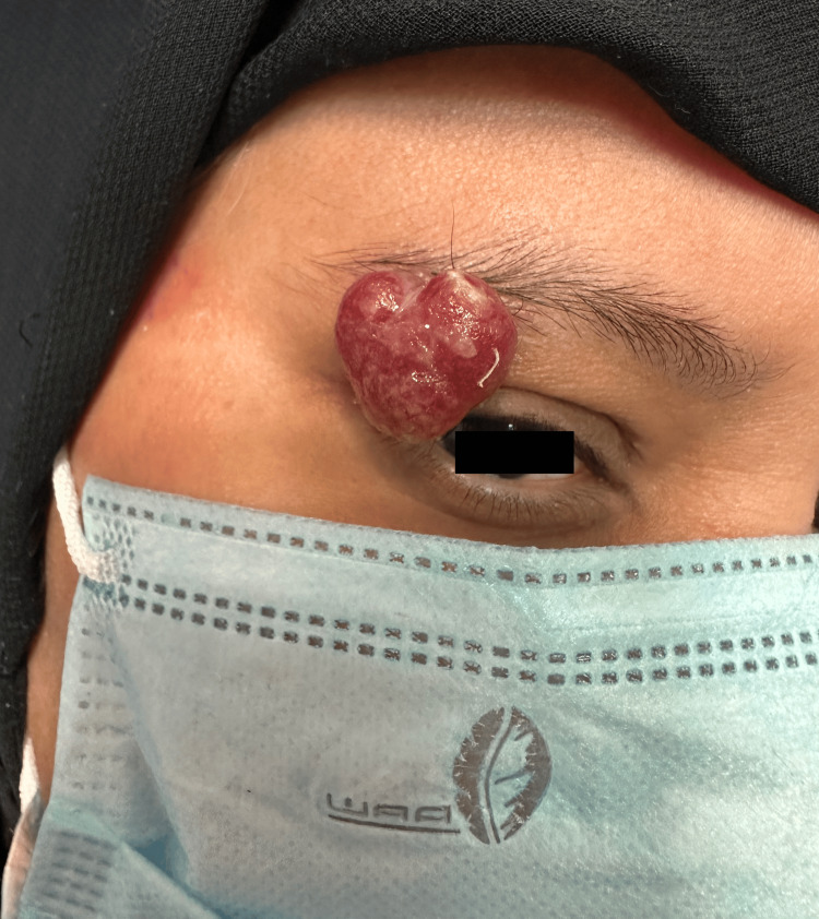 Figure 1
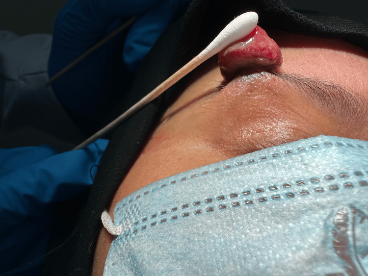 Figure 2
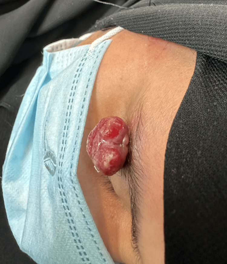 Figure 3
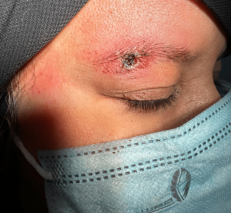 Figure 4
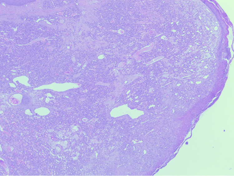 Figure 5
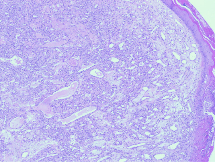 Figure 6
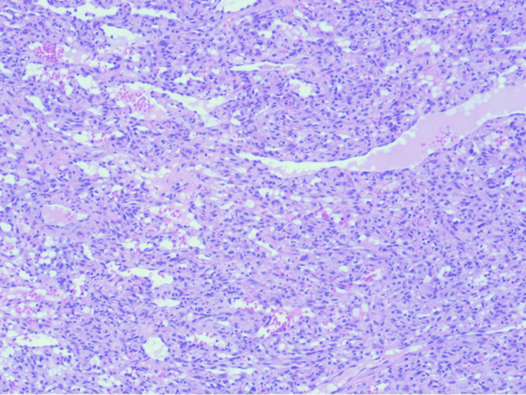 Figure 7
Peer Reviews
No public reviews on file for this paper yet. If you reviewed it on a platform where reviews are public (OpenReview, ICLR, NeurIPS, ICML), you can paste yours below so the community can read it here.
Videos
No videos yet. Explain this paper in a talk, walkthrough, or lecture? Add one.
Taxonomy
TopicsVascular Malformations and Hemangiomas · Neurofibromatosis and Schwannoma Cases · Vascular Malformations Diagnosis and Treatment
Introduction
Pyogenic granuloma (PG), also referred to as lobular capillary hemangioma, is a benign vascular lesion that develops in the skin and mucosal surfaces [1]. Despite its historical terminology, the condition is neither infectious nor granulomatous in nature; histologically, it is composed of clusters of proliferating capillaries separated by fibrous septa [2,3]. Clinically, PG usually presents as a solitary, reddish, pedunculated growth that expands quickly and bleeds with minimal trauma [2,3]. It occurs most often in younger individuals, children, and pregnant women, commonly affecting the gingiva, lips, digits, and other facial sites [4-6]. Involvement of less typical sites is distinctly uncommon, and eyebrow localization has been described only sporadically in the literature [1,2]. These rare presentations may imitate other vascular or malignant lesions such as amelanotic melanoma, angiosarcoma, or bacillary angiomatosis, thereby complicating the diagnostic process [1]. Presenting unusual cases contributes to the clinical understanding and supports accurate recognition. Here, we report the case of a 50-year-old woman with a pyogenic granuloma arising from the eyebrow, an anatomical location rarely encountered in practice.
Case presentation
A 50-year-old female patient with no prior medical conditions presented to the dermatology clinic with a painless, easily bleeding lesion over the right eyebrow that had persisted for 4 months. This was her first episode of such a lesion. She had previously attended several private clinics, where she was prescribed topical treatments, though she was unaware of their exact type and did not experience any benefit from them. Later, at a private clinic, she underwent an electrocautery procedure; however, the lesion subsequently recurred, enlarged, and caused repeated episodes of bleeding that required direct pressure to stop. In the few weeks leading up to her visit to our clinic, the lesion had shown progressive enlargement compared to earlier episodes, and it began to interfere with her daily activities by obscuring her vision due to its large size and downward extension over the right eye. A complete medical history was obtained and was unremarkable for trauma at the site or family history of similar lesions. The patient was not taking any medications. On the other hand, dermatological examination demonstrated a solitary, well-defined, exophytic, pedunculated, red to white, friable, lobulated nodule is seen arising from the right eyebrow region, measuring 2x1.5x1 cm. The surface appears smooth to slightly eroded and glistening (Figures 1-3).
Pyogenic granuloma over the right eyebrow, obscuring the eyelid.
Pyogenic granuloma on the right eyebrow elevated with a cotton swab.
Pyogenic granuloma on the right eyebrow
A provisional diagnosis of pyogenic granuloma was made. Under aseptic technique, local anesthesia with 1% lidocaine was administered, followed by shave excision of the lesion using a flexible DermaBlade (Vedo Trade LLC, Las Vegas, USA). Electrocautery was also applied to minimize bleeding during the procedure (Figure 4).
Post-shave excision site of the pyogenic granuloma on the right eyebrow.
The excised tissue was submitted for histopathological evaluation. The histopathological findings revealed a polypoid lesion with surface ulceration covered by fibrinous exudate (Figure 5). The lesion shows small capillaries arranged in a lobular fashion (Figure 6). Endothelial cells of the lesion show no atypia (Figure 7), which confirmed the diagnosis of pyogenic granuloma. The patient was followed clinically for one year with no recurrence, after which she was discharged from the dermatology clinic.
Low-power view of lobular capillary hemangioma showing a polypoid lesion comprised of lobules separated by fibrous septa. The lesion is covered by a focally ulcerated epidermis (hematoxylin-eosin stain, original magnifications x20).
The lobules are formed of capillary-sized vascular channels with several large feeding vessels. The stroma is edematous with few mixed inflammatory cells (hematoxylin-eosin stain, original magnifications x40).
The vascular channels are lined by a single layer of bland endothelial cells, with rare typical mitotic figures (hematoxylin-eosin stain, original magnifications x100).
Discussion
Pyogenic granuloma is considered a reactive vascular tumor-like lesion with multifactorial origins, including minor trauma, hormonal fluctuations, drug exposure, and, in some cases, genetic alterations [2,6]. Certain pharmacologic agents such as retinoids (including isotretinoin) and BRAF inhibitors have been implicated in its development [5-8]. More recent molecular research has identified *BRAF *and *RAS *mutations in a subset of patients, reinforcing the concept of aberrant angiogenesis as a key mechanism [9]. This evolving evidence suggests that PG is not only reactive but may also involve molecular drivers of vascular proliferation.
Anatomically, PGs most often involve the gingiva, lips, and fingers, whereas eyebrow presentations are extremely uncommon [2]. In one retrospective review of 155 cases, only five lesions were located in the eyebrow region [2]. Likewise, Giblin et al. evaluated 408 cases and observed that most lesions occurred on oral and acral surfaces, while uncommon sites such as the eyebrow remained exceptional and required histological confirmation to exclude malignancies [7]. Similarly, Wollina et al. noted the rarity of periocular and eyebrow cases despite the otherwise broad anatomical distribution of PG [6]. A recurrent eyebrow PG with satellitosis has also been documented, further demonstrating the atypical potential of lesions in this region [1].
Histopathologically, PG is characterized by lobular capillary proliferation with endothelial cells lacking atypical changes [2,5]. This description was consistent with the findings in our case. Because PG can mimic malignant or infectious vascular disorders, biopsy is necessary for a definitive diagnosis [1,6].
Surgical excision remains the most effective treatment approach, with recurrence rates substantially lower compared with other methods [2,6,7]. Giblin et al. reported excellent results with excision across hundreds of cases [7]. Nevertheless, other treatment modalities - such as curettage, cryotherapy, electrocautery, sclerotherapy, and topical or systemic beta-blockers like timolol and propranolol - have also been explored, especially in cosmetically sensitive or pediatric cases [3,5,6,10]. Reviews, including that of Plachouri and Georgiou, emphasize tailoring therapy to lesion size, site, and patient preference [10].
In our patient, shave excision followed by electrocautery was selected, balancing effectiveness with cosmetic outcomes and the need for prompt resolution. Unlike most eyebrow PGs, which are reported in younger individuals [1,2], our patient was 50 years old. Additionally, her lesion caused significant functional impairment by obstructing vision. These features distinguish this case from the majority of previously published reports.
Conclusions
Although PG is a frequent benign vascular lesion, eyebrow involvement is rarely encountered. This case underscores the importance of including PG in the differential diagnosis of unusual vascular nodules, particularly in cosmetically and functionally significant regions. Histological assessment remains essential for confirmation, and complete excision ensures both diagnostic certainty and excellent therapeutic results with minimal recurrence. Moreover, documenting such atypical presentations raises awareness among clinicians and highlights the need for careful examination of uncommon sites. It also emphasizes the value of individualized treatment approaches that consider both medical efficacy and cosmetic outcomes.
The reference list from the paper itself. Each links out to its DOI / PubMed record.
- 1Red-pink papules around a scar on the eyebrow Medicine Today Ho G Phan TA 6971222021 https://medicinetoday.com.au/dermatology-quiz/redpink-papules-around-scar-eyebrow
- 2Pyogenic granuloma: a retrospective analysis of cases treated over a 10-year Arch Craniofac Surg Koo MG Lee SH Han SE 16201820172891329710.7181/acfs.2017.18.1.16PMC 5556737 · doi ↗ · pubmed ↗
- 3Case report: pyogenic granuloma-just salt, a simple and pain-free treatment Dermatol Ther Bernales Salinas A Toro Sepúlveda A Meier Pincheira H Sánchez Castillo R 035202210.1111/dth.1519434750919 · doi ↗ · pubmed ↗
- 4Pyogenic granuloma in a 6-year-old boy: a rare case report Int J Contemp Pediatr Gera D Tanwar A Nigam AG Jain S Sharma V 607610102023
- 523-Year-old male with multiple giant facial pyogenic granulomas being treated with combined topical timolol and steroid: a case report Ann Med Surg (Lond) Kheder K Khaddam H Almohi Alsaid Mushaweh H Nabhan R 1045448220223626833310.1016/j.amsu.2022.104544 PMC 9577453 · doi ↗ · pubmed ↗
- 6Pyogenic granuloma - a common benign vascular tumor with variable clinical presentation: new findings and treatment options Open Access Maced J Med Sci Wollina U Langner D França K Gianfaldoni S Lotti T Tchernev G 423426520172878532310.3889/oamjms.2017.111PMC 5535648 · doi ↗ · pubmed ↗
- 7Pyogenic granuloma - the quest for optimum treatment: audit of treatment of 408 cases J Plast Reconstr Aesthet Surg Giblin AV Clover AJ Athanassopoulos A Budny PG 103010356020071747813510.1016/j.bjps.2006.10.018 · doi ↗ · pubmed ↗
- 8Pyogenic granuloma association with isotretinoin treatment for acne Australas J Dermatol Simmons BJ Chen L Hu S 0557201610.1111/ajd.1241827885661 · doi ↗ · pubmed ↗