Bilateral Persistent Pupillary Membrane
Hassan Asadigandomani, Mohammad Soleimani

TL;DR
A 31-year-old man with bilateral persistent pupillary membranes was found to have no vision impairment due to a spared central pupil area.
Contribution
This case highlights that extensive PPMs may not require surgery if the visual axis is unobstructed and the patient is beyond the amblyogenic period.
Findings
Bilateral PPMs were present but did not impair vision due to a 1.5 mm central pupil zone remaining clear.
The patient's age and unobstructed visual axis supported a non-interventional management approach.
PPMs were confirmed to be asymptomatic and not affecting visual development.
Abstract
A 31‐year‐old man presented for a routine eye exam with bilateral asymptomatic persistent pupillary membranes, with the central 1.5 mm zone of the pupil spared, preventing vision impairment. Persistent pupillary membranes (PPMs) are common congenital remnants that typically do not interfere with visual development and rarely lead to amblyopia. Consequently, surgical intervention is seldom required. In this case, despite extensive bilateral PPMs, the spared visual axis and the patient's age beyond the amblyogenic period justified an observation‐only approach.
Genes, proteins, chemicals, diseases, species, mutations and cell lines named across the full text — each resolved to its canonical identifier and authoritative record.
Click any figure to enlarge with its caption.
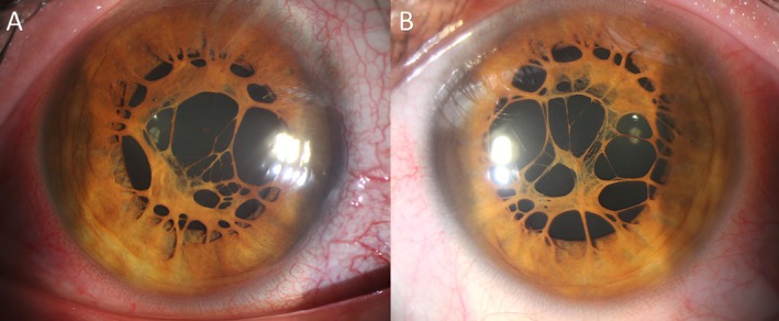 FIGURE 1
Peer Reviews
No public reviews on file for this paper yet. If you reviewed it on a platform where reviews are public (OpenReview, ICLR, NeurIPS, ICML), you can paste yours below so the community can read it here.
Videos
No videos yet. Explain this paper in a talk, walkthrough, or lecture? Add one.
Taxonomy
TopicsSympathectomy and Hyperhidrosis Treatments · Glaucoma and retinal disorders · Nasolacrimal Duct Obstruction Treatments
Case Presentation
1
A persistent pupillary membrane (PPM) is a congenital ocular anomaly, representing remnants of the anterior tunica vasculosa lentis, which nourishes the developing lens during embryogenesis [1, 2]. Most PPMs regress within the first year of life and require no intervention [2].
A 31‐year‐old healthy man presented for a routine eye examination. Uncorrected visual acuity (UCVA) was 20/20 in both eyes using a Snellen chart at 6 m, and best corrected visual acuity (BCVA) remained 20/20 bilaterally. Near visual acuity was Jaeger 1 (J1) in both eyes. Refraction was plano −0.75° × 120° for the right eye and plano −0.5° × 80° for the left eye. Biomicroscopy demonstrated transparent corneas, normal anterior chambers, and the absence of synechiae bilaterally. The gonioscopy findings and intraocular pressures (13 mmHg right eye, 14 mmHg left eye) were within normal limits. Slit‐lamp examination revealed extensive, radially oriented, and asymptomatic PPMs in both eyes (Figure 1A,B). The membranes measured approximately 2–3 mm in length, extending from the iris collarette toward the center but sparing the central 1.5 mm visual axis in both eyes. The dilated fundus examination was normal and unremarkable.
Slit‐lamp examination revealed extensive persistent pupillary membranes in the right (A) and the left eye (B). Fortunately, the 1.5 mm central zone of the pupil was not involved, preventing the development of amblyopia.
Importantly, the central 1.5 mm zone of the pupil, corresponding to the visual axis, was completely spared, resulting in preserved visual acuity and no signs of amblyopia. The sparing of the central 1.5 mm visual axis in both eyes prevented form‐deprivation amblyopia during the critical period of visual development. This underscores the importance of carefully evaluating the extent and location of PPMs, as peripheral involvement without axis obstruction may require no intervention [3].
Author Contributions
Hassan Asadigandomani: data curation, writing – original draft, writing – review and editing. Mohammad Soleimani: conceptualization, data curation, writing – review and editing.
Funding
The authors have nothing to report.
Consent
Written informed consent was obtained, ensuring patient confidentiality and de‐identification.
Conflicts of Interest
The authors declare no conflicts of interest.
The reference list from the paper itself. Each links out to its DOI / PubMed record.
- 1M. Gavriş , I. Horge , E. Avram , R. Belicioiu , I. A. Olteanu , and H. Kedves , “Persistent Pupillary Membrane or Accessory Iris Membrane?,” Romanian Journal of Ophthalmology 59, no. 3 (2015): 184–187.26978889 PMC 5712966 · pubmed ↗
- 2J. V. Forrester , A. D. Dick , P. G. Mc Menamin , F. Roberts , and E. Pearlman , The Eye: Basic Sciences in Practice (Elsevier Health Sciences, 2015).
- 3R. K. Bafna , M. Tripathi , S. Kumari , et al., “The Enigma of Subnormal Vision in Persistent Pupillary Membrane,” Medical Hypotheses 148 (2021): 110514.33549962 10.1016/j.mehy.2021.110514 · doi ↗ · pubmed ↗