Optimising Gallium-68 (⁶⁸Ga) DOTATATE PET/CT Reconstruction in Neuroendocrine Tumours: A Paired Comparison of Penalised-Likelihood (BSREM/Q.Clear) and Ordered Subset Expectation Maximisation Algorithms
James A Temple, Rachna Prem

TL;DR
This study compares two image reconstruction methods for Ga-68 DOTATATE PET scans in neuroendocrine tumors, finding that the Bayesian Penalised Likelihood method improves image quality and signal metrics.
Contribution
The study evaluates the Bayesian Penalised Likelihood algorithm for Ga-68 DOTATATE PET scans in neuroendocrine tumors, identifying optimal parameters for improved image quality and quantitative metrics.
Findings
BPL reconstruction increased lesion SUVmax and SNR compared to VPFX across all β values.
Qualitative image quality was best at β = 800 with lower artefact scores.
BPL is a viable alternative for Ga-68 DOTATATE PET reconstruction in neuroendocrine tumors.
Abstract
Objective The aim was to determine whether “Q.Clear” (GE Healthcare, Bayesian Penalised Likelihood (BPL) reconstruction algorithm) of Gallium-68 (⁶⁸Ga) DOTATATE PET scans at different penalisation factors (β) could improve qualitative and quantitative image parameters compared with the standard Ordered Subsets Expectation Maximisation (OSEM), VPFX reconstruction. Methods Twenty-five PET/CT scans performed 60 minutes after injection of 110-224 MBq (activity 153 MBq/kg) of ⁶⁸Ga-DOTATATE on a GE Discovery 710 PET/CT scanner were reconstructed using VPFX (2 iterations, 24 subsets) and Q.Clear with β values ranging from 200-1200. A representative neuroendocrine tumour (NET) lesion and three reference regions (liver, spleen, and L3 bone marrow) were measured for standardised uptake values (SUVₘₐₓ/mean/peak/SD), signal-to-noise ratio (SNR = SUVₘₐₓ/liver SUVSD), and signal-to-background…
Genes, proteins, chemicals, diseases, species, mutations and cell lines named across the full text — each resolved to its canonical identifier and authoritative record.
Click any figure to enlarge with its caption.
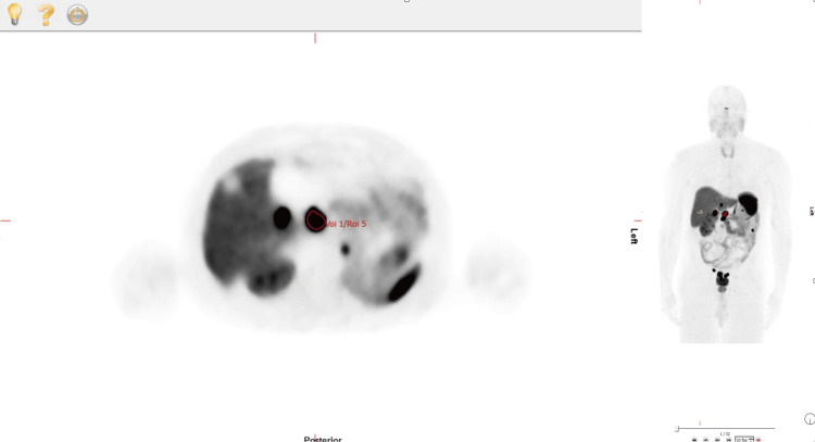 Figure 1
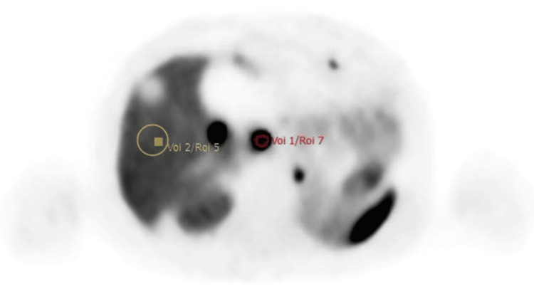 Figure 2
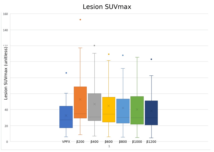 Figure 3
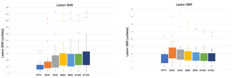 Figure 4
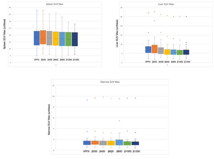 Figure 5
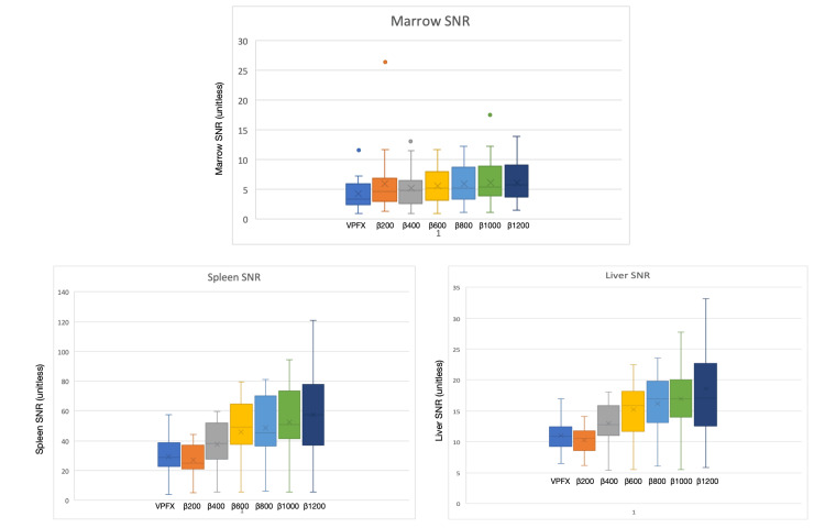 Figure 6
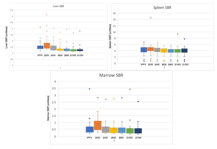 Figure 7
Peer Reviews
No public reviews on file for this paper yet. If you reviewed it on a platform where reviews are public (OpenReview, ICLR, NeurIPS, ICML), you can paste yours below so the community can read it here.
Videos
No videos yet. Explain this paper in a talk, walkthrough, or lecture? Add one.
Taxonomy
TopicsMedical Imaging Techniques and Applications · Radiopharmaceutical Chemistry and Applications · Radiomics and Machine Learning in Medical Imaging