From loops to caps: discriminating peptide binding to distinct G-quadruplex tetrads using 5-furyl-2′-deoxyuridine fluorescent probes
Jack Barr, Tayler D. Prieto Otoya, Christine Cardin, Enrico Cadoni

TL;DR
A fluorescent probe helps track how peptides bind to specific parts of G-quadruplex structures.
Contribution
A new method using 5FU fluorescent probes distinguishes peptide binding to specific G-quadruplex tetrads.
Findings
5FU labels at G4 caps enable fluorescence turn-on for tetrad-specific binding detection.
The method reveals tetrad preference and binding affinities of RHAU23 peptides.
The approach works on T95-2T and other parallel G-quadruplex structures.
Abstract
We report the use of 5-furyl-2′-deoxyuridine (5FU) as a fluorescent probe to distinguish ligand binding to distinct G-quadruplex tetrads. Site-specific incorporation of 5FU into T95-2T at the capping regions enabled discrimination of peptide-binding events via fluorescence turn-on, providing insights into tetrad preference and binding affinities using the 5FU-G4/RHAU peptide system. Site-specific 5-furyl-2′-deoxyuridine (5FU) labels at G-quadruplex (G4) caps generate fluorescence turn-on readouts that resolve tetrad-specific binding of peptide RHAU23, enabling tetrad affinity determination on T95-2T and other parallel G4s.
Genes, proteins, chemicals, diseases, species, mutations and cell lines named across the full text — each resolved to its canonical identifier and authoritative record.
Click any figure to enlarge with its caption.
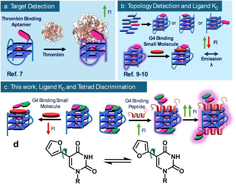 Figure 1
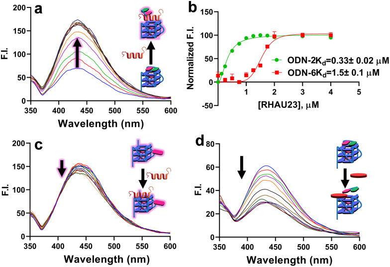 Figure 2
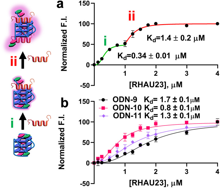 Figure 3
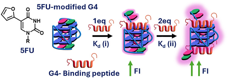 Figure 4
 Figure 5
Peer Reviews
No public reviews on file for this paper yet. If you reviewed it on a platform where reviews are public (OpenReview, ICLR, NeurIPS, ICML), you can paste yours below so the community can read it here.
Videos
No videos yet. Explain this paper in a talk, walkthrough, or lecture? Add one.
Taxonomy
TopicsDNA and Nucleic Acid Chemistry · Chemical Synthesis and Analysis · Click Chemistry and Applications