Renal capsule patient-derived xenograft model for gastric cancer: establishment and MRI characterization
Zhenyu Yin, Qian Liu, Ewetse Paul Maswikiti, Zhuanfang Wang, Yuping Bai, Lin Xiang, Yuhan Wang, Bin Ma, Lei Gao, Jianming Shi, Hao Chen

TL;DR
This study establishes a gastric cancer patient-derived xenograft model under the mouse kidney and identifies optimal conditions for tumor growth and MRI imaging techniques.
Contribution
The study identifies optimal tumor fragment size and transplantation timing for successful engraftment and evaluates MRI parameters for monitoring gastric cancer xenografts.
Findings
2×2×2 mm tumor fragments transplanted within 2 hours had the highest engraftment success rate.
T2-weighted MRI was most effective for detecting and measuring tumor size in xenografts.
PDX tumors retained histological and Ki67 expression features of primary tumors across generations.
Abstract
Identify factors associated with the engraftment of gastric cancer patient-derived xenograft (GC PDX) in the renal capsule and explore optimal MRI sequence parameters for observing renal capsule PDX. Tumor tissues from 33 gastric cancer patients were cut into fragments of 1×1×1 mm, 2×2×2 mm, and 3×3×3 mm, then transplanted beneath the renal capsule of NOD/SCID mice within 2, 8 and 24 hours. Depending on tissue availability, tumor samples from each patient were implanted into 1–4 mice, totaling 73 mice. Clinical data were collected. Tumor growth was monitored weekly via MRI. T1WI, contrast-enhanced T1WI, T2WI was used to measure tumor length. After euthanasia (10g/L sodium pentobarbital, 180mg/kg, intraperitoneal), tumors were excised, and caliper-measured were compared with MRI results. The xenografts were serially passage into new mice for three generations. Histopathological (H&E),…
Genes, proteins, chemicals, diseases, species, mutations and cell lines named across the full text — each resolved to its canonical identifier and authoritative record.
Click any figure to enlarge with its caption.
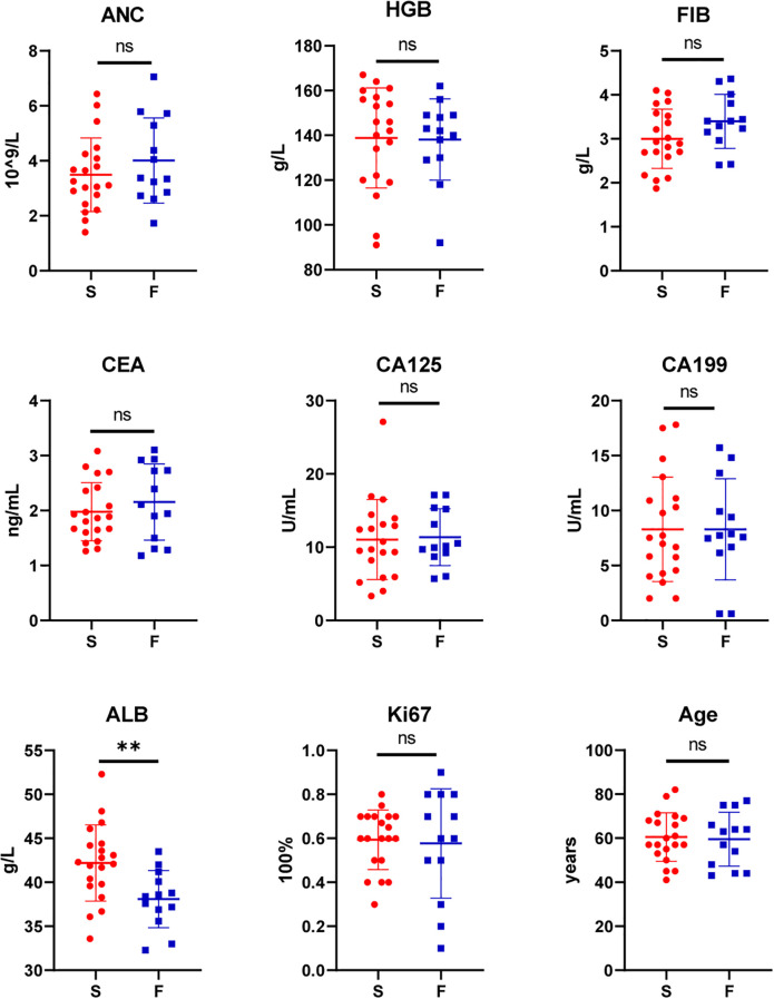 Figure 1
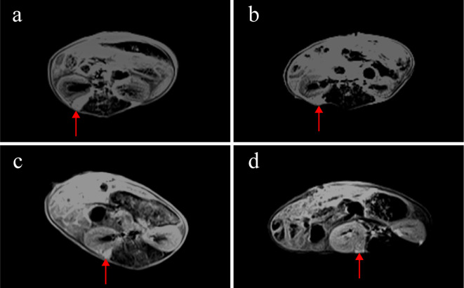 Figure 2
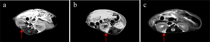 Figure 3
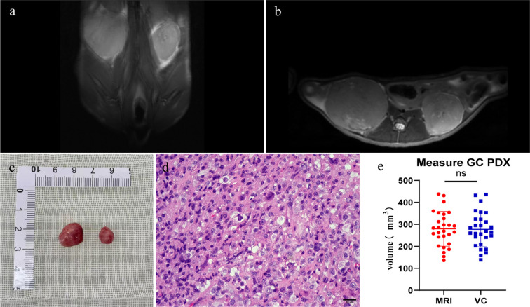 Figure 4
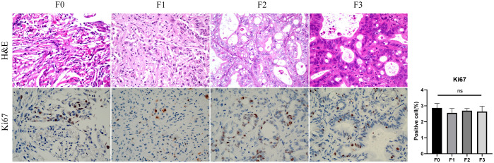 Figure 5
Peer Reviews
No public reviews on file for this paper yet. If you reviewed it on a platform where reviews are public (OpenReview, ICLR, NeurIPS, ICML), you can paste yours below so the community can read it here.
Videos
No videos yet. Explain this paper in a talk, walkthrough, or lecture? Add one.
Taxonomy
TopicsGastric Cancer Management and Outcomes · MRI in cancer diagnosis · Esophageal Cancer Research and Treatment