Shape analysis of the amygdala, hippocampus and thalamus in former American football players
Omar John, Alana Wickham, Leonard B Jung, Anya S Mirmajlesi, Jared Stearns, Katherine Breedlove, Nicholas Kim, Daniel H Daneshvar, Tashrif Billah, Ofer Pasternak, Arushi Chamaria, Michael J Coleman, Yorghos Tripodis, Charles H Adler, Charles Bernick, Laura J Balcer

TL;DR
This study finds that former American football players have structural brain changes in the hippocampus, amygdala, and thalamus, linked to early exposure and head impacts.
Contribution
The study introduces surface-based shape metrics as potential in vivo markers for brain changes due to repetitive head impacts in athletes.
Findings
Former football players showed bilateral surface area contractions in the hippocampus and amygdala compared to controls.
Earlier age of first football exposure correlated with surface contractions in the thalamus and left hippocampus.
Greater cumulative linear acceleration was linked to hippocampal surface contractions and reduced thalamic thickness.
Abstract
Repetitive head impacts are common in contact and collision sports and are linked to structural brain changes and an elevated risk of neurodegenerative diseases such as Chronic Traumatic Encephalopathy. Identifying early in vivo structural markers remains challenging. Although diagnosis currently requires post-mortem confirmation, clinical symptoms, including cognitive impairment and behavioural changes, are reflected in the diagnosis of Traumatic Encephalopathy Syndrome. These symptoms align with dysfunction in key brain regions—amygdala, hippocampus and thalamus—which support memory, emotion and behaviour and commonly show tau pathology in Chronic Traumatic Encephalopathy. This study uses shape analysis to examine structural differences in these regions between former American football players and unexposed asymptomatic controls and evaluates the influence of age, head impact exposure…
Genes, proteins, chemicals, diseases, species, mutations and cell lines named across the full text — each resolved to its canonical identifier and authoritative record.
Click any figure to enlarge with its caption.
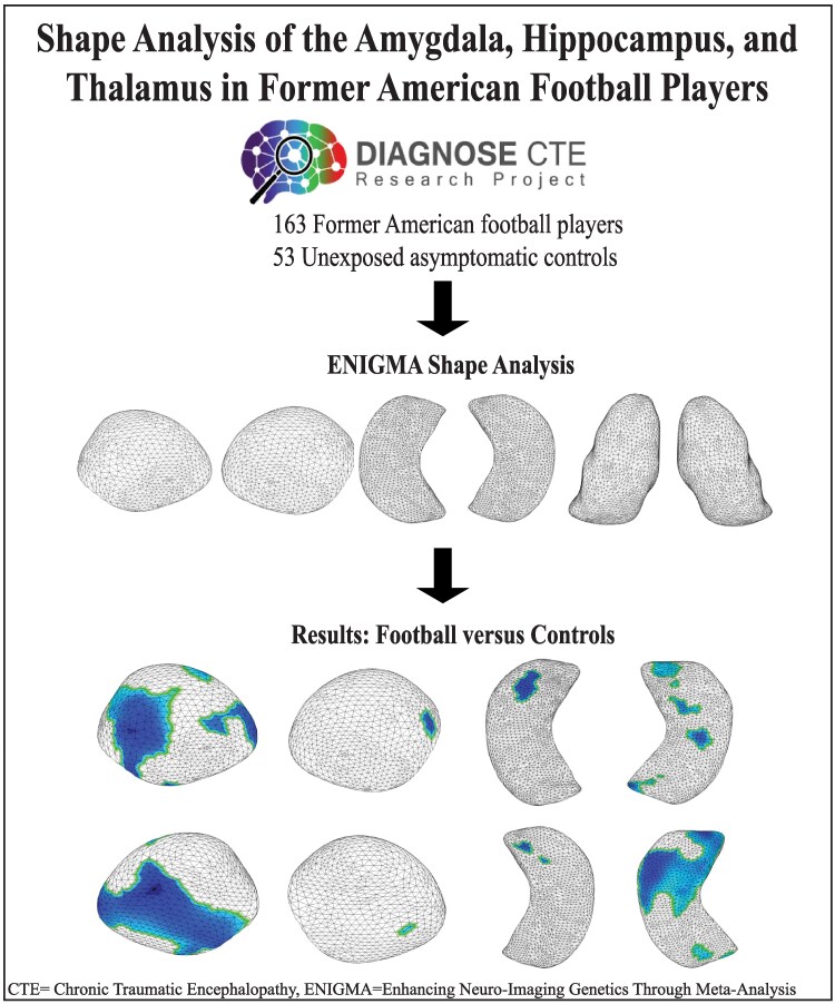 Figure 1
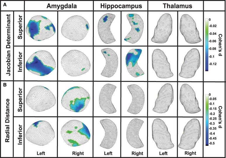 Figure 2
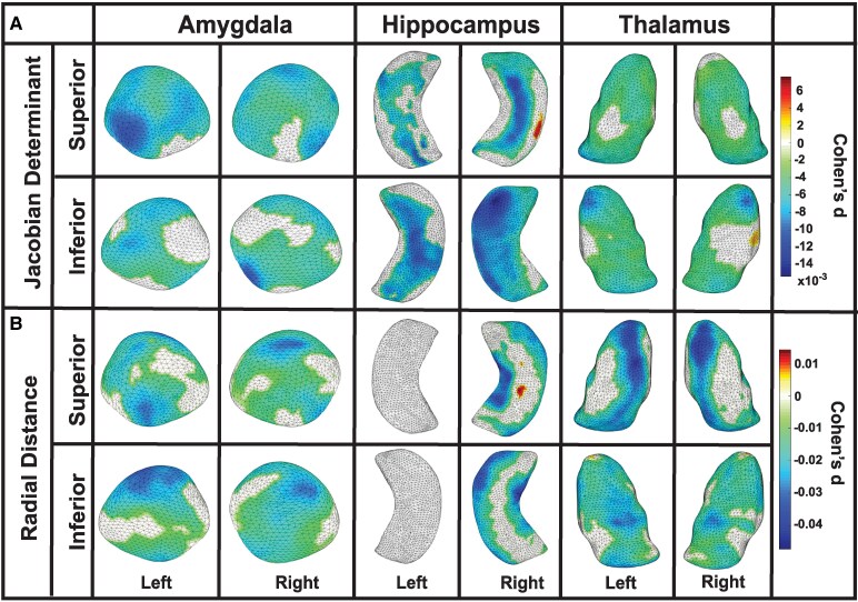 Figure 3
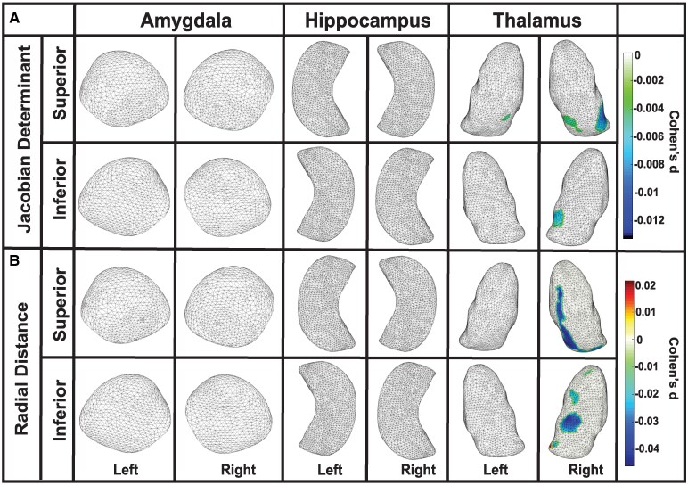 Figure 4
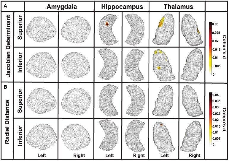 Figure 5
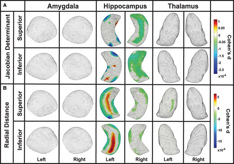 Figure 6
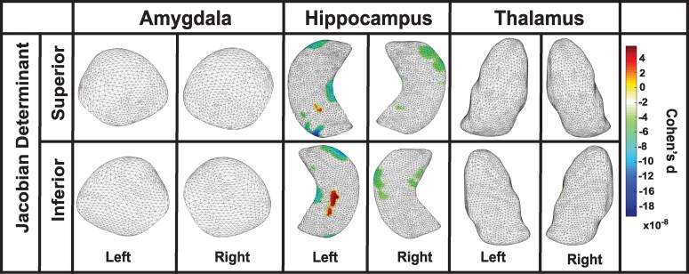 Figure 7
Peer Reviews
No public reviews on file for this paper yet. If you reviewed it on a platform where reviews are public (OpenReview, ICLR, NeurIPS, ICML), you can paste yours below so the community can read it here.
Videos
No videos yet. Explain this paper in a talk, walkthrough, or lecture? Add one.
Taxonomy
TopicsTraumatic Brain Injury Research · Advanced Neuroimaging Techniques and Applications · Functional Brain Connectivity Studies