Multispectral optoacoustic tomography of salivary glands in patients with clinically suspected Sjögren’s disease: A pilot study
Rik de Jong, Milou E. Noltes, Hendrika Bootsma, Gooitzen M. van Dam, Andor W.J.M. Glaudemans, Schelto Kruijff, Riemer H.J.A. Slart, Alja Stel, Max J.H. Witjes, Konstantina Delli, Jasper Vonk

TL;DR
This pilot study explores using multispectral optoacoustic tomography (MSOT) as a non-invasive imaging method to diagnose Sjögren’s disease by analyzing salivary gland hemoglobin signals.
Contribution
The study introduces a novel MSOT scoring system based on hemoglobin signals for non-invasive SjD classification.
Findings
MSOT achieved 92% sensitivity and 100% specificity for SjD classification using predefined cut-off values.
SjD patients showed significantly higher hemoglobin-related signals compared to non-SjD patients.
MSOT outperformed established ultrasound scoring systems and other diagnostic tests in this pilot study.
Abstract
Sjögren’s disease (SjD) is a systemic auto-immune disease characterized by salivary gland inflammation and glandular dysfunction. Diagnosis is challenging due to its heterogeneity and currently relies on a variety of tests that are present in the ACR-EULAR classification criteria. These include invasive and resource-intensive tests, highlighting the unmet need for a single, accurate, non-invasive diagnostic modality. Multispectral optoacoustic tomography (MSOT), enabling functional imaging of hemoglobin-related parameters, may address this gap. This pilot study evaluates MSOT's potential for salivary gland imaging in patients suspected of SjD. This study included 20 patients clinically suspected of SjD. Which underwent MSOT imaging of the major salivary glands, alongside the full ACR-EULAR diagnostic workup, including serology, salivary gland biopsy, and sialometry, alongside salivary…
Genes, proteins, chemicals, diseases, species, mutations and cell lines named across the full text — each resolved to its canonical identifier and authoritative record.
Click any figure to enlarge with its caption.
 Figure 1
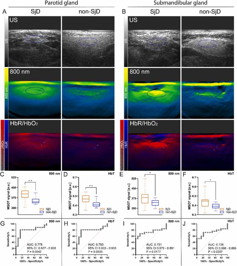 Figure 2
Peer Reviews
No public reviews on file for this paper yet. If you reviewed it on a platform where reviews are public (OpenReview, ICLR, NeurIPS, ICML), you can paste yours below so the community can read it here.
Videos
No videos yet. Explain this paper in a talk, walkthrough, or lecture? Add one.
Taxonomy
TopicsPhotoacoustic and Ultrasonic Imaging · Salivary Gland Disorders and Functions · Advanced X-ray and CT Imaging