Microbubble Contrast Agent Use During Invasive Coronary Microvascular Assessment
Rahul Bahl, Samay Mehta, Henry Seligman, Christopher A. Rajkumar, Suraya Gafore, Sayan Sen, Sukhjinder S. Nijjer, Rasha Al-Lamee, Daniel Chamié, Daniel Bandeira, Xiaowei Zhou, Matthew Shun Shin, Meng-Xing Tang, Ricardo Petraco

TL;DR
This study shows that using microbubble contrast agents can improve the quality of coronary flow measurements during invasive assessments, potentially making the technique more reliable and widely used.
Contribution
The novel finding is that microbubble contrast agents significantly enhance Doppler signal quality in cases with poor baseline signals during invasive coronary flow reserve assessment.
Findings
Microbubble contrast agents significantly improved signal-to-noise ratio, Doppler envelope quality, and reduced coefficient of variation in poor baseline signal cases.
Administration of microbubbles did not induce significant coronary hyperemia.
No improvements were observed in cases with already good baseline Doppler signal quality.
Abstract
Coronary microvascular function assessment with invasive Doppler-derived coronary flow reserve (CFR) has challenges related to Doppler signal quality, limiting clinical adoption. We investigated whether the use of ultrasound microbubble contrast agents can improve coronary flow signals and reduce measurement variability during invasive CFR measurement. Participants underwent assessment of coronary flow velocity using a Doppler sensor-tipped coronary wire. Baseline signal quality was determined by expert clinicians, and signals were categorized into good or poor quality. Doppler quality was then assessed at predetermined time periods after administration of SonoVue (Bracco): baseline, first pass, and subsequent discrete 20-second time periods, following this up to 260 seconds after first pass. For each time period, several parameters were assessed: signal-to-noise ratio, Doppler…
Genes, proteins, chemicals, diseases, species, mutations and cell lines named across the full text — each resolved to its canonical identifier and authoritative record.
Click any figure to enlarge with its caption.
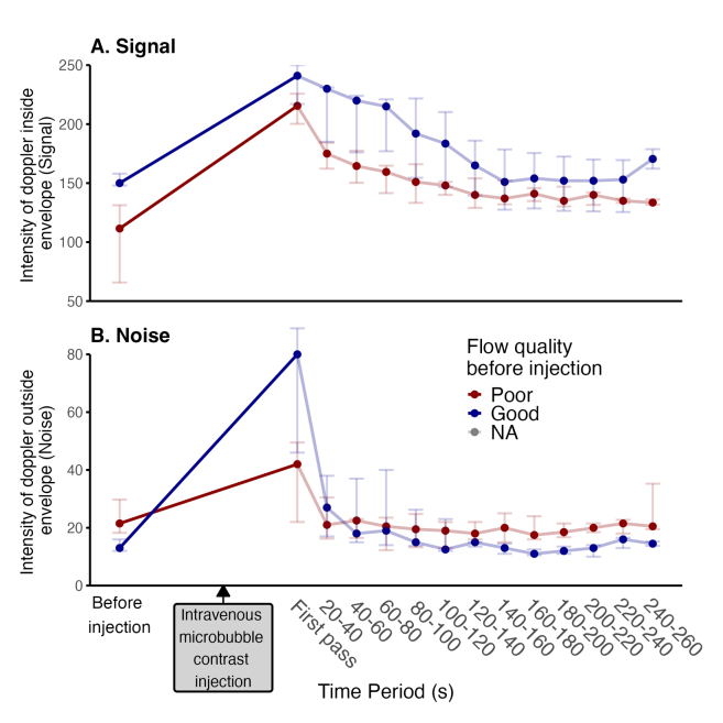 Figure 1
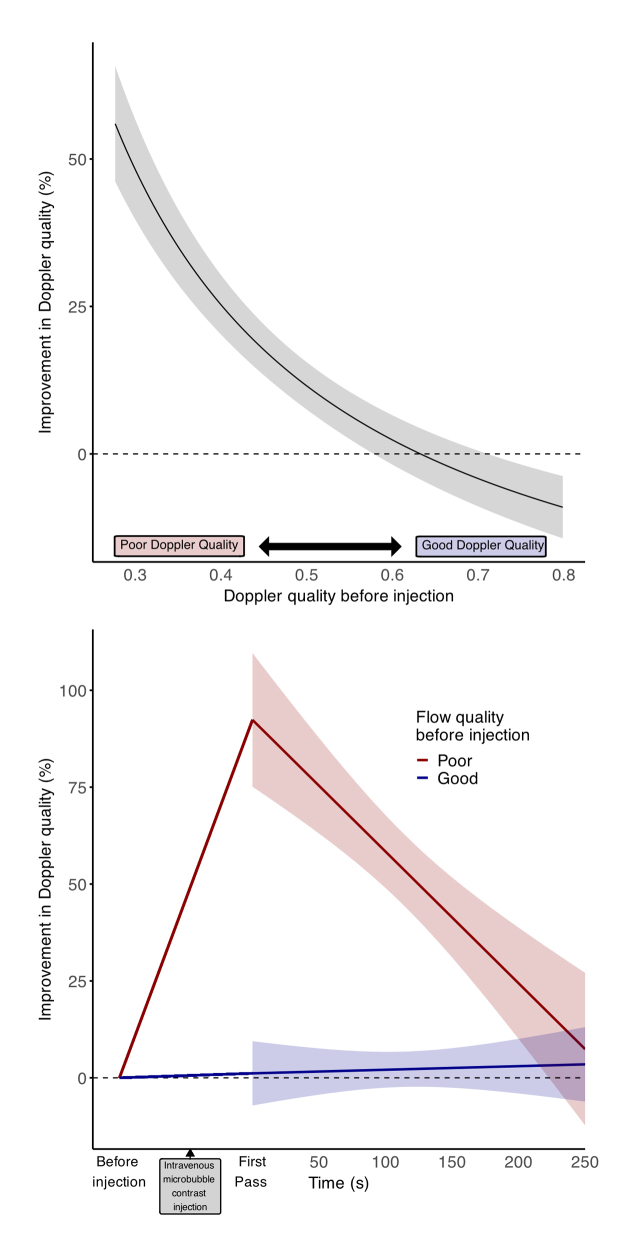 Figure 2
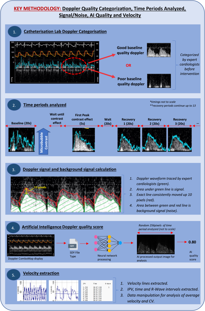 Figure 3
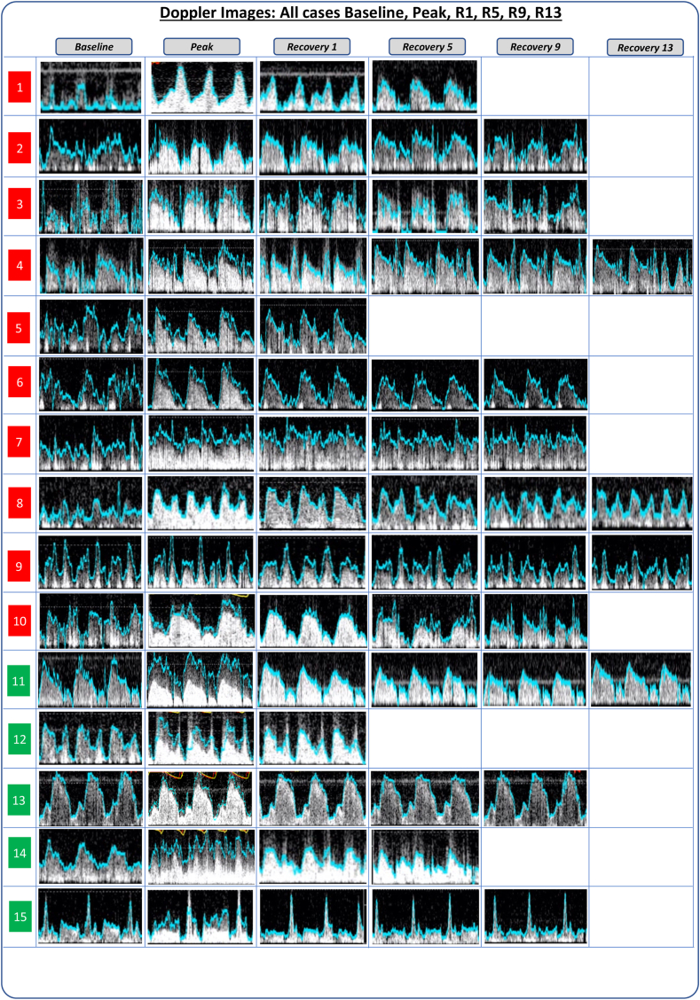 Figure 4
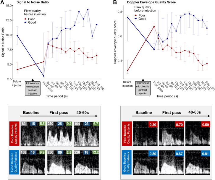 Figure 5
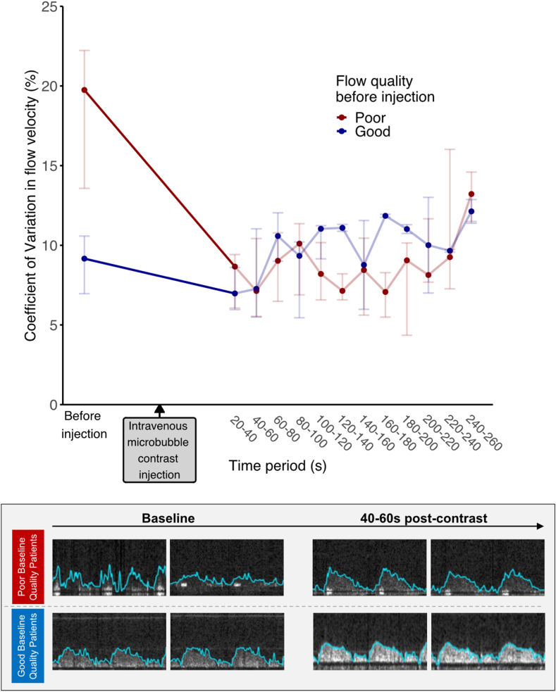 Figure 6
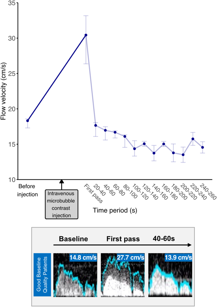 Figure 7
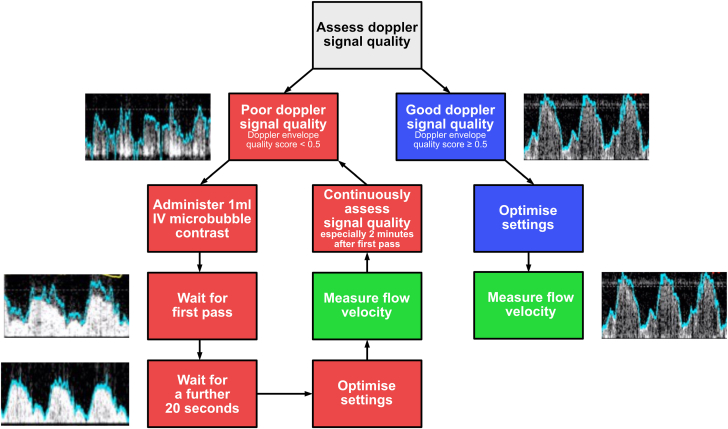 Figure 8
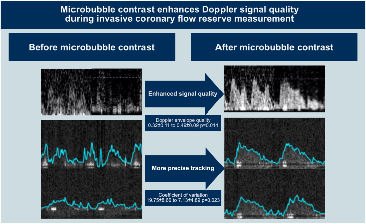 Figure 9
Peer Reviews
No public reviews on file for this paper yet. If you reviewed it on a platform where reviews are public (OpenReview, ICLR, NeurIPS, ICML), you can paste yours below so the community can read it here.
Videos
No videos yet. Explain this paper in a talk, walkthrough, or lecture? Add one.
Taxonomy
TopicsCardiac Imaging and Diagnostics · Ultrasound and Hyperthermia Applications · Venous Thromboembolism Diagnosis and Management