Systematic review of the prevalence of Gastrointestinal helminths in ruminants in Mexico
Roberto González-Garduño, Rosa Isabel Higuera-Piedrahita, Jorge Alfredo Cuéllar-Ordaz, Abel Villa-Mancera, Pedro Mendoza-de Gives, J. Felipe Torres-Acosta

TL;DR
This paper reviews the prevalence of gastrointestinal helminths in ruminants in Mexico over 37 years to inform control measures.
Contribution
A systematic review of helminth prevalence in Mexican ruminants using PRISMA guidelines and multiple databases.
Findings
Haemonchus contortus had a prevalence of 38.8% in ruminants.
Mecistocirrus prevalence in cattle was estimated at 40.5%.
Cooperia and Strongyloides were the most prevalent nematodes in cattle's small intestine.
Abstract
In Mexico, the abundance of endoparasites that affect the health of ruminants and the economy of the farmer, so the objective of this review was to determine the prevalence of the main genera and species of ruminant helminths that have been identified in Mexico in the last 37 years. A systematic search was carried out following the PRISMA 2020 guidelines and supported by the Elsevier platform (Scopus and ScienceDirect), Google Scholar, Redalyc and Scielo tools. In addition, information was sought in the proceedings Congress of the Buiatrics and Veterinary Parasitologists. A database was created with 36 documents containing information on the prevalence of gastrointestinal parasites in Mexico. Research studies that included treatments that affected prevalence were not included, nor were those of an experimental nature that did not aim to study prevalence. The prevalence for Haemonchus…
Genes, proteins, chemicals, diseases, species, mutations and cell lines named across the full text — each resolved to its canonical identifier and authoritative record.
Click any figure to enlarge with its caption.
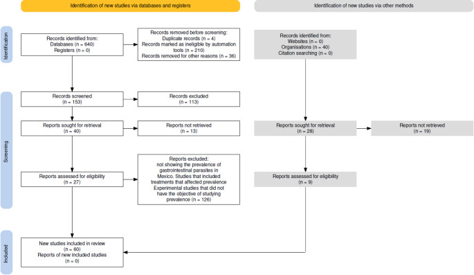 Figure 1
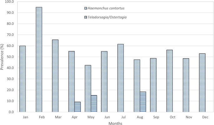 Figure 2
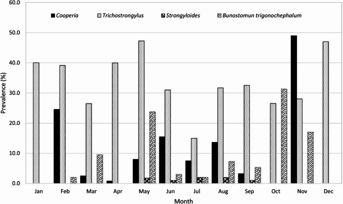 Figure 3
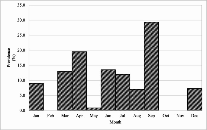 Figure 4
- —Dirección general de investigación y posgrado de la universidad autónoma chapingo
Peer Reviews
No public reviews on file for this paper yet. If you reviewed it on a platform where reviews are public (OpenReview, ICLR, NeurIPS, ICML), you can paste yours below so the community can read it here.
Videos
No videos yet. Explain this paper in a talk, walkthrough, or lecture? Add one.
Taxonomy
TopicsHelminth infection and control · Mollusks and Parasites Studies · Parasites and Host Interactions
Introduction
Identification of the genus and species of gastrointestinal parasites (GIP) affecting ruminants help to establish appropriate measures for parasite control on farms. In addition to coproparasitoscopic diagnosis, identification of adult parasites can be performed after necropsy of dead or slaughtered animals (González-Garduño et al. 2014). To obtain a diagnosis of the gastrointestinal nematodes (GIN) species involved, eggs are initially detected using a flotation technique or quantified using the McMaster technique (Thienpont et al. 2003; Cringoli et al. 2004), of which there are currently a number of variants such as the FLOTAC technique (Cringoli et al. 2010) and FECPAK^G2^ (Boelow et al. 2022). However, these techniques are limited in that they generally identify only the parasite family or, in some cases, the genus—but rarely allow for species-level identification among(Thienpont et al. 2003), therefore, one possibility for accurate diagnosis is the identification of infective larvae obtained from larval cultures to determine the genus and in some cases the species (van Wyk and Mayhew 2013). But when studies require the identification of parasites present in the infectious process, post-mortem studies must be performed that allow the recovery of adult parasites (López Ruvalcaba et al. 2013) and thus direct parasite control programs according to the species present in a region. Another possibility is the identification of the species through molecular biology techniques that are available in many research institutions (Amarante et al. 2017). In small ruminants, the importance of diagnosing gastrointestinal nematodes is essential because there are some that endanger the lives of animals. One of the most important is Haemonchus contortus, a highly prevalent and virulent species in sheep and goats. Its blood-feeding behavior often results in severe anemia, physical deterioration, and death in heavily infected or susceptible animals. In Mexico, climatic diversity causes an abundance of endoparasites that affect the health of ruminants and the economy of the farmer, so the objective of this review is to determine the prevalence of the main genera and species of ruminant helminths that have been identified in Mexico in the last 37 years.
The importance of diagnosing gastrointestinal nematodes (GINs) is paramount in small ruminants, as some species not only compromise productivity but also endanger the lives of animals. Among the most pathogenic is Haemonchus contortus, a blood-sucking nematode of the abomasum with high prevalence and virulence in sheep and goats. Its blood-feeding habit leads to severe anemia, hypoproteinemia, edema, and death in susceptible animals, particularly under conditions of high parasitic load (Besier et al. 2016; Adduci et al. 2022a, b; Kapó et al. 2024). Beyond its impact on animal health, haemonchosis is a primary constraint for goat production in regions such as East Africa (Githiori et al. 2006; Perry et al. 2002), and it is the most prevalent GIN in countries like Uganda (Nsereko et al. 2015; Kalule et al. 2023). Other GINs, such as species of the genus Trichostrongylus, also have a significant economic impact by affecting appetite and nutrient absorption or utilization in the small intestine (Francis et al. 2020). Furthermore, some Trichostrongylus species are of zoonotic importance, with a broad geographical distribution, particularly in the Middle East, Far East, and some African regions (Abbas and Hildreth 2022; Ghasemikhah et al. 2011). A high prevalence and diversity of trichostrongylid species in ruminants has been reported, as observed in northern Iran, where the total infection prevalence was 74.6% in sheep and 84.6% in goats (Hosseinnezhad et al. 2021). The control of these parasites is further complicated by the emerging issue of anthelmintic resistance, particularly to benzimidazoles, as genetically documented in H. contortus via mutations in the β-tubulin gene in countries like Uganda and Sudan (Kalule et al. 2023; Mohammedsalih et al. 2020).”
Materials and methods
To carry out this study, a systematic search was carried out following the procedure using the flow diagram (Fig. 1) of the Preferred Reporting Items for Systematic reviews and Meta-Analyses (PRISMA) 2020 website (Page et al. 2021).Fig. 1. Flow diagram for the search for information on the prevalence of helminths in Mexico according to PRISMA
Manuscripts were searched using the Elsevier platform (Scopus and ScienceDirect), Clarivate (Web of Science), Google Scholar, Redalyc and Scielo. Information was also sought in the proceedings of the congresses of the Mexican Association of Veterinarian Specializing in Bovines AC (Buiatría 2003, 2012, 2014, 2015, 2019, 2022) and the Mexican Association of Veterinary Parasitologists (AMPAVE 2012, 2017, 2019). Of 640 records displayed in the search engines that included articles, theses, books, meta-analyses and other references, only 36 were cited with information on the prevalence of gastrointestinal parasites in Mexico. Previously, 153 reviewed manuscripts were related to veterinary parasitology topics in ruminants, of which 113 were discarded as they did not indicate the prevalence of gastrointestinal parasites in Mexico and only the documents that addressed the prevalence of the main gastrointestinal nematodes (Haemonchus,* Ostertagia*,* Teladorsagia*,* Cooperia*), which infect ruminants in Mexico, were selected, so the scientific names were used as keywords and searched in English and Spanish, in addition the following words were used combined with the scientific names: prevalence, frequency, gastrointestinal nematodes, small ruminants, sheep, goats, buffalos, cows, cattle and the word Mexico was included in all searches. Research studies that included treatments with which the prevalences were affected or of experimental nature that did not have the objective of studying the prevalence were not included in the review (Table 1).Table 1. Gastrointestinal nematodes that parasitize the abomasum, small intestine, cecum and colon of cattle, sheep and goats according to the authors of prevalence studies in MexicoSample originAbomasumSmall intestineCecum and colonSpeciesNHacOstMecCooTricStronBunToxOesChaTrichAutorCattleSlaughterhouse100ӁӜӜ֍(Mejía García and Orozco de Gortari 1987)SheepFecal40ӁӜӜӜӜ֍(George Sánchez and Quiroz Romero 1993)CattleFecal1908ӁӁӜӜӜӜӜ֍֍(Domínguez Alpízar et al. 1993)RuminantsFecal3827Ӝ֍(Rodríguez-Vivas et al. 2001)SheepFecal312ӁӜ֍(Nahed-Toral et al. 2003)GoatsFecal132ӁӁӜӜӜ֍֍֍(Avelino et al. 2003)GoatsFecalNaӁӁӜӜӜӜ(Aguilar et al. 2003)GoatsFecal8ӁӜӜ֍(Torres-Acosta et al. 2004)CattleSlaughterhouse5ӁӜӜ(Vásquez-Prats et al. 2004)CattleFecal336ӁӜӜ֍(Olivares-Pérez et al. 2006)SheepFecal219ӁӜӜӜ֍֍(Rojas-Hernández et al. 2007)SheepSlaughterhouse242ӁӜӜӜӜ֍֍(González-Garduño et al. 2011)GoatsSlaughterhouse10ӁӜӜӜ֍(Olivares-Perez et al. 2012)SheepGrasslands30ӁӜӜ(Flota-Bañuelos et al. 2013)SheepFecal50ӁӁӜӜӜӜ֍(Acevedo-Ramírez et al. 2013)CattleSlaughterhouse68ӁӁ(von Son-de Fernex et al. 2014)CattleFecal214ӁӁӜӜӜ֍֍(Felipe et al. 2014)CattleFecal50ӁӜӜ(Landeros et al. 2014)SheepFecal29ӁӁӜӜӜ֍֍(Acevedo et al. 2014)CattleFecal214ӁӜӜӜ֍֍(Fernández-Figueroa et al. 2015)SheepGrasslands117ӁӁӜӜӜӜ֍֍֍(Sánchez Brito 2017)BuffaloesFecal383Ӝ֍(Ojeda-Robertos et al. 2017)SheepFecal90ӁӁӜӜ֍֍(Acevedo-Ramírez et al. 2017)CattleFecal100ӁӜӜ֍(Ortiz-Timoteo et al. 2017)GoatsFecal101ӁӜӜ(Figueroa Antonio et al. 2018)GoatsFecal371ӁӁӜ(Olivas-Salazar et al. 2018)GoatsSlaughterhouse499ӁӜӜӜ֍(Munguía-Xóchihua et al. 2018)GoatsFecal262Ӝ֍(Barrón et al. 2019)SheepFecal177ӁӁӜӜ֍֍(Castellanos et al. 2019)CattleFecal411ӁӁӜӜ(Castillo et al. 2019)CattleFecal32ӜӜ֍(Hernández et al. 2019)SheepFecalNaӁӜӜ(Pérez et al. 2019)SheepFecal216ӁӜӜӜ֍(Camacho Ronquillo et al. 2021)CattleFecal185Ӝ֍(Ortiz-Muñoz et al. 2021)SheepFecal300ӁӜӜӜӜ(García et al. 2022)CattleFecal190ӁӁӜ֍֍(Salazar et al. 2022)SheepFecal126ӁӜӜ֍(Valladares-Carranza et al. 2024)Total authors281322726199721811N Number of samples. Hac Haemonchus, Ost Ostertagia, Mec Mecistocirrus, Coo Cooperia, Tric Trichostrongylus, Stron Strongyloides, Bon Bunostomum, Tox Toxocara, Oes Oesophagostomum, Cha Chabertia, Trich Trichuris. Na not available
For the analysis of the information, the JASP program (JASP Team, 2024) was used to obtain measures of central tendency and dispersion from the database formed in EXCEL, in which the prevalence averages were obtained and analyzed together with independent variables such as the species of ruminant, the type of study (fecal, slaughterhouse or pasture), the state of the republic and the month of the year, so the model used was analyzed in SAS (SAS 2017):
\documentclass[12pt]{minimal} \usepackage{amsmath} \usepackage{wasysym} \usepackage{amsfonts} \usepackage{amssymb} \usepackage{amsbsy} \usepackage{mathrsfs} \usepackage{upgreek} \setlength{\oddsidemargin}{-69pt} \begin{document}$$y_{ijklm}=\mu+\gamma_i+\gamma\tau_{i\left(j\right)}+\delta_k+\theta_1+\varepsilon_{ijklm}$$\end{document}where \documentclass[12pt]{minimal} \usepackage{amsmath} \usepackage{wasysym} \usepackage{amsfonts} \usepackage{amssymb} \usepackage{amsbsy} \usepackage{mathrsfs} \usepackage{upgreek} \setlength{\oddsidemargin}{-69pt} \begin{document}$$\:{\boldsymbol{y}}_{\boldsymbol{i}\boldsymbol{j}\boldsymbol{k}\boldsymbol{l}}=$$\end{document} Prevalence, µ = general mean, \documentclass[12pt]{minimal} \usepackage{amsmath} \usepackage{wasysym} \usepackage{amsfonts} \usepackage{amssymb} \usepackage{amsbsy} \usepackage{mathrsfs} \usepackage{upgreek} \setlength{\oddsidemargin}{-69pt} \begin{document}$$\:{\boldsymbol{\gamma\:}}_{\boldsymbol{i}}$$\end{document} = effect of the i-th specie (Buffalo, Cattle, Sheep, Goats), \documentclass[12pt]{minimal} \usepackage{amsmath} \usepackage{wasysym} \usepackage{amsfonts} \usepackage{amssymb} \usepackage{amsbsy} \usepackage{mathrsfs} \usepackage{upgreek} \setlength{\oddsidemargin}{-69pt} \begin{document}$$\:{\boldsymbol{\tau\:}}_{\boldsymbol{i}\left(\boldsymbol{j}\right)}$$\end{document} = effect of sample type nested in the i-th specie (fecal, slaughterhouse or pasture), \documentclass[12pt]{minimal} \usepackage{amsmath} \usepackage{wasysym} \usepackage{amsfonts} \usepackage{amssymb} \usepackage{amsbsy} \usepackage{mathrsfs} \usepackage{upgreek} \setlength{\oddsidemargin}{-69pt} \begin{document}$$\:{\boldsymbol{\delta\:}}_{\boldsymbol{k}}$$\end{document} = effect of k-th state of the republic (1,2,3, … 14), θl = effect of l-th month of year (l=January, February, … December), and \documentclass[12pt]{minimal} \usepackage{amsmath} \usepackage{wasysym} \usepackage{amsfonts} \usepackage{amssymb} \usepackage{amsbsy} \usepackage{mathrsfs} \usepackage{upgreek} \setlength{\oddsidemargin}{-69pt} \begin{document}$$\varepsilon$$\end{document} ijklm= experimental error.
Results
The study of gastrointestinal parasites in Mexico for 37 years (1987–2024) has considered Platyhelminthes and roundworms (Table 2) as the main agents that affect the health of ruminants, although there are other studies in which protozoa are also described as highly prevalent in Mexico (García et al. 2022; Valladares-Carranza et al. 2024). However, this review focuses primarily on nematodes; however, due to its high prevalence, the cestode Moniezia is also included.Table 2. Prevalence (%) of Gastrointestinal nematodes in Mexico, according to sample type collectedHaemonchusTeladorsagia/OstertagiaMecistocirrusSample typeN.N2PrevalenceSDN.N2PrevalenceSDN.PrevalenceCattleEggs/Larvae (Feces)71633.423.85526.441.3Slaughterhouse(worms)2516.010.3119.6-240.5GoatsEggs/Larvae (Feces)2520.819.964.33.9Slaughterhouse(worms)2763.621.91212.011.3SheepEggs/Larvae (Feces)102350.225.0487.86.7Slaughterhouse(worms)1137.2.Larvae (Pasture)32250.338.42335.031.4N Number or authors, N2 number of records, SD Standard deviation
The taxonomic classification of the prevalent nematodes in Mexico has been taken from a recent study of the classification of the Phylum Nematoda Cobb, 1932 (Hodda 2022) Therefore the order and family of each of them is listed below:
- Order Rhabditida, Family Trichostrongylidae Leiper, 1908: Haemonchus,* Cooperia*,* Trichostrongylus*,* Mecistocirrus. Ostertagia*,* Teladorsagia*.
- Order Rhabditida. Family Strongylidae Baird, 1853. Oesophagostomum,* Chabertia*.
- Order Rhabditida Family Ancylostomatidae Looss, 1905. Bunostoth highmum.
- Order Panagrolaimida. Family Strongyloididae Chitwood & MacIntosh, 1934. Strongyloides.
- Order Trichocephalida. Family Trichuridae Ransom, 1911. Trichuris.
- Order Spirurida, Family Ascarididae Baird, 1853. Toxocara.
Abomasal nematodes
The genus Haemonchus Cobb 1898, is the most studied due to its economic and veterinary importance. Many publications have been dedicated to describing this genus, such as the book edited by Gasser and Von Samsom-Himmelstjerna (Gasser and Von Samson-Himmelstjerna 2016) and many other authors who have described it in sheep (Flay et al. 2022; Mohamed et al. 2024), cattle (Jabbar et al. 2014; Haider et al. 2024), goat (Adduci et al. 2022a, b; Kapo et al. 2024) y South American camelids (Jabbar et al. 2013; Zahid et al. 2024). Of the 14 species currently recognized (Hodda 2022), Haemonchus contortus has been described as an abomasal parasite in sheep and other ruminants (Zarlenga et al. 2016). H. contortus and Haemonchus placei are also known as barber pole worms. They are the two main species of this parasite that is found in small ruminants and cattle, respectively, however, hybrids produced by crossing H. placei and H. contortus can be found, although they have low fertility or are sterile due to meiotic abnormalities (Amarante et al. 2017).
Another genus of abomasal nematode is Mecistocirrus Railliet & Henry, 1912 (Hodda 2022) with four species, which has a high prevalence in southeastern Mexico and has been little studied. Since 1965, there has been extensive knowledge of the life cycle of M. digitatus and of larval development and anatomical aspects of larvae and adults (Fernando 1965). In Mexico the discovery was reported for the first time in 1987 (Mejía García and Orozco de Gortari 1987), in that same year the third stage larva of the parasite was described(García Ortiz and Mejía García 1987) and since then only one study indicated that M. digitatus was present in the humid tropical regions but they did not report its prevalence (Vásquez-Prats et al. 2004). Later, in 2013, a morphological description of this species observed in the state of Tabasco and Chiapas was made (González-Garduño et al. 2013). In the following year its presence was also indicated, and the identification was made by electron microscopy and by PCR in the Mexican tropics (von Son-de Fernex et al. 2014). In 2017, M. digitatus was identified from one animal at necropsy in a study group treated with netobimin under an organic milk production system (Ortiz Pérez et al. 2017).
In the subtribu Ostertaginii Skryabin & Schulz, 1937 (Hodda 2022), 10 species have been reported to be present in cattle, sheep and goats in North America. The species described include Ostertagia ostertagi and O. lyrata in cattle and Teladorsagia circumcincta, T. trifurcata and T. davtiani in sheep and goats with a worldwide distribution and are the main causative agents of ostertagiosis (Lichtenfels et al. 1988). Polymorphism has also been described among males, generating identification keys for nine species (Lichtenfels and Hoberg 1993). In addition, a comprehensive review has been conducted including life cycle characteristics and implantation rate of Ostertagia (Verschave et al. 2014). In recent studies, the identification of nematodes of the Teladorsagia genus from ruminants has been carried out with the help of species-specific markers based on ITS2 rDNA, with which it is possible to accurately detect eggs and larvae of this genus (Ibrokhimov et al. 2023). In Mexico, O. ostertagi was reported in a slaughterhouse study (González-Garduño et al. 2011) and more recently the importance and prevalence of Ostertagia has been addressed in cattle in southern Mexico (Villa-Mancera et al. 2018).
Prevalence of abomasal nematodes in Mexico
The average prevalence for H. contortus, the main abomasal nematode of ruminants in the different regions of Mexico was 38.8% (Table 2), mentioned by at least 28 authors. The prevalence by identifying larvae obtained by fecal cultures has been estimated at 40%. Similar values have been observed in studies with slaughtered animals obtaining adult parasites (43%). While studies using the recovery of larvae in pastures contaminated with GIN during grazing have shown a 50% prevalence.
In the case of the brown abomasal worm or Teladorsagia/Ostertagia, the values found in the literature indicate that cattle have the highest prevalence with identification of larvae and only one study indicates the recovery of larvae in pasture. In cattle and goats, the presence of Teladorsagia/Ostertagia identified in adults is reported. In the case of Mecistocirrus, only four studies indicate the prevalence of Mecistocirrus in cattle (Mejía García and Orozco de Gortari 1987; von Son-de Fernex et al. 2014), which was estimated at 40.5% (Table 2), in another study in the tropics of Mexico the presence of this nematode is also indicated, but without mentioning its prevalence (González-Garduño et al. 2013). On the other hand, in Ovis canadensis, a prevalence of 10 to 20% of M. digitatus has been indicated (García et al. 2022).
The distribution of H. contortus in cattle, sheep and goats in Mexico had the highest prevalence in southeastern states (51.4, 52.3 and 55.7% respectively). In the central states of Mexico, the prevalence in cattle and sheep is in second place (19.2 and 39.9% respectively) and only in the north of the country, the prevalence of this species in goats is in second place (29.6%) at the national level. It can also be noted that the largest number of studies have been carried out in sheep in which the prevalence is 39.7% and the lowest number of studies has been in goats with a prevalence of 35.9% (Table 3).Table 3. Prevalence (%) of Haemonchus contortus by ruminant species and state of the Mexican RepublicStateCattleGoatSheepNMeanSDNMeanSDNMeanSDEstado de México119.2239.613.4Morelos2040.330.7Puebla28.69.0Tlaxcala440.09.2Coahuila14.012.0Nayarit226.08.5Sinaloa217.26.815.6Sonora24.92.9355.26.8Chiapas180.0Guerrero185.1470.028.3132.0Tabasco137.2Veracruz1029.519.21187.811.9Yucatán339.628.3241.58.1Sureste433.04.6Average31.735.939.7N Number of studies, SD Standard deviation
The prevalence of Teladorsagia/Ostertagia in cattle was 6.6 to 13% in Sonora, Veracruz and Yucatan and exceptionally in the State of Mexico a study indicates a 100% prevalence (Castillo et al. 2019). In goats, these nematodes were reported in Guerrero and Puebla with 4 to 8% prevalence, while in sheep the highest prevalence was 35.5% in the State of Mexico, while in Morelos, Sinaloa and Veracruz the maximum values reached were 13%.
The distribution of H. contortus throughout all months of the year indicates that this species has a wide distribution without seasonality and with consistently high prevalence, likely attributable to its prolific reproductive rate, which can reach up to 1,295 eggs per female per day (Saccareau et al. 2017) (Fig. 2).
Fig. 2. Monthly prevalence of Haemonchus contortus and Teladorsagia/Ostertagia in Mexico
Nematodes of the small intestine
Cooperia
The genus Cooperia Ransom, 1907 has 33 species (Hodda 2022). In America, Cooperia curticei is the main species in sheep, while in temperate climates is C. oncophora, and the tropical species C. pectinata and C. punctata are the ones that parasitize and affect the health of cattle (Stewart 1954; Francis et al. 2020). These nematodes have a detrimental effect on both appetite and nutrient absorption or utilization by residing in the small intestine. Differentiation between C. pectinata and C. punctata is not possible in larvae, and it is very difficult to differentiate them from C. oncophora; the only reliable identification of a species is achieved by morphological analysis of adults, but this requires post-mortem analysis of the small intestine and is restricted to research settings (Francis et al. 2020), Therefore, molecular identification studies are more reliable. In Tabasco, Mexico, the presence of C. curticei and C. punctata was reported in 2013 and morphologically described in the small intestine of sheep and cattle, respectively (González-Garduño et al. 2014). Work has also been carried out on resistance to natural infection by Cooperia spp. in young Zebu x Holstein crossbred cattle in the tropics (García-Ruíz et al. 2019).
Trichostrongylus
The genus Trichostrongylus Looss, 1905 has 53 species (Hodda 2022), it is one of the most important zoonotic nematodes with wide geographic distribution in the world (Ghasemikhah et al. 2011; Abbas and Hildreth 2022). Trichostrongylus was widely described during 1932 and its presence in humans and ruminants is highlighted. The main species described were: T. retortaeformis Looss, 1905. T. colubriformis Ransom, 1911. T. capricola Ransom, 1907. T. axei Railliet and Henry, 1909. T. vitrinus Looss, 1905. T. falculatus Ransom, 1911. T. rugatus Monnig, 1925. T. tenuis Shipley, 1909. T. pergracilis Shipley, 1909. T. affinis Graybill, 1924. T. calcaratus Ransom, 1911. T. probolurus Looss, 1905. T. orientalis Jimbo, 1914. Due to the large number of Trichostrongylus species, molecular identification has become important, and a large number of studies have been published that identify this species molecularly (Zarlenga et al. 2001; Bakooie Katrimi et al. 2022).
Strongyloides
The genus Strongyloides Grassi, 1879 has 63 species (Hodda 2022). Although Strongyloides have been reported in different species of animals including ruminants and pigs and even birds and amphibians, the discovery was in humans with S. stercoralis. A broad description of this genus was made in 1925 (Sandground 1925) and later a study carried out in 1966 (Little 1966) established the criteria for the identification of Strongyloides describing parasitic species such as S. stercoralis, S. fiilleborni,* S. cebus*,* S. myopotami*,* S. venezuelensis* y S. ratti. For the determination of Strongyloides, PCR has currently been used (Dorris et al. 2002; Kramme et al. 2011; Saugar et al. 2015).
Prevalence of small intestine nematodes in Mexico
Of the small intestine nematodes in Mexico, those with the highest prevalence in cattle were Cooperia and Strongyloides, which were also mentioned in the largest number of records. In buffaloes, Strongyloides was also the nematode with the highest prevalence and the most reported. In goats and sheep, the nematode with the highest prevalence was Trichostrongylus, although Toxocara had a prevalence of 67% in a single study in goats, while in sheep only the reported prevalence was 6% (Table 4).Table 4. Prevalence (%) of the main genera of small intestine helminths in ruminants in MexicoCattleBuffalosGoatSheepGenusNMeanSDNMeanSDNMeanSDNMeanSDCooperia1746.25.51021.07.6348.01.8Trichostrongylus139.73.61554.68.44830.34.0Strongyloides2116.62.5441.86.9719.03.71814.94.9Bunostomun74.52.5110.0.219.42.4Toxocara42.00.913.9.167.4.16.1.Moniezia135.71.1319.24.899.44.9614.66.0N Number of studies, SD Standard deviation
According to the stage at which prevalence was determined, Trichostrongylus was the GIN with the highest prevalence identified as an adult parasite obtained in a slaughterhouse. It was followed by Cooperia, and only one study indicated the presence of Strongyloides in a slaughterhouse (Table 5). The larvae recovered from copro-cultures corresponded to the highest prevalence of Cooperia in cattle and Trichostrongylus in goats. Only in sheep a prevalence study recovering larvae in pasture.Table 5. Prevalence (%) of the three main genera of gastrointestinal nematodes of the small intestine in ruminants in MexicoSpecies and sample typeCooperiaTrichostrongylusStrongyloidesN.N2PrevalenceSDN.N2PrevalenceSDN.N2PrevalenceSDBuffalos Eggs/Larvae (Feces)441.813.9Cattle Eggs/Larvae (Feces)81645.623.16139.712.862116.611.7 Slaughterhouse (worms)1157.0Goats Eggs/Larvae (Feces)367.87.541142.629.85719.09.8 Slaughterhouse (worms)1440.827.81487.55.0Sheep Eggs/Larvae (Feces)8188.110.7112421.519.371515.822.4 Slaughterhouse (worms)1136.01125.2113.3 Larvae (Pasture)3155.97.932339.832.52214.317.6Average28.737.713.8N Number or authors, N2 number of records, SD Standard deviation
In the genera with the lowest prevalence, only in one case a high prevalence value was observed in Toxocara in goats with 67%, while the second highest value corresponded to the cestode Moniezia expansa in sheep (Table 6).Table 6. Prevalence (%) of the genera of gastrointestinal parasites of the small intestine with lower prevalence in ruminants in MexicoBunostomumToxocaraMonieziaSpecies and sample typeN.N2PrevalenceSDN.N2PrevalenceSDN.N2PrevalenceSDBuffalos Eggs/Larvae (Feces)113.91319.28.4Cattle Eggs/Larvae (Feces)2210.312.6332.42.06116.63.7 Slaughterhouse(worms)252.21.8111.0221.11.3Goats Eggs/Larvae (Feces)1167.4599.414.7 Slaughterhouse(worms)1110.0Sheep Eggs/Larvae (Feces)5123.24.13410.39.2 Slaughterhouse(worms)110.8116.2 Larvae (Pasture)1819.910.3116.11140.4Average7.719.212.3N Number or authors, N2 number of records, SD Standard deviation
In the monthly distribution it was noted that Trichostrongylus sp was reported in all months of the year with a prevalence higher than 20% except in July, while Cooperia sp had no records from October to January and its prevalence was less than 25% except in November when it almost reached 50% prevalence. Bunostomum also showed high prevalence in May and October and Strongyloides has been reported with a prevalence less than 3% (Fig. 3).Fig. 3. Prevalence of the main species of nematodes that parasitize the small intestine of ruminants in Mexico
Parasites of the cecum and colon
Oesophagostomum
In the colon, the genus Oesophagostomum Molin, 1861 has 47 species (Hodda 2022). In the literature is described Oesophagostomum columbianum in sheep, with a life cycle like other species such as O. dentatum,* O. quadrispinulatum*,* O. radiatum y O. venulosum* in domestic ruminants (Dash 1973). PCR identification has been required to characterize species of the subfamily Chabertiinae by means of the second internal transcribed spacer rDNA sequence (Newton et al. 1998). In Mexico several studies directed by Olivares (Olivares et al. 2001; Olivares-Orozco 2002) were carried out in 2000 and in 2019 they published the morphology of O. columbianum through observations with scanning electron microscopy (Olivares-Orozco and Gregorio Rodríguez-Diego 2019).
Trichuris
In the cecum, the genus Trichuris Roederer, 1761, has 107 species (Hodda 2022). This genus parasitizes the caecum of different hosts. The specific differentiation of the Trichuris genus has been controversial, for example, between Trichuris trichiura and T. suis. Morphological features may vary depending on environmental conditions and host-related factors, making species-level identification unreliable (García-Sánchez et al. 2019). The morphology of T. suis, T. trichiura, T. colobae y T. ursinus has been described for a long-time parasitizing primates and pigs. In sheep, the species T. ovis has been indicated (Gobind and Suresh 1954) and molecular differentiation has also been used with respect to Trichuris discolor (Vejl et al. 2017).
Prevalence of cecum and colon nematodes in Mexico
The main parasites of the cecum and colon indicated in the studies carried out in Mexico include Oesophagostomum and Chabertia in the colon and Trichuris in the cecum, identified in larvae and worms in slaughterhouses (Cuadro 7). The average prevalence was highest for Oesophagostomum (21.6%) and the highest value was observed in goats in slaughterhouses (44.5%). In the case of Trichuris, the highest value is reported in sheep feces identified as eggs and in the case of Chabertia, the highest prevalence is reported in larvae in goats (Table 7).Table 7. Prevalence (%) of the three main genera of Gastrointestinal nematodes parasitic to the cecum and colon in ruminants in MexicoSpecies and sample typeOesophagostomumTrichurisChabertiaN.N2PrevalenceSDN.N2PrevalenceSDN.N2PrevalenceSDBuffalos Eggs/Larvae (Feces)316.110.7Cattle Eggs/Larvae (Feces)72210.513.451112.89.9332.11.9 Slaughterhouse(worms)1119.2.112.4.Goats Eggs/Larvae (Feces)239.56.8454.66.12632.419.0 Slaughterhouse(worms)1444.526.1Sheep Eggs/Larvae (Feces)91822.627.74425.113.1333.63.8 Slaughterhouse(worms)2329.333.5110.4. Larvae (Pasture)1115.8.1119.0.118.3.Average2321.611.511.6N Number or authors, N2 number of records, SD Standard deviation
In the monthly distribution, Oesophagostomum showed a high prevalence in September while in February, October and November, no prevalence was found. In the case of Trichuris and Chabertia there were no references regarding prevalence by month (Fig. 4).Fig. 4. Monthly prevalence of Oesophagostomum in ruminants in Mexico
In addition to the most frequent genera of gastrointestinal parasites that have been reported in Mexico, the prevalence of other genera and species has been indicated, such as: in the peritoneum Setaria cervi (11%) in cattle (Mejía García and Orozco de Gortari 1987), Nematodirus battus, N. spathiger, Muellerius capillaris (George Sánchez and Quiroz Romero 1993), Dictyocaulus filaria (Nahed-Toral et al. 2003; Valladares-Carranza et al. 2024), Mammomonogamus spp. (Domínguez Alpízar et al. 1993), Bunostomum phlebotomum (Vásquez-Prats et al. 2004), B. trigonocephalum (González-Garduño et al. 2011), Skrajabinema caprae (Munguía-Xóchihua et al. 2018).
Conclusions
According to the review described in this manuscript, it can be shown that the parasite with the highest prevalence is Haemonchus contortus, although other helminths such as Moniezia and Oesophagostomum are also indicated with high prevalence in Mexico. The high prevalence of gastrointestinal nematodes in ruminants makes it essential to consider control measures to reduce their prevalence. Although necropsy-based examinations remain valuable for diagnosing GIN, molecular tools, especially when applied to cultured larvae offer greater accuracy and diagnostic precision. However, we acknowledge the reviewer’s valuable point that the application of these molecular techniques can be limited in clinical and field settings by constraints of time and cost, particularly on extensive or small ruminant farms. In such cases, as we have discussed in previous work, a combination of rapid diagnostic methods and clinical observation remains a crucial and practical approach for the timely management of acute infections, such as haemonchosis.
The reference list from the paper itself. Each links out to its DOI / PubMed record.
- 1Gasser R, Von Samson-Himmelstjerna G (2016) Advances in Parasitology. Haemonchus contortus and Haemonchosis- Past, Present and Future Trends, First. Academic Press, Elsevier, Cambridge, MA 02139, USA
- 2Jabbar A, Cotter J, Lyon J, Koehler AV, Gasser RB, Besier B (2014) Unexpected occurrence of Haemonchus placei in cattle in southern Western Australia. Infect Genet Evol 21:252-258. 10.1016/j.meegid.2013.10.025
- 3JASP Team (2024) JASP (Version 0.19.3) [Computer software]. University of Amsterdam. https://jasp-stats.org/
- 4Olivares J, Rodríguez-Diego J, Herrera H, Cortés S, González O (2001) Infestación experimental de Oesophagostomum columbianum en ovinos. Rev Salud Anim 23:118–123
- 5Vejl P, Nechybová S, Peřinková P, Melounová M, Sedláková V, Vašek J et al (2017) Reliable molecular differentiation of Trichuris ovis and Trichuris discolor from sheep (Ovis orientalis aries) and roe deer (Capreolus capreolus) and morphological characterisation of their females: morphology does not work sufficiently. Parasitol Res 116:2199–2210. 10.1007/s 00436-017-5524-9
- 6Villa-Mancera A, Pastelín-Rojas C, Olivares-Pérez J, Córdova-Izquierdo A, Reynoso-Palomar A (2018) Bulk tank milk prevalence and production losses, spatial analysis, and predictive risk mapping of Ostertagia ostertagi infections in Mexican cattle herds. Parasitol Res 117(5):1613-1620. 10.1007/s 00436-018-5845-3